TiO2 immobilized zein microspheres: a biocompatible adsorbent for effective dye decolourisation

S. Babitha and Purna Sai Korrapati*
Biomaterials Department, CSIR – Central Leather Research Institute, Chennai, India-600 020. E-mail: purnasaik.clri@gmail.com; Tel: +91-44-24437263 Tel: +91-44-24453491

Received 5th January 2015 , Accepted 4th March 2015

First published on 4th March 2015


Abstract

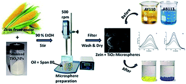
Natural protein polymers and metal oxide nanoparticles individually possess potential efficiency for dye binding and photocatalytic dye degradation, respectively. To attain these multifunctional properties, we have developed a composite zein–TiO2 microsphere, characterized it and evaluated its dye adsorption and photocatalytic efficiency. The phase structure, morphology, elemental composition and porous nature of the microspheres were characterized by using X-ray diffraction (XRD), Fourier transform-infra red (FTIR) spectroscopy, field emission-scanning electron microscopy (FE-SEM) with energy dispersive X-ray spectroscopy (EDAX), and nitrogen adsorption–desorption isotherm measurements. Adsorption experiments were performed by varying different parameters like contact time, initial dye concentration and adsorbent dosage, and the various adsorption isotherms and kinetics were tested. The results revealed that the Langmuir and Tempkin isotherms were more appropriate and followed a pseudo-first order model for the adsorption of acid yellow (AY110) and acid blue (AB113) dyes. The fluorescence probe method was used to confirm the generation of ˙OH radicals and elucidated the photodegradation mechanism. Therefore, metallo-polymer based adsorbents provide new scope in the selection of ideal adsorbents for efficient separation and remediation technologies in tannery effluent management.


Introduction

Dyes are widely used in textiles, leather, printing, pulp mills, plastics, and cosmetics industries. Owing to their high fastness and low foaming level, acid dyes are comprehensively used in leather dyeing, in spite of their resistance to decomposition, photodegradation and oxidizing agents. Therefore, the discharge of effluent from these industries poses a great threat to the aquatic environment by obstructing light penetration and disturbing biological metabolism.1 Due to the mutagenic potential of the aromatic amines of dyes, the metabolites are classified as cytotoxic as well as being human carcinogens.2 Several physical, chemical, biological and electrochemical processes have been employed for the treatment of dye effluent and most of them are inadequate. Toxic by-products and sludge generated from these techniques are difficult to dispose of. To overcome these limitations, unconventional methods were investigated and a number of studies have been reported.

The adsorption mode of dye decolourisation is found to be superior among the other treatment methods due to its low cost, flexibility in design and ease of operation.3 An ideal adsorbent should be biodegradable, inexpensive, and have high affinity for a wide range of dyes. Many adsorbents, such as activated carbon,4 clay,5 chitin,6 silica,7 fly ash,8 zeolites,9 coir pith10 and natural polymers,11 have been extensively studied for the removal of dyes from aqueous solutions. However, there is still a need to find more effective, economical and easily accessible ideal adsorbents. Zein, a by-product of the bio-ethanol industry, is widely used for its biodegradable and biocompatible properties.12 However, only a few reports are available for its use as an adsorbent for environmental cleanup. Different types of nano-structured materials, like titanium dioxide,13 bi-metallics,14 carbon nanotubes15 and magnetic nanoparticles,16 have been investigated for use in environmental treatments. Metal and metal oxide nanoparticles gained enormous significance owing to their unique properties in regard to sorption behaviours, magnetic activity, chemical reduction, and ligand sequestration, amongst others.17 The excellent chemical stability, non-toxicity and photo-catalytic activities of TiO2 nanoparticles have attracted more attention towards self-cleaning, anti-bacterial and UV protection applications. They have also proved to be useful in environmental applications, like organic waste and waste water treatment processes.18 These effects are mainly due to the formation of hydroxyl radicals (OH) and superoxide ion radicals (O2) in the presence of light.19 Further, surface modification20 or conjugation with a charge transfer catalyst, doping with metal and non-metal ions, blending with polymers or metal oxides and coupling with semiconductors preclude aggregation and result in a uniform distribution and enhanced photo-response and photocatalytic dye degradation efficiency.21 Therefore, attempts are continuously being made to develop a hybrid nano-composite with improved durability, robust mechanical strength, and excellent sorption characteristics for a multitude of applications.22–24 Hybrid nanocomposites, such as Ag and Ti doped BiOI,25 chitosan-ZnO,26,27 magnetic chitosan composite,28 AgNP-coated activated carbon,29 polyaniline–iron oxide composite,30 and ZnO polymeric hybrid beads,31 have been reported for dye adsorption. The combination of zein and TiO2, which individually possess excellent dye adsorption and degradation properties, would prove an efficient alternative as a composite material for environmental cleanup.

In this study, we have attempted to tailor zein–TiO2 microspheres as an adsorbent with high adsorption and photocatalytic dye degradation capacities by combining the advantages of inorganic metal oxides (large surface area, inertness, cheapness) and natural polymers (thermal stability, biodegradability). The effects of initial dye concentration, contact time, adsorbent dosage, dye removal efficiency and adsorption equilibrium, the isotherm and kinetics of dye adsorption and the photo-degradation mechanism reveal an ideal metallo-polymer composite for removing toxic dyes from tannery effluents.

Results and discussion

Physico-chemical characterization of the adsorbent

The X-ray diffraction analysis revealed the nature of the synthesized microspheres. The broad peak at 19.44° corresponds to the native zein structure32 and the diffraction angles at 25.15°, 37.38°, 47.99° and 62.92°, matching the planes of 101, 004, 200 and 204, confirmed the presence of the anatase phase of TiO2 nanoparticles in the zein microspheres (Fig. 1).
image file: c5ra00167f-f1.tif
Fig. 1 X-ray diffraction pattern of zein–TiO2 microspheres.

The immobilization of the TiO2 nanoparticles on the zein microspheres were confirmed by the FTIR analysis. The FTIR spectrum of the zein microspheres on their own (Fig. 2a) showed characteristic peaks at 1658 cm−1, 1537 cm−1, and 1238 cm−1 corresponding to amide I, II, and III, respectively. The contribution of TiO2 dominated the spectrum of the zein–TiO2 microspheres (Fig. 2b). The decrease in the bands related to primary amine groups at 1450–1650 cm−1, 3300–3500 cm−1, and 700–800 cm−1 revealed the immobilization of TiO2 on the surface of the zein microspheres. The peak located at 1088 cm−1, assigned to C–O stretching, shifted to a lower wavelength of 1038 cm−1. The surface modification of TiO2 nanoparticles by APTMS resulted in asymmetrical and symmetrical stretching vibrations of the C–H bonds of the methylene groups at 2928 and 2870 cm−1. Furthermore, the peak corresponding to the Si–O–Si bond was observed at around 1040 cm−1, indicating the condensation reaction between silanol groups. The peak of the Si–O–Si bond was much stronger, and the N–H bending vibration of the primary amine (NH2) was observed as a broad band in the region 1605–1560 cm−1. The appearance of these bands demonstrated that amine functional groups from the organosilane were grafted onto the modified particle surface.


image file: c5ra00167f-f2.tif
Fig. 2 FT-IR spectra of (a) zein microspheres, (b) zein–TiO2 microspheres and (c) silanized TiO2 nanoparticles.

The surface morphology and the elemental characterization at a specific position of the zein–TiO2 microspheres was evident from the FE-SEM and EDAX images. The effective dye adsorption can be attributed to the irregular and porous structure of the microspheres (Fig. 3a and b).


image file: c5ra00167f-f3.tif
Fig. 3 Field emission-scanning electron micrograph of zein–TiO2 microspheres (a) and EDAX analysis (b).

The nitrogen adsorption–desorption isotherm of the zein–TiO2 microspheres, shown in Fig. 4, belongs to the type IV profile according to the IUPAC classification, with a pronounced hysteresis for partial pressure P/P0 > 0.45. This is an indicative of the presence of a mesoporous structure and multilayer adsorption on the outer surface of the particles.33 The specific surface area of the prepared zein–TiO2 microspheres is 37.37 ± 0.457 m2 g−1, which was calculated using the BET method. According to the desorption isotherm, the average pore volume and pore diameter are 0.132 cm3 g−1 and 22 nm based on the BJH model.


image file: c5ra00167f-f4.tif
Fig. 4 Nitrogen adsorption–desorption isotherm (a), and pore size distribution curve of zein–TiO2 microspheres (b).

Effect of contact time

The UV-Vis spectra showed the effect of the microspheres on dye degradation in terms of the absorbance data over a wavelength range for two different dyes. The characteristic absorption peaks of AY110 and AB113 are at 420 and 565 nm, respectively. The greater the intensity of the peak, the lower the adsorption rate of the dye to the adsorbent. The increased degradation rate resulted in a lower absorbance reading. The peak became gradually smoother with increased contact time, signifying that adsorption and sufficient photocatalytic reaction had been achieved to remove the maximum amount of dye from the solution, which is clearly evident from Fig. 5a and b.
image file: c5ra00167f-f5.tif
Fig. 5 Effect of contact time on the removal of (a) A110 and (b) AB113 by zein–TiO2 microspheres.

The loading of TiO2 nanoparticles on the zein microspheres serves as an important factor for dye removal by affecting the number of active sites on the surface and leading to OH generation when exposed to light. Under UV or visible light irradiation, the TiO2-incorporated zein microspheres generate hydroxyl radicals on the surface, which initiates various oxidation reactions, thereby enhancing the degradation rate of dye pollutants. This has been elucidated in Fig. 6 using a fluorescence probe method. Hence, an increase in TiO2 loading resulted in excellent photo-catalytic activity and an enhanced dye degradation rate.


image file: c5ra00167f-f6.tif
Fig. 6 Fluorescence analysis of reactive oxygen species (hydroxyl radicals) generated on the surface of zein–TiO2 microspheres.

Effect of initial dye concentration

The effect of initial dye concentration and the percentage of dye removal are shown in Fig. 7a and b. The adsorption efficiencies for AY110 and AB113 were evaluated by determining the percentage decrease in the absorbance at 420 and 565 nm, respectively. It is apparent that the higher the initial dye concentration, the lower the percentage of dye adsorbed. The percentage dye removal efficiencies reached maximum levels of 96% and 89% at a lower concentration (10 mg L−1) and decreased to 77% and 61% at a higher concentration (100 mg L−1) of AY110 & AB113, respectively. Owing to the availability of free binding sites on the adsorbent, a low initial dye concentration leads to higher dye removal efficiency and a high concentration results in low adsorption efficiency due to the binding sites already being occupied.
image file: c5ra00167f-f7.tif
Fig. 7 Effect of initial dye concentration on dye removal of (a) AY110 and (b) AB113 by zein–TiO2 microspheres.

Effect of adsorbent dosage

The effect of zein–TiO2 microsphere dosage on dye removal was studied by adding dye solution with an initial concentration of 100 mg L−1 at room temperature with a constant stirring speed of 100 rpm. Different amounts of zein–TiO2 microspheres (0.5, 1, 1.5 and 2g L−1) were added to test the dye removal competency. After equilibrium, the solution samples were centrifuged and the concentration of dye in the supernatant was analyzed. The plot of dye removal (%) vs. adsorbent dosage (g L−1) is shown in Fig. 8.
image file: c5ra00167f-f8.tif
Fig. 8 Effect of adsorbent (zein–TiO2 microspheres) dosage on the adsorption of dyes.

Adsorption isotherms

The interaction behaviour of the adsorbate and the adsorbent can be expressed using several adsorption isotherm models. The data were analyzed using the following equilibrium adsorption isotherms: Langmuir, Freundlich and Tempkin isotherms. The Langmuir isotherm has been successfully applied to a number of sorption methods to explain the adsorption mechanism. The basic assumption is that sorption takes place at specific sites on the surface of the adsorbent where the adsorbate is strongly attracted.34 This can be expressed as follows:
image file: c5ra00167f-t1.tif
where Ce is the equilibrium concentration of the dye solution (mg L−1), Qe is the amount of dye adsorbed on the zein–TiO2 microspheres at equilibrium (mg g−1), KL is the Langmuir constant (L g−1) and Q0 is the maximum adsorption capacity (mg g−1).

The essential characteristic term of the Langmuir equation can be expressed by RL, the dimensionless separation factor which is represented as:

image file: c5ra00167f-t2.tif
where C0 is the initial dye concentration (mg L−1), and RL indicates the type of isotherm, whether it is favourable (0 < RL < 1), unfavourable (RL > 1), linear (RL = 1) or irreversible (RL = 0).

On the other hand, the Freundlich isotherm takes into account a heterogeneous system which is not restricted to monolayer adsorption.35 This can be expressed as:

image file: c5ra00167f-t3.tif
where KF is the adsorption capacity at unit concentration (mg L−1), and 1/n is the adsorption intensity.

The value of 1/n indicates the nature of the isotherm. If 1/n < 0 then the isotherm is irreversible, while it is more heterogeneous if 1/n = 0, has a favourable surface heterogeneity if (0 < 1/n < 1), and displays cooperative adsorption if 1/n > 1.

The Tempkin model assumes that the rate of adsorption in the layer decreases linearly with coverage due to adsorbent–adsorbate interactions and is characterized by a uniform distribution of binding energies, up to a maximum binding energy.36 The isotherm is given as follows:

Qe = B1[thin space (1/6-em)]ln[thin space (1/6-em)]KT + B1[thin space (1/6-em)]ln[thin space (1/6-em)]Ce
where KT is the equilibrium binding constant (L mol−1) corresponding to the maximum binding energy and B1 is a constant related to the heat of adsorption.

The correlation coefficients of the Langmuir, Freundlich and Tempkin isotherms are given in Table 1. The correlation coefficients imply that the Langmuir and Tempkin isotherms are more appropriate for the adsorption of AY110 and AB113 on zein–TiO2 microspheres. Moreover, the dimensionless constants RL (0.14 & 0.193) revealed that the adsorption is favourable and irreversible and KT (234 & 295) showed the maximum binding energies.

Table 1 Linearised isotherm coefficients
Langmuir isotherm Freundlich isotherm Tempkin isotherm
Q0 KL RL r12 KF n r22 KT B1 r32
AY110
25 0.057 0.14 0.9948 7.1343 1.15 0.94 234 48.78 0.96
AB113
12.04 0.041 0.193 0.9908 12.50 0.44 0.97 295 63.45 0.99


Adsorption kinetics

The mechanism of solute sorption onto a sorbent can be expressed using several adsorption kinetic models. The characteristic constants have to be determined in order to predict the rate at which a solute is removed from the aqueous solution using pseudo-first order37 and pseudo-second order38 kinetic models according to the following equations:
ln(QeQt) = ln[thin space (1/6-em)]Qek1t

image file: c5ra00167f-t4.tif
where Qe and Qt are the amount of dye adsorbed at equilibrium (mg g−1) and at time t (mg g−1), respectively, and k1 and k2 are the equilibrium rate constants for pseudo-first order kinetics (min−1) and pseudo-second order kinetics (g mg−1 min−1), respectively.

The adsorption kinetic plots are shown in Fig. 9 and 10 and the kinetic parameters are listed in Table 2. The calculated correlation coefficients are closer to unity for the pseudo-first order kinetic model than for the second order kinetics (0.99 vs. 0.94) and therefore the rate of sorption is pseudo-first order with good correlation.


image file: c5ra00167f-f9.tif
Fig. 9 Pseudo-first order kinetic model for adsorption of (a) AY110 & (b) AB113 on zein–TiO2 microspheres.

image file: c5ra00167f-f10.tif
Fig. 10 Pseudo-second order kinetic model for adsorption of (a) AY110 & (b) AB113 on zein–TiO2 microspheres.
Table 2 Kinetic constants for pseudo-first and pseudo-second order model
  Pseudo-first order Pseudo-second order
Qe k1 (min−1) R2 Qe k2 (g mg−1 min−1) R2
AY110 129.19 0.020 0.9905 13.36 0.0021 0.9464
AB113 120.8 0.021 0.9954 34.48 0.0009 0.9690


Intraparticle diffusion

The intraparticle diffusion model is used to determine the common functional relationship to the adsorption process, where uptake varies almost proportionally with t1/2 rather than with contact time t:
Qt = Kidt1/2 + C
where Kid is the intraparticle diffusion rate constant and C is the intercept. The intercept gives an idea of the thickness of the boundary layer, i.e. the larger the intercept, the greater the boundary layer effect.39 It also indicates that there is some degree of boundary layer control as it does not pass through the origin.40 The diffusion rate constants are shown in Table 3. The value of the intraparticle diffusion rate constant clearly showed that the boundary layer has a significant effect on the diffusion mechanism of dye uptake by the zein–TiO2 microspheres.
Table 3 Intraparticle diffusion rate constants
  Intra particle diffusion
Kid C r2
AY110 9.78 41.36 0.9828
AB113 7.25 28.54 0.9763


Experimental

Materials

Zein, (3-aminopropyl)trimethoxy silane (APTMS), and dimethyl sulfoxide (DMSO) were from Sigma-Aldrich. TiO2 nanoparticles, sesame oil, Span 80, and petroleum ether were used without any pre-treatment. The leather dyes, Acid Yellow (AY110) and Acid Blue (AB113) were obtained from the tannery department of CLRI and were used as model pollutants.

Surface modification of TiO2 nanoparticles

The titanium dioxide nanoparticles were synthesised using microbial methods as described by Babitha et al.41 The agglomeration of nanoparticles can be prevented by the surface functionalisation of TiO2 nanoparticles, which would further enhance the durability, dispersion and compatibility of the nanoparticles. The surface modification was carried out using a silane coupling agent, (3-aminopropyl)trimethoxysilane (APTMS), without disturbing the inherent properties of the nanoparticles.42 The synthesised TiO2 nanoparticles were dispersed in 50 ml dimethyl sulfoxide (DMSO) and ultra-sonicated for 1 hour. Then, the silane coupling agent (APTMS) was added to the dispersion and refluxing maintained for about 3 hours. The dispersed particles were separated out from the solvent by centrifugation (10 min at 10[thin space (1/6-em)]000 rpm). The excess silanes were removed by washing thrice with ethanol and water, alternatively. The pellet was re-dispersed in fresh solvent and sonicated for about 10 minutes to ensure uniform dispersion before centrifugation. The modified particles were dried in an oven at 60 °C overnight and cooled in a vacuum chamber for 1 h at room temperature.

Preparation of zein–TiO2 microspheres

The microspheres were prepared in accordance to the method of Karthikeyan et al.43 Solution blending is carried out to attain a molecular level of mixing. Directly, the silanized nanoparticles and polymer were mixed in solvent (90% ethanol) and kept under stirring for about 30 min. The solution was added to the continuous phase (sesame oil) containing 0.5% span 80 as an emulsifying agent. The mixture was then agitated using a homogenizer with a rotating speed of 500 rpm. The dispersed TiO2 nanoparticle and zein solution was immediately transformed into fine droplets, which was subsequently solidified into rigid microspheres due to solvent evaporation. The particles were finally collected by filtration, washed extensively with petroleum ether, and desiccated under vacuum for 24 hours.

Physico-chemical characterization of adsorbent

The powder X-ray diffraction (XRD) patterns were evaluated using an X-ray diffractometer with CuKα radiation (λ = 1.5405 Å) over a wide range of Bragg angles (10° ≤ 2θ ≥ 70°). The scanning was carried out at 0.02° min−1 with a time constant of 2 s.

Fourier transform infrared (Perkin Elmer – Spectrum One) spectra of silanized TiO2 nanoparticles and zein–TiO2 microspheres were obtained by mixing samples with potassium bromide, compressed into a pellet by applying 1 ton per unit. Spectral scanning was carried out over the range of 4000–400 cm−1.

The surface morphology of the zein–TiO2 microspheres was examined using scanning electron microscopy (SEM, Hitachi SU-6600). The microspheres were vacuum dried, mounted onto a brass stub, sputter coated with gold under an argon atmosphere and analysed at a voltage of 15 kV. The elemental composition of the microspheres at a specific position was analyzed using energy dispersive X-ray spectroscopy.

The nitrogen adsorption–desorption isotherms were measured at −196 °C using a Micromeritics ASAP 2020 analyzer. The sample was degassed prior to characterization. The specific surface area was calculated according to the BET44 (Brunauer–Emmet–Teller) model, whereas the pore size and average pore volume were calculated using the BJH45 (Barrett–Joyner–Halenda) formula based on the desorption data of the isotherms.

Fluorescence was employed to detect the reactive oxygen species (mainly ˙OH radicals) using 2,7-dichlorodihydrofluorescein (DCFH) as a fluorescence probe molecule.46 The probe solution was prepared by dispersing 0.5 ml of 1 mM DCFH-DA solution in ethanol in 2 ml of 0.01 M aqueous NaOH solution and allowing it to stand in the dark for 30 min to generate DCFH. Then the above mixture was diluted to 5 μM with 0.1 M PBS buffer to obtain the final probe solution. The zein–TiO2 microspheres were dispersed in the above solution to form a 50 μg ml−1 suspension. The suspension was then exposed to UV-Visible light irradiation for 20 min. After irradiation, the fluorescence was observed using a Leica fluorescence microscope with the excitation wavelength at 488 nm.47

Adsorption studies

The effects of contact time, initial dye concentration and adsorbent dosage for the removal of acid dyes, AY110 and AB113, from aqueous solutions were evaluated. The dye adsorption measurements were conducted by mixing an optimum amount of zein–TiO2 microspheres (1 mg ml−1) in varied concentrations (10, 50 and 100 mg L−1) of dye solution, with an agitation speed of 100 rpm. The changes in the absorbance of all samples were monitored and determined at different time intervals during the adsorption process. At the end of the experiments, the solution samples were centrifuged and the dye concentrations were measured using a UV-Visible spectrophotometer at 420 nm and 565 nm, respectively. The amount of adsorbed dye, Q (mg g−1), was calculated according to
image file: c5ra00167f-t5.tif
where C0 is the initial concentration (mg L−1), Ce is the equilibrium concentration (mg L−1), V is the volume of dye solution (L) and W is the weight (g) of the adsorbent.
image file: c5ra00167f-t6.tif

The dye removal efficiencies under different conditions were also calculated from the difference between the initial and equilibrium concentrations of the solution. The results were verified using the adsorption isotherms (Langmuir, Freundlich and Tempkin) and kinetics (pseudo first order, pseudo second order and intraparticle diffusion model).

Conclusions

Biocompatible and biodegradable zein–TiO2 microspheres were synthesized and characterized and their dye adsorption properties were investigated. The XRD, FTIR, FE-SEM and EDAX results confirmed the presence of anatase TiO2 NPs on the zein microspheres. The porosity measurements using the N2 adsorption–desorption technique revealed the pore size distribution and the mesoporous nature of the material. Adsorption isotherms and kinetic studies were performed and the results showed that the equilibrium data followed the Langmuir and Tempkin isotherms for AY110 and AB113, respectively. The adsorption kinetics followed the pseudo-first-order model. Being a low-cost and eco-friendly adsorbent with the combined advantages of TiO2 nanoparticles and the agro-industrial by-product zein, the zein–TiO2 microspheres showed maximum adsorption and photocatalytic dye degradation efficiency. The degradation mechanism is also elucidated using the fluorescence probe method. These promising results showed that the microspheres could be used as an effective alternative for the removal of acid dyes from aqueous solutions. Therefore, this study opens up new methods for the efficient treatment of tannery effluent to reduce dye toxicity.

Acknowledgements

The authors thank the Director, CSIR – CLRI for his constant support. The authors also thank Dr NK Chandra Babu, Chief Scientist, Leather Processing Department, CSIR – CLRI for providing the dye samples. The first author gratefully acknowledges the DST – INSPIRE, New Delhi for the fellowship (IF110583).

References

  1. V. K. Gupta and Suhas, J. Environ. Manage., 2009, 90, 2313–2342 CrossRef CAS PubMed.
  2. N. Mathur, P. Bhatnagar and P. Sharma, Univers. J. Environ. Res. Technol., 2012, 2, 1–18 CAS.
  3. G. Crini, Bioresour. Technol., 2006, 97, 1061–1085 CrossRef CAS PubMed.
  4. V. Meshko, L. Markovska, M. Mincheva and A. Rodrigues, Water Res., 2001, 35, 3357–3366 CrossRef CAS.
  5. G. Atun, G. Hisarli, W. S. Sheldrick and M. Muhler, J. Colloid Interface Sci., 2003, 261, 32–39 CrossRef CAS.
  6. S. A. Figueiredo, J. M. Loureiro and R. A. Boaventura, Water Res., 2005, 39, 4142–4152 CrossRef CAS PubMed.
  7. Y. Badr, M. G. Abd El-Wahed and M. A. Mahmoud, J. Hazard. Mater., 2008, 154, 245–253 CrossRef CAS PubMed.
  8. P. Janos, H. Buchtová and M. Rýznarová, Water Res., 2003, 37, 4938–4944 CrossRef CAS PubMed.
  9. S. Babel and T. A. Kurniawan, J. Hazard. Mater., 2003, 97, 219–243 Search PubMed.
  10. C. Namasivayam and D. Kavitha, Dyes Pigm., 2002, 54, 47–58 CrossRef CAS.
  11. G. Z. Kyzas, M. Kostoglou, N. K. Lazaridis and D. N. Bikiaris, in Eco-Friendly Textile Dyeing and Finishing, 2013, pp. 177–206 Search PubMed.
  12. A. R. Patel and K. P. Velikov, Curr. Opin. Colloid Interface Sci., 2014, 19, 450–458 CrossRef CAS PubMed.
  13. C. C. Chen, C. S. Lu, Y. C. Chung and J. L. Jan, J. Hazard. Mater., 2007, 141, 520–528 CrossRef CAS PubMed.
  14. P. Wilhelm and D. Stephan, J. Photochem. Photobiol., A, 2007, 185, 19–25 CrossRef CAS PubMed.
  15. T. A. Saleh, in Synthesis and Applications of Carbon Nanotubes and Their Composites, 2013, pp. 479–493 Search PubMed.
  16. N. M. Mahmoodi, J. Abdi and D. Bastani, J. Environ. Sci. Health, Part A: Toxic/Hazard. Subst. Environ. Eng., 2014, 12, 96 Search PubMed.
  17. J. Zhang, Z. Xiong and X. S. Zhao, J. Mater. Chem., 2011, 21, 3634 RSC.
  18. W.-C. Lin, W.-D. Yang and S.-Y. Jheng, J. Taiwan Inst. Chem. Eng., 2012, 43, 269–274 CrossRef CAS PubMed.
  19. J. Zhang and Y. Nosaka, J. Phys. Chem. C, 2014, 118, 10824–10832 CAS.
  20. B. Gradzik, M. E. L. Fray, E. Wiśniewska and W. Pomeranian, Chemik, 2011, 2, 4–6 Search PubMed.
  21. H. Arora, C. Doty, Y. Yuan, J. Boyle, K. Petras and B. Rabatic, Titanium Dioxide Nanocomposites, 2010, vol. 8 Search PubMed.
  22. S. Sarkar, E. Guibal, F. Quignard and A. K. SenGupta, J. Nanopart. Res., 2012, 14, 715 CrossRef.
  23. C. Shen, S. Song, L. Zang, X. Kang, Y. Wen, W. Liu and L. Fu, J. Hazard. Mater., 2010, 177, 560–566 CrossRef CAS PubMed.
  24. G. Zaccariello, E. Moretti, L. Storaro, P. Riello, P. Canton, V. Gombac, T. Montini, E. Rodríguez-Castellón and A. Benedetti, RSC Adv., 2014, 4, 37826 RSC.
  25. Y. S. Yohan Park, Y. Na, D. Pradhan and B.-K. Min, CrystEngComm, 2014, 16, 3155–3167 RSC.
  26. R. Salehi, M. Arami, N. M. Mahmoodi, H. Bahrami and S. Khorramfar, Colloids Surf., B, 2010, 80, 86–93 CrossRef CAS PubMed.
  27. W. S. Wan Ngah, L. C. Teong and M. A. K. M. Hanafiah, Carbohydr. Polym., 2011, 83, 1446–1456 CrossRef CAS PubMed.
  28. L. W. Peng Wang and T. Yan, BioResources, 2013, 8, 6026–6043 Search PubMed.
  29. J. Pal, M. K. Deb, D. K. Deshmukh and D. Verma, Appl. Water Sci., 2013, 3, 367–374 CrossRef CAS PubMed.
  30. S. A. Jebreil, Int. J. Chem. Nucl. Metall. Mater. Eng., 2014, 8, 1336–1341 Search PubMed.
  31. H. Shokry Hassan, M. F. Elkady, A. H. El-Shazly and H. S. Bamufleh, J. Nanomater., 2014, 2014, 1–14 CrossRef PubMed.
  32. H. Lai, P. H. Geil and G. W. Padua, J. Appl. Polym. Sci., 1998, 71, 1267–1281 CrossRef.
  33. Y. Zhang, S. Zhang, K. Wang, F. Ding and J. Wu, J. Nanomater., 2013, 2013, 1–7 Search PubMed.
  34. I. Langmuir, J. Am. Chem. Soc., 1916, 252, 2221–2295 CrossRef.
  35. M. B. Desta, J. Thermodyn., 2013, 1–6 CrossRef PubMed.
  36. Y. Kim, C. Kim, I. Choi, S. Rengaraj and J. Yi, Environ. Sci. Technol., 2004, 38, 924–931 CrossRef CAS.
  37. C. Lee, H. Kim, I. Jang, J. Im and N. Park, ACS Appl. Mater. Interfaces, 2011, 3, 1953–1957 CAS.
  38. G. M. Y. S. Ho, Chem. Eng. J., 1998, 70, 115–124 CrossRef.
  39. A. Ozcan and A. S. Ozcan, J. Hazard. Mater., 2005, 125, 252–259 CrossRef PubMed.
  40. W. Plazinski and W. Rudzinski, J. Phys. Chem. C, 2009, 113, 12495–12501 CAS.
  41. S. Babitha and P. S. Korrapati, Mater. Res. Bull., 2013, 48, 4738–4742 CrossRef CAS PubMed.
  42. J. Zhao, M. Milanova, M. M. C. G. Warmoeskerken and V. Dutschk, Colloids Surf., A, 2012, 413, 273–279 CrossRef CAS PubMed.
  43. K. Karthikeyan, R. Lakra, R. Rajaram and P. S. Korrapati, AAPS PharmSciTech, 2012, 13, 143–149 CrossRef CAS PubMed.
  44. S. Brauner, P. H. Emmett and E. Teller, J. Am. Chem. Soc., 1938, 60, 309–319 CrossRef.
  45. E. P. Barrett, L. G. Joyner and P. P. Halenda, J. Am. Chem. Soc., 1951, 73, 373–380 CrossRef CAS.
  46. A. Gomes, E. Fernandes and J. L. F. C. Lima, J. Biochem. Biophys. Methods, 2005, 65, 45–80 CrossRef CAS PubMed.
  47. L. Sun, Y. Qin, Q. Cao, B. Hu, Z. Huang, L. Ye and X. Tang, Chem. Commun., 2011, 47, 12628–12630 RSC.

This journal is © The Royal Society of Chemistry 2015
Click here to see how this site uses Cookies. View our privacy policy here.