Determination of benzoylurea insecticides in environmental water and honey samples using ionic-liquid-mingled air-assisted liquid–liquid microextraction based on solidification of floating organic droplets

Miyi Yang, Xuefei Xi, Xiaoling Yang, Lizhen Bai, Runhua Lu, Wenfeng Zhou, Sanbing Zhang and Haixiang Gao*
Department of Applied Chemistry, China Agricultural University, Yuanmingyuan West Road 2#, Haidian District, Beijing 100193, China. E-mail: hxgao@cau.edu.cn; Fax: +86 010 62731991; Tel: +86 010 62731991

Received 5th January 2015 , Accepted 5th March 2015

First published on 5th March 2015


Abstract

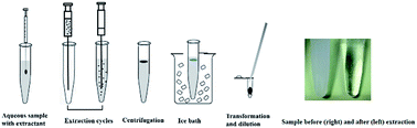
A novel and simple ionic-liquid-mingled air-assisted liquid–liquid microextraction based on solidification of floating organic droplets combined with high performance liquid chromatography was developed for the determination of six benzoylureas (BUs) in water and honey samples. In this method, a mixture of low-density and low melting point extraction solvents and aqueous sample solutions was rapidly sucked up and injected several times using a glass syringe. The influence of the main factors on the efficiency of this procedure is studied. Under the optimal conditions, the enrichment factors (EFs) for BUs were acquired in the range of 144 to 187, limits of detection (LODs) were between 0.01 and 0.1 μg L−1 and limits of quantitation (LOQs) were changed in the range of 0.03 and 0.33 μg L−1. The obtained extraction recoveries ranged from 84.03% to 109.20% with intra-day lower than 4.5%, and inter-day precision lower than 6.5%. The method is successfully applied to determine the BUs in environmental water and honey samples with recoveries in the range of 78.57–109.72%, which proved the potential use of this method in real samples.


Introduction

Benzoylureas (BUs) were introduced by Bayer of Germany in 1978.1 In agricultural production, BUs are widely applied to many crops to control numerous pest species2 and they are usually used as insect growth regulators that interfere with chitin synthesis in the target pests and cause their death.3,4 The direct toxicity of BUs is very low to mammals because they do not synthesize chitin.5 However, according to several organizations and institutions, such as the European Food Safety Authority (EFSA),6,7 the European Chemical Agency (ECHA),8 the US Environmental Protection Agency (USEPA),9 and the WHO,10 BUs are toxic to several organisms and the insecticides present the risk of contamination of natural products and waters due to their persistence in the environment.11 Honey is a valuable natural food product. It is widely used as a natural sweetener and food additive due to its significant nutritional and quality properties.12 During production and harvesting, honey may be contaminated with pesticides applied in agriculture and forestry and distributed in the environment by being carried to the hive on bee bodies or by foragers.13 Pesticide residues cause chronic exposure and long-term toxicity effects throughout the food chain.14 According to European Union (EU) regulations, the residues of BUs in honey must be less than the maximum residue limits (MRLs) (the default value is 0.01 mg kg−1).15 Therefore, it is of paramount importance to determine the presence of BUs at trace levels in both water and honey using a sensitive, green, rapid analytical method.

Extraction and preconcentration procedures are critical steps in the determination of BUs by gas chromatography (GC),16 high-performance liquid chromatography (HPLC),3,14,17,18 GC-mass spectrometry (GC-MS),19 and HPLC-triple quadrupole mass spectrometry (HPLC-MS/MS).20 Conventional sample preparation techniques, such as liquid–liquid extraction (LLE),21 solid–phase extraction (SPE),22 single drop microextraction (SDME),23,24 dispersive solid–phase microextraction (DSPME),25,26 solid–phase microextraction (SPME),27 and dispersive liquid–liquid microextraction (DLLME),28 have been developed for residue analysis.

LLE and SPE usually require large amounts of sample and organic solvents and are time-consuming and expensive, and the materials used in the experiments are difficult to recycle.29 SDME is a simple and inexpensive sample preparation method that can be applied to liquid, gaseous and solid samples.30,31 However, disadvantages of this technique include the difficulty of maintaining a stable organic drop, the ready formation of air bubbles, and the length of time required to reach equilibrium.32 DSPME and SPME are widely used sample preparation methods. These methods are simple and solvent-free and preconcentrate analytes with high enrichment.29,33–35 However, most commercial extraction sorbents are relatively expensive, fragile, have restricted lifetimes and are limited in terms of available polarities, which reduces the selectivity of the extraction process.36 As to the self-restraint sorbents, the preparation process is intricate.37 DLLME is a useful and efficient liquid phase microextraction method that was first applied by Berijani et al. to determine 13 OPPs in river water, well water and farm water.38 In DLLME, analytes are extracted using a dispersion of the extraction solvent in an aqueous sample.39 This technology is generally based on a ternary component solvent system. Because the extraction and disperser solvents are immiscible, a cloudy solution is formed upon injection of the solvents into an aqueous sample. Extraction equilibrium is achieved quickly because the surface contact between the droplets of the extraction solvent and the aqueous sample is high. Extraction solvents with densities greater than water, such as carbon tetrachloride and tetrachloroethylene, will sediment at the bottom of the tube after centrifugation and can be removed with a microsyringe before instrument detection.40 The advantages of this technique are obvious and include easy operation, speed, low cost, high recovery and high enrichment.41 However, the large volume of dispersive solvent consumption is environmentally unfriendly, and the polarities of the dispersive solvents are lower than that of the aqueous solution, resulting in incomplete analyte removal and reduced extraction efficiency.42

Besides the use of disperser solvents, disadvantages of the traditional DLLME procedure include (i) the requirement that the extraction solvent have a density greater than that of water to permit simple separation of the extraction phase after centrifugation; (ii) the use of hazardous solvents, such as halogenated hydrocarbons, in the vast majority of examples; and (iii) the requirement that the supernatant be removed to obtain the extraction phase.

Several disperser-solvent-free techniques have been developed to address these problems in DLLME, such as ultrasound-assisted emulsification microextraction (USAEME), which was introduced by Regueiro in 2008;43 vortex-assisted liquid–liquid microextraction (VALLME), which was developed by Yiantzi;44 and air-assisted liquid–liquid microextraction (AALLME), which was first presented by Farajzadeh in 2012.42 USAEME and VALLME require additional energy from the ultrasound and vortexing. In addition, the cloudy solution in these two method is not as easily formed as in DLLME.45 In most AALLME, a small volume of extraction solvent is transferred into the aqueous solution, and the mixture is then repeatedly withdrawn into a syringe and injected into a tube. The analytes exchange into the extraction solvent via the bolus flow formed during the process of aqueous sample withdrawal and ejection.

And new techniques based on may overcome the above difficulties on extraction solvents.46,47 These methods use an extraction solvent with a density lower than that of water and with a melting point near or below room temperature (10–30 °C). After extraction, the solvent can be solidified by exposure to low temperatures, facilitating its removal as a droplet of floating solvent by centrifugation prior to analysis.

The most commonly used extraction solvents for the microextraction based on solidification of floating organic droplets such as liquid phase microextraction based on solidification of floating organic droplets (LPME-SFO) and dispersive liquid–liquid microextraction based on solidification of floating organic droplets (DLLME-SFO) are long-chain alcohols or halogenated solvents.48–50 While, in this work, trihexyl (tetradecyl) phosphonium hexafluorophosphate ([P14,6,6,6]PF6) was used for extracting. The [P14,6,6,6]PF6 is an ionic liquid (IL) with low density (ρ = 1.013 kg m−3) and a low melting point (39.5 °C). In addition, during the extraction process, ILs can function not only as an extractant but also as a surfactant to reduce the interfacial tension between two immiscible phases by adsorption at the liquid–liquid interface.51,52 However, [P14,6,6,6]PF6 is not suitable as the sole extractant because its density is very close to that of water, which makes it difficult to separate from aqueous samples. In addition, the viscosity of [P14,6,6,6]PF6 is quite high, causing it to adhere to the tube instead of forming a cloudy solution. The combined use of an organic solvent and [P14,6,6,6]PF6 could avoid these problems and increase analyte recovery. Therefore, a new method named IL-mingled air-assisted liquid–liquid microextraction based on solidification of floating organic droplets (ILAALLME-SFOD) was first suggested by our group as an improvement of the common AALLME technique.

In this study, for the first time, a novel, highly efficient, and environmentally friendly technique, ILAALLME-SFOD is presented to determine six BUs in environmental water and honey combined with HPLC. The effects of some experimental parameters, including the type of extraction solvent, volume of the extraction solvent, extraction cycles and salt addition were investigated and optimized. Finally, the optimized method was applied to real samples.

Experimental

Reagents and materials

Several insecticide standards (diflubenzuron, hexaflumuron, flufenoxuron, lufenuron, chlorfluazuron, diafenthiuron) with purities ranging from 97% to 98% were obtained from the Agricultural Environmental Protection Institution (Tianjin, China). 1-Dodecanol was purchased from Ouhe Technology (Beijing, China). Trihexyl (tetradecyl) phosphonium hexafluorophosphate ([P14,6,6,6]PF6) was purchased from the Center for Green Chemistry and Catalysis, LICP, CAS (Lanzhou, China). HPLC-grade methanol and acetonitrile were obtained from Dikma Technologies (Lake Forest, CA, USA). Analytical-grade sodium chloride (NaCl) was purchased from Beijing Chemical Factory (Beijing, China). Deionized water was purified using a Milli-Q SP Reagent Water System (Millipore, Bedford, MA, USA). Stock standard solutions were prepared in acetonitrile at 100 mg L−1 and stored in the dark at 4 °C. Working standard solutions were prepared by diluting the stock standard solutions to various concentrations in acetonitrile. Solutions of 0.1 mol L−1 sodium hydroxide and concentrated hydrochloric acid were used to adjust the pH of the samples. Glassware was thoroughly cleaned by soaking in nitric acid (10%, v/v) for at least 24 h, followed by rinsing with ultra-high-purity deionized water.

Instrumental and analytical conditions

HPLC-UV analysis was performed using an Agilent 1200 series high-performance liquid chromatograph (CT, USA) equipped with a binary high-pressure pump, a column oven, an auto-sampler and a variable-wavelength detector (VWD). Agilent Chem-Station software was used for the HPLC-UV system operation and data analysis. The separations were performed on Spursil C18 columns (5 μm, 4.6 × 250 mm, Dikma Limited) with Spursil C18 Guard Cartridges (5 μm, 2.1 × 10 mm, Dikma Limited) using an acetonitrile–water (75[thin space (1/6-em)]:[thin space (1/6-em)]25, v/v) solution mixture as the mobile phase. The flow rate was set as 1 mL min−1, and the column temperature was maintained at 25 °C. The UV absorbance of the samples was measured at 254 nm, and the sample injection volume was 10 μL. The analytes were weighed using a Mettler-Toledo AL104 electronic balance (Shanghai, China). A low-speed, refrigerated centrifuge (Baiyang 52A, Baoding, China) and 0.22 μm micropore membranes (Agla, USA) were used for sample treatment. To perform the microextraction procedure, a 10 mL homemade glass tube, 10 mL Pasteur pipette, 10 mL glass syringe, syringe needle (0.9 × 100 mm), 100 μL microsyringe (Anting, Shanghai, China) and ultrasonication equipment (KQ3200DE, Kunshan, China) were used.

Preparation of the real samples

The water samples included river water (Xiaoyuehe, Haidian District, Beijing, China) and underground water (Cold spring village, Haidian District, Beijing, China). Before analysis, a 50.0 mL water sample spiked to a target concentration was centrifuged at 4000 rpm for 10 min, followed by filtration through a 0.22 μm membrane.

Honey samples (milk vetch honey and acacia honey) were purchased from a local market (Beijing, China). For sample preparation, 10 g honey samples were diluted with 100 mL of ultrapure water and spiked with a standard solution of pesticides. The solution was then centrifuged at 4000 rpm for 10 min, and the supernatant was filtered through a 0.22 μm membrane. Blank samples were prepared in the same manner but were not spiked with compound. All samples were stored at 4 °C until use.

ILAALLME-SFOD procedure

First, 6 mL aliquots of the aqueous samples spiked with different concentrations of BUs were placed in 10 mL glass tubes with conical bottoms. Then, 30 μL of 1-dodecanol and 10 μL of [P14,6,6,6]PF6 were sequentially added to the aqueous samples as extraction solvents using microsyringe. The mixture solvent was transparent and suspended on the surface of sample solution. Then, the mixture was rapidly withdrawn using a 10 mL glass syringe and ejected into the tube 10 times using the same syringe. All operations were performed in a 45 °C water bath. After centrifugation of the mixture at 4500 rpm for 12 min, the glass test tube was placed in an ice bath for 5 min. The solidified extraction solvent floating on the solution surface was collected into a 0.5 mL Eppendorf tube using manicured straws and then diluted with 40 μL of ethanol. Next, 10 μL of the solvent was auto-injected into the HPLC system for analysis. The ILAALLME-SFOD procedure is shown in Fig. 1.
image file: c5ra00140d-f1.tif
Fig. 1 The scheme of microextraction.

Calculation

The experimental parameters affecting the microextraction procedure were investigated. The performances are expressed using the enrichment factors (EFs) and the extraction recovery (R%), which can be calculated as follows:
 
image file: c5ra00140d-t1.tif(1)
 
image file: c5ra00140d-t2.tif(2)
where Cc and C0 are the analyte concentration in the final collected organic extract and the initial analyte concentration in the sample aqueous phase, respectively, and Vc and Vaq are the volumes of the organic phase and sample solution. The standard calibration curve for each insecticide was obtained by fitting the concentrations of each pesticide to the HPLC peak areas using a linear regression model, and various Cc values in the samples were calculated based on the standard calibration curves.

Results and discussion

Optimization of ILAALLME-SFOD

Selection of the extraction device. A critical aspect of AALLME is the dispersal of the organic extraction solvents into aqueous and muddy samples via airflow to enhance the contact area of the analytes and extractant. Pasteur pipets and syringes similarly enable sample mixing with the extraction solvent via air exchange. We compared the extraction ability of these two devices (Fig. S1). The sample volume was 6 mL, which is the maximum volume of the 10 mL Pasteur pipet (A) can hold. The withdrawal and injection mixture produced by B was more turbid than that of A, indicating that B enables superior exchange of the sample solution and extraction solvent.
Effect of extraction solvent type and volume. The extraction solvent for this method must have certain properties, including low density, low toxicity, low volatility, low water solubility, good chromatographic behavior and a low melting point (m.p.) near room temperature (in the range of 10–30 °C). In our study, 1-bromohexadecane (m.p. = 17–18 °C) and 1-dodecanol (m.p. = 23–25 °C) were evaluated for the extraction of BUs. Higher extraction efficiency and easier solidification were obtained with the use of 1-dodecanol, which was therefore selected as the extraction solvent.

To examine the effect of the extraction solvent volume on ILAALLME-SFOD performance, different volumes of 1-dodecanol (20–60 μL) were studied. As shown in Fig. 2, the recovery for the six BUs was maximal at an extraction volume of 30 μL. However, for the majority of analytes, recovery decreased as the volume of 1-dodecanol increased further from 40 to 60 μL. The volume of the collected organic phase increased with increasing extraction solvent volume, leading to a decrease in the peak area and the EFs. Therefore, 30 μL of 1-dodecanol was used in the subsequent experiments.


image file: c5ra00140d-f2.tif
Fig. 2 Effect of extraction solvent volume on extraction efficiency. Extraction conditions: analyte concentration, 50 μg L−1; IL volume, 10 μL; extraction temperature, 40 °C; number of extraction cycles, 10; centrifugation time, 15 min; centrifugation speed, 4500 rpm; no salt. The error bars correspond to the relative standard deviation of the mean recovery for n = 3 replicates.
Effect of the addition of IL. IL addition is an important extraction parameter. Due to their outstanding properties, ILs have been popular in analytical chemistry not only as direct extractant53 but also as cosolvent54–56 to improve extraction ability. The ILs most commonly used in conventional microextraction are imidazolium ILs, which have a density greater than water and a high melting point. However, [P14,6,6,6]PF6 with low density and room temperature melting point is suitable for ILAALLME-SFOD. Different volumes of [P14,6,6,6]PF6 were investigated, from 0 to 24 μL with 30 μL 1-dodecanol adding. The results are shown in Fig. 3. The recoveries of all analytes increased significantly as the IL volume increased up to 10 μL. When IL volumes greater than 10 μL, the recoveries of hexaflumuron, diafenthiuron and diflubenzuron remained nearly unchanged, while the recoveries of flufenoxuron, chlorfluazuron and lufenuron decreased. The results show that when the volume ratio of IL and 1-dodecanol was 1[thin space (1/6-em)]:[thin space (1/6-em)]3, the mixture extractant will achieve a satisfactory extraction performance which owe to the combined effect of those two extraction solvents. Therefore, 10 μL of IL was selected for use in subsequent studies.
image file: c5ra00140d-f3.tif
Fig. 3 Effect of IL volume on extraction efficiency. Extraction conditions: analyte concentration, 50 μg L−1; 1-dodecanol volume, 30 μL; extraction temperature, 40 °C; number of extraction cycles, 10; centrifugation time, 15 min; centrifugation speed, 4000 rpm; no salt. The error bars correspond to the relative standard deviation of the mean recovery for n = 3 replicates.
Effect of temperature. Temperature is a vital factor in the extraction process because high temperature can accelerate the mass transfer rate and increase the contact area between target analytes and aqueous solution.57 A temperature range of 30 to 50 °C was investigated, and the results are shown in Fig. 4. The maximum extraction efficiency was obtained at 45 °C; the extraction efficiency decreased at higher temperatures, most likely because higher temperatures enhance the movement of analytes, including transfer into and migration out of the extraction phase. Thus, a temperature of 45 °C was used in subsequent experiments.
image file: c5ra00140d-f4.tif
Fig. 4 Effect of temperature on extraction efficiency. Extraction conditions: analyte concentration, 50 μg L−1; 1-dodecanol volume, 30 μL; IL volume, 10 μL; number of extraction cycles, 10; centrifugation time, 15 min; centrifugation speed, 4000 rpm; no salt. The error bars correspond to the relative standard deviation of the mean recovery for n = 3 replicates.
Effect of centrifugation. Centrifugation is a useful process that rapidly separates extractant droplets from the aqueous phase. To optimize the centrifugation speed and time, centrifugation time of 6–21 min and speed of 3000–5000 rpm were evaluated. As shown in Fig. S2, when the centrifugation speed was increased from 3000 to 4500 rpm, the extraction efficiencies of the BUs increased dramatically. When the centrifugation was increased further to 5000 rpm, the extraction efficiencies of the target analytes did not change. The amount of the collected organic phase increased with increasing centrifugation speed, and the maximum volume of extraction droplets was obtained at a centrifugation speed of 4500 rpm. Centrifugation time ranging from 5 to 30 min were evaluated (Fig. S3) to determine the influence of centrifugation time on the recovery efficiency for mass transfer between the two phases in the extraction, which is time-dependent. The extraction performance of the BUs slightly increased with time up to 12 min and remained constant or slightly decreased at longer time. Therefore, centrifugation at 4500 rpm for 12 min was selected for this extraction method.
Effect of salt addition. The salting out effect is an important parameter in microextraction because salt addition decreases the solubility of analytes in aqueous samples and increases their partitioning into extraction solvents. In this study, the effect of ionic strength on extraction performance was evaluated by adding different concentrations of NaCl [0–10% (m/v)], as shown in Fig. 5. As the ionic strength was increased from 0% to 2%, the recovery efficiencies of the BUs decreased, with the exception of hexaflumuron, the recovery of which remained nearly unchanged. In addition, as the NaCl concentration in the sample solution increased, the recoveries of all BUs decreased, which may be attributable to the increase in viscosity induced by the addition of salt. Increased viscosity would reduce the transport of the BUs from the aqueous solution to the extraction solvent. Therefore, no salt was added to ensure the extraction performance of the proposed method.
image file: c5ra00140d-f5.tif
Fig. 5 Effect of salt addition on extraction efficiency. Extraction conditions: analyte concentration, 50 μg L−1; 1-dodecanol volume, 30 μL; IL volume, 10 μL; extraction temperature, 45 °C; number of extraction cycles, 10; centrifugation time, 12 min; centrifugation speed, 4500 rpm. The error bars correspond to the relative standard deviation of the mean recovery for n = 3 replicates.
Effect of ultrasonication time and number of extraction cycles. In this study, the extraction process involved the mixing of the extraction solvent and the sample solution by rapid suction into a 10 mL glass syringe, followed by injection into a glass test tube. The number of suction–injection cycles was defined as the “number of extraction cycles”. Ultrasound is also commonly used to increase the recovery performance of microextraction.58 To clarify the effect of ultrasonication and the number of extraction cycles on the extraction efficiency, a series of experiments were performed with ultrasonication time of 0, 0.5, 1, 2, 3 and 4 min and extraction cycles of 4, 6, 8, 10, 12 and 14. For all 25 (5 × 5) treatments, ultrasonication was performed during the extraction. Fig. 6 shows the total recovery of the BUs with different ultrasonication times and extraction cycles. Appropriate ultrasonication clearly improved recovery when 4 and 6 extraction cycles were used. However, for 8 or more extraction cycles, ultrasonication was ineffective and even reduced the total recovery of BUs. Therefore, no ultrasonication was performed in subsequent experiments. In Fig. S4, the recoveries of the BUs with 4–14 extraction cycles were studied. Recovery increased up to 10 extraction cycles and slightly decreased as the number of extraction cycles increased further, which is likely attributable to significant vaporization of the extraction solvent. Therefore, the number of extraction cycles was set at 10.
image file: c5ra00140d-f6.tif
Fig. 6 Effect of ultrasonication time and number of extraction cycles on the total extraction efficiency. Extraction conditions: analyte concentration, 50 μg L−1; 1-dodecanol volume, 30 μL; IL volume, 10 μL; extraction temperature, 45 °C; centrifugation time, 12 min; centrifugation speed, 4500 rpm.
Effect of sample pH. The pH value of sample solution is another factor that should be considered because of the effect of this parameter on sample preconcentration and cleanup. The pH value of the solution will affect the existing form of the analytes, and the possible extraction efficiency of the target analytes can be determined. Fig. 7 demonstrates the effects of the pH value on the extraction performance in the range of pH 3–11. The pH value of the water in our experiment was 7.2 which is close to neutral. Obviously, the best extraction recoveries of BU pesticides were achieved when sample was neutral. When pH of the sample was below 7, the extraction efficiencies were acceptable. However, lower extraction efficiencies were achieved when the sample was alkaline. According to Gil-Garcia's report,59 the reason for this result may be that the amido bonds in the molecular structures will breakdown through hydrolysis, which makes the BUs unstable in strong acidic and alkaline environments. Therefore, the pH value was not need additional control in the following experiments.
image file: c5ra00140d-f7.tif
Fig. 7 Effect of sample pH. Extraction conditions: analyte concentration, 50 μg L−1; 1-dodecanol volume, 30 μL; IL volume, 10 μL; extraction temperature, 45 °C; centrifugation time, 12 min; number of extraction cycles, 10; centrifugation speed, 4500 rpm. The error bars correspond to the relative standard deviation of the mean recovery for n = 3 replicates.

Method validation

Efficient separation and high recoveries were achieved under the optimized conditions. For analytical methods, the limit of detection (LOD), the limit of quantification (LOQ), precision and linear range are important parameters. A series of experiments were designed to evaluate these factors. Ultrapure water spiked with working standard solutions at concentration levels in the range of 0.1–500 μg L−1 were used to prepare working calibration curves. The characteristic calibration data are summarized in Table 1. Linearity was observed in the range of 0.5–500 μg L−1 for all BUs, with correlation coefficients (R2) greater than 0.996. The LOD is defined as the lowest detectable concentration with an S/N = 3 for LOQ is calculated as the concentration giving an S/N = 10. The precision was studied as the percent of relative standard deviation (RSD%) within a day and among days. RSD% values were for all BUs below 4.5% and below 6.5% for intra-day precision and inter-day precision, respectively. The LOQs of the target analytes are lower than 0.33 μg L−1. The LODs of the BUs were in the range of 0.02–0.1 μg L−1, which are lower than the MRLs in the European Union. These results demonstrate that the developed method has high sensitivity and precision. The EF and recovery are useful parameters to assess the extraction ability of the proposed ILAALLME-SFOD. The EF and recovery of the six BUs were in the range of 144–187 and 84.03–109.2%, respectively. Note that diflubenzuron has a lower EF and recovery than the other BUs, which may be due to its lower polarity (log[thin space (1/6-em)]Kow = 3.86), which results in a weaker interaction between diflubenzuron and the extraction solvent.
Table 1 The performance characteristics of the ILAALLME-SFOD method combined with HPLC-VWD
BUs Linearity equations Linearity (μg L−1) R2 Intra-day precisiona (RSD, %) Inter-day precisionb (RSD, %) Enrichment factors LOD (μg L−1) LOQ (μg L−1) Recovery (%)
a Method precision within a day (for every concentration n = 3).b Method precision among two days (for every concentration n = 3).
Diflubenzuron Y = 1.825X + 5.915 0.5–500 0.9998 1.8 6.4 144 0.01 0.03 84.03
Hexaflumuron Y = 1.537X + 4.468 0.5–500 0.9999 2.4 5.9 180 0.06 0.20 104.84
Flufenoxuron Y = 1.131X + 5.487 0.5–500 0.9988 2.1 6.1 155 0.05 0.17 90.73
Lufenuron Y = 1.183X + 10.65 0.5–500 0.9967 3.2 5.0 187 0.03 0.10 109.20
Chlorfluazuron Y = 1.571X + 6.111 0.5–500 0.9991 2.3 5.2 156 0.02 0.07 93.45
Diafenthiuron Y = 2.066X + 4.900 0.5–500 0.9995 4.4 6.1 151 0.10 0.33 87.80


Analysis of real samples

The performance of the proposed method was evaluated by determining six BUs in water samples (river water and underground water) and honey samples (milk vetch honey and acacia honey) (Table 2). No target pesticides were detected in the blank samples using ILAALLME-SFOD. In addition, the recoveries of the six BUs were evaluated by spiking real samples at two concentrations (50 and 200 μg L−1) before analysis. Satisfactory recoveries were obtained in the range of 85.05–109.72% for the water samples and 78.57–104.40% for the honey samples. In addition, the RSD% values for these two types of real samples varied from 0.84 to 5.99 and 1.1 to 5.91, respectively. The high recovery and low RSD% values demonstrated that the matrix effect is negligible on ILAALLME-SFOD. The results also demonstrated adequate accuracy of the proposed method. Therefore, the ILAALLME-SFOD method is reliable and potentially useful for the detection of BUs in water and honey samples. Fig. 8 shows the typical chromatograms of the ground water samples.
Table 2 Analytical performance of the method for real samples (n = 3)
Sample Analytes Spiked level (μg L−1) Recovery ± RSD (%) Sample Analytes Spiked level (μg L−1) Recovery ± RSD (%)
River water Diflubenzuron 50 88.61 ± 1.30 Ground water Diflubenzuron 50 88.44 ± 2.68
200 85.42 ± 2.33 200 85.51 ± 2.57
Hexaflumuron 50 97.54 ± 5.17 Hexaflumuron 50 93.42 ± 3.92
200 89.76 ± 5.5 200 86.2 ± 2.91
Flufenoxuron 50 88.92 ± 2.04 Flufenoxuron 50 87.25 ± 4.63
200 86.84 ± 3.75 200 85.62 ± 3.49
Lufenuron 50 104.73 ± 5.55 Lufenuron 50 109.72 ± 3.81
200 104.14 ± 5.65 200 100.95 ± 1.95
Chlorfluazuron 50 94.34 ± 2.38 Chlorfluazuron 50 103.81 ± 4.12
200 90.48 ± 5.99 200 89.73 ± 5.76
Diafenthiuron 50 89.47 ± 3.23 Diafenthiuron 50 90.70 ± 5.70
200 85.05 ± 3.03 200 86.9 ± 0.84
Milk vetch honey Diflubenzuron 50 81.96 ± 1.29 Acacia honey Diflubenzuron 50 83.6 ± 1.15
200 78.57 ± 1.99 200 80.31 ± 5.04
Hexaflumuron 50 90.43 ± 1.03 Hexaflumuron 50 90.56 ± 2.87
200 85.99 ± 2.07 200 86.01 ± 5.41
Flufenoxuron 50 85.12 ± 5.67 Flufenoxuron 50 89.29 ± 4.07
200 84.69 ± 1.44 200 84.98 ± 5.81
Lufenuron 50 104.4 ± 4.98 Lufenuron 50 101.29 ± 5.29
200 92.6 ± 5.91 200 103.17 ± 5.7
Chlorfluazuron 50 89.74 ± 3.51 Chlorfluazuron 50 86.7 ± 4.18
200 81.19 ± 2.94 200 99.22 ± 4.1
Diafenthiuron 50 84.95 ± 2.94 Diafenthiuron 50 84.3 ± 3.17
200 80.3 ± 1.1 200 83.17 ± 1.4


image file: c5ra00140d-f8.tif
Fig. 8 Chromatograms obtained for the ground water samples extracted using the proposed ILAALLME-SFOD method. Peak identification: (1) diflubenzuron; (2) hexaflumuron; (3) flufenoxuron; (4) lufenuron; (5) chlorfluazuron; and (6) diafenthiuron.

Comparison of ILAALLME-SFOD with other methods

We compared the presented ILAALLME-SFOD method with several published methods for the determination of BUs, and the results are listed in Table 3. This comparison reveals that the proposed method is superior in several aspects. (i) The consumption of organic solvents is much lower, 30 μL of 1-dodecanol and 10 μL of IL, increasing the environmental friendliness of the analytical procedure. (ii) Extraction via suction-injection using a syringe saves time and is easily operated for ten extraction cycles. Ten cycles are complete within 1 min, and glass syringes are readily available. (iii) The LOD values, analytical ranges and accuracy are suitable for the detection of pesticide residues. In conclusion, the ILAALLME-SFOD method is simple, green, and effective and can be widely used in different matrices.
Table 3 Comparison of ILAALLME-SFOD with other methods for the determination of BUs
Method Extraction solvent Experiment time Sample Organic solvent in process Analytical ranges (μg L−1) RSD% LOD (μg L−1) Ref.
SPE-HPLC-MS/MS 73 min Oolong tea 30 mL of acetonitrile and toluene mixture + 1.0 mL of methanol 5–250 2.3–8.3 0.03–1.00 20
Column extraction-GC-MS >45 min Oolong tea 11 mL of n-hexane/acetone mixture (50[thin space (1/6-em)]:[thin space (1/6-em)]50, v/v) + 40 mL of ethyl acetate/petroleum ether (30[thin space (1/6-em)]:[thin space (1/6-em)]70, v/v) 50–2000 1.3–5.6 6.4–12.8 19
PLE-LC-MS Methanol 31 min Food 7.5 mL of methanol 0.01–100 mg kg−1 3–21 0.7–3.4 μg kg−1 1
FDME-HPLC 1-Dodecanol 30 min Peach juice 8 μL of 1-dodecanol 10–1000.0 2.04–3.47 5–10 60
NWPP-VALLME-HPLC [C8MIM][NTF2] 20 min Water 80 μL of IL + 90 μL of acetonitrile 5–500 0.8–3.5 0.73–5.0 17
ILAALLME-SFOD-HPLC 1-Dodecanol, [P14,6,6,6]PF6 13 min Water and honey 30 μL of 1-dodecanol + 10 μL of IL + 40 μL of ethanol 0.5–500 1.8–4.4 0.01–0.1 In this work


Conclusion

This study described the first use of IL-mingled air-assisted liquid–liquid microextraction based on solidification of floating organic droplets combined with HPLC as a sample preparation method to detect BUs. The proposed technique is an efficient, rapid, economical and green procedure for the detection of trace analytes. One advantage of the procedure is that the dispersive and extraction processes are performed in a glass syringe simultaneously. Therefore, no dispersive solvents are required, reducing the extraction time. In addition, the organic phase can be easily separated from the aqueous phase after solidification. The good performance in real sample analysis further supports the appropriateness of this procedure as a valuable alternative for the analysis of real environmental and food samples.

Acknowledgements

This work was supported by the Chinese Universities Scientific Fund (Project no. 2014FG010) and the National Natural Science Foundation of China (Project no. 21277172 and 21377163).

References

  1. M. Brutti, C. Blasco and Y. Pico, J. Sep. Sci., 2010, 33, 1–10 CrossRef CAS PubMed.
  2. British Crop Protection Council and The Royal Society of Chemistry, The pesticide manual, British Crop Protection Council and The Royal Society of Chemistry, London, UK, 10th edn, 1994 Search PubMed.
  3. M. M. Galera, M. D. Garcia and R. S. Valverde, Talanta, 2008, 76, 815–823 CrossRef CAS PubMed.
  4. G. Piccirilli, G. Escandar, F. Canada, I. Meras and A. Munozdelapena, Talanta, 2008, 77, 852–857 CrossRef CAS PubMed.
  5. M. D. Gil Garcia, M. Martinez Galera and R. Santiago Valverde, Anal. Bioanal. Chem., 2007, 387, 1973–1981 CrossRef CAS PubMed.
  6. L. Wang, W. Li, P. Li, M. Li, S. Chen and L. Han, Environ. Monit. Assess., 2014, 186, 7793–7799 CrossRef CAS PubMed.
  7. Y. Xia, M. Cheng, F. Guo, X. Wang and J. Cheng, Anal. Chim. Acta, 2012, 724, 47–53 CrossRef CAS PubMed.
  8. The European Chemical Agency, Document ECHA/RAC/CLH-O-0000001741-79-01/A1, http://echa.europa.eu/documents/10162/c70b5fe8-f8d0-49aa-8ed8-ea400b9c50fb, 2011.
  9. The USEPA, Document EPA-738-F-97-008, http://www.epa.gov/oppsrrd1/REDs/factsheets/0144fact.pdf, 1997.
  10. The WHO, Who specifications and evaluations for public health pesticides, http://www.who.int/whopes/quality/diflubenzuron%20eval%20march%202006.pdf, 2006 Search PubMed.
  11. J. K. Jozef Tekel', J. Chromatogr. A, 1993, 643, 291–303 CrossRef.
  12. F. H. Salami and M. E. Queiroz, J. Chromatogr. Sci., 2013, 51, 899–904 CAS.
  13. M. W. Kujawski and J. Namieśnik, Food Control, 2011, 22, 914–919 CrossRef CAS PubMed.
  14. Q. Zhou, G. Wang and G. Xie, J. Sep. Sci., 2013, 36, 2323–2329 CrossRef CAS PubMed.
  15. M. A. Farajzadeh, M. R. Mogaddam and H. Ghorbanpour, J. Chromatogr. A, 2014, 1347, 8–16 CrossRef CAS PubMed.
  16. C. D.-M. Manuela Correia and A. Alves, J. Chromatogr. A, 2000, 889, 59–67 CrossRef.
  17. L. Hu, S. Li, P. Zhang, X. Yang, M. Yang, R. Lu and H. Gao, J. Sep. Sci., 2014, 37, 2545–2551 CrossRef CAS PubMed.
  18. J. Zhang, M. Li, M. Yang, B. Peng, Y. Li, W. Zhou, H. Gao and R. Lu, J. Chromatogr. A, 2012, 1254, 23–29 CrossRef CAS PubMed.
  19. L. Chen, L. ShangGuan, Y. Wu, L. Xu and F. Fu, Food Control, 2012, 25, 433–440 CrossRef CAS PubMed.
  20. L. Chen, J. Chen, Y. Guo, J. Li, Y. Yang, L. Xu and F. Fu, Food Chem., 2014, 143, 405–410 CrossRef CAS PubMed.
  21. W. Zhao, J. Li, T. Wu, P. Wang and Z. Zhou, J. Sep. Sci., 2014, 37, 2599–2604 CrossRef CAS PubMed.
  22. C. Rimayi, D. Odusanya, F. Mtunzi and S. Tsoka, Chemosphere, 2014, 118, 35–43 CrossRef PubMed.
  23. J. P. dos Anjos and J. B. de Andrade, Microchem. J., 2014, 112, 119–126 CrossRef PubMed.
  24. A. Salemi, R. Rasoolzadeh, M. M. Nejad and M. Vosough, Anal. Chim. Acta, 2013, 769, 121–126 CrossRef CAS PubMed.
  25. J. L. Arias, C. Rombaldi, S. S. Caldas and E. G. Primel, J. Chromatogr. A, 2014, 1360, 66–75 CrossRef CAS PubMed.
  26. N. Li, Z. Wang, L. Zhang, L. Nian, L. Lei, X. Yang, H. Zhang and A. Yu, Talanta, 2014, 128, 345–353 CrossRef CAS PubMed.
  27. R. Flores-Ramirez, M. D. Ortiz-Perez, L. Batres-Esquivel, C. G. Castillo, C. A. Ilizaliturri-Hernandez and F. Diaz-Barriga, Talanta, 2014, 123, 169–178 CrossRef CAS PubMed.
  28. N. Campillo, P. Viñas, G. Férez-Melgarejo and M. Hernández-Córdoba, Talanta, 2014, 116, 376–381 CrossRef PubMed.
  29. M. Polo, M. Llompart, C. Garcia-Jares and R. Cela, J. Chromatogr. A, 2005, 1072, 63–72 CrossRef CAS PubMed.
  30. A. A. Rincon, V. Pino, J. H. Ayala and A. M. Afonso, Talanta, 2011, 85, 1265–1273 CrossRef CAS PubMed.
  31. A. Garbi, V. Sakkas, Y. C. Fiamegos, C. D. Stalikas and T. Albanis, Talanta, 2010, 82, 1286–1291 CrossRef CAS PubMed.
  32. Y. B. Pakade and D. K. Tewary, J. Sep. Sci., 2010, 33, 3683–3691 CrossRef CAS PubMed.
  33. P. P. K. Luks-Betlej, B. Janoszka and H. Paschke, J. Chromatogr. A, 2001, 938, 93–101 CrossRef.
  34. K. Holadova, G. Prokupkova, J. Hajslova and J. Poustka, Anal. Chim. Acta, 2007, 582, 24–33 CrossRef CAS PubMed.
  35. X. Cheng, H. Yan, X. Wang, N. Sun and X. Qiao, Food Chem., 2014, 162, 104–109 CrossRef CAS PubMed.
  36. M. Tankiewicz, J. Fenik and M. Biziuk, Talanta, 2011, 86, 8–22 CrossRef CAS PubMed.
  37. A. M. Mohammad Ovais Aziz-Zanjani, Microchim. Acta, 2014, 181, 1169–1190 CrossRef.
  38. S. Berijani, Y. Assadi, M. Anbia, M. R. Milani Hosseini and E. Aghaee, J. Chromatogr. A, 2006, 1123, 1–9 CrossRef CAS PubMed.
  39. A. Zgoła-Grześkowiak and T. Grześkowiak, TrAC, Trends Anal. Chem., 2011, 30, 1382–1399 CrossRef PubMed.
  40. A. V. Herrera-Herrera, M. Asensio-Ramos, J. Hernández-Borges and M. Á. Rodríguez-Delgado, TrAC, Trends Anal. Chem., 2010, 29, 728–751 CrossRef CAS PubMed.
  41. M. I. Leong, M. R. Fuh and S. D. Huang, J. Chromatogr. A, 2014, 1335, 2–14 CrossRef CAS PubMed.
  42. M. A. Farajzadeh and M. R. Mogaddam, Anal. Chim. Acta, 2012, 728, 31–38 CrossRef CAS PubMed.
  43. J. Regueiro, M. Llompart, C. Garcia-Jares, J. C. Garcia-Monteagudo and R. Cela, J. Chromatogr. A, 2008, 1190, 27–38 CrossRef CAS PubMed.
  44. E. Yiantzi, E. Psillakis, K. Tyrovola and N. Kalogerakis, Talanta, 2010, 80, 2057–2062 CrossRef CAS PubMed.
  45. J. Cheng, Y. Xia, Y. Zhou, F. Guo and G. Chen, Anal. Chim. Acta, 2011, 701, 86–91 CrossRef CAS PubMed.
  46. M. R. Khalili Zanjani, Y. Yamini, S. Shariati and J. A. Jonsson, Anal. Chim. Acta, 2007, 585, 286–293 CrossRef CAS PubMed.
  47. H. Xu, Z. Ding, L. Lv, D. Song and Y. Q. Feng, Anal. Chim. Acta, 2009, 636, 28–33 CrossRef CAS PubMed.
  48. S. Jia, Y. Ryu, S. W. Kwon and J. Lee, J. Chromatogr. A, 2013, 1282, 1–10 CrossRef CAS PubMed.
  49. G. Li, D. Wu, W. Xie, Y. Sha, H. Lin and B. Liu, J. Chromatogr. A, 2013, 1296, 243–247 CrossRef CAS PubMed.
  50. H. Wang, H. Yan, C. Wang, F. Chen, M. Ma, W. Wang and X. Wang, J. Chromatogr. A, 2012, 1253, 16–21 CrossRef CAS PubMed.
  51. T. L. Merrigan, E. D. Bates, S. C. Dorman and J. H. Davis Jr, Chem. Commun., 2000, 2051–2052 RSC.
  52. S. Gao, J. You, X. Zheng, Y. Wang, R. Ren, R. Zhang, Y. Bai and H. Zhang, Talanta, 2010, 82, 1371–1377 CrossRef CAS PubMed.
  53. A. Martín-Calero, V. Pino and A. M. Afonso, TrAC, Trends Anal. Chem., 2011, 30, 1598–1619 CrossRef PubMed.
  54. J. Gao, L. Chen and Z. C. Yan, Sep. Purif. Technol., 2014, 124, 107–116 CrossRef CAS PubMed.
  55. Q. Yang, H. Xing, B. Su, K. Yu, Z. Bao, Y. Yang and Q. Ren, Chem. Eng. J., 2012, 181–182, 334–342 CrossRef CAS PubMed.
  56. N. L. Mai, S. H. Ha and Y.-M. Koo, Process Biochem., 2014, 49, 1144–1151 CrossRef CAS PubMed.
  57. F. Kamarei, H. Ebrahimzadeh and Y. Yamini, Microchem. J., 2011, 99, 26–33 CrossRef CAS PubMed.
  58. C. Cortada, L. C. dos Reis, L. Vidal, J. Llorca and A. Canals, Talanta, 2014, 120, 191–197 CrossRef CAS PubMed.
  59. M. Gil-Garcia, M. Martínez-Galera, T. López-López, J. Martínez-Vidal, M. Mahedero and F. Salinas, Talanta, 2001, 53, 915–925 CrossRef CAS.
  60. J. Zhou, R. Liu, G. Song and M. Zhang, Anal. Lett., 2009, 42, 1805–1819 CrossRef CAS.

Footnote

Electronic supplementary information (ESI) available. See DOI: 10.1039/c5ra00140d

This journal is © The Royal Society of Chemistry 2015
Click here to see how this site uses Cookies. View our privacy policy here.