Rational design of 5-HT6R ligands using a bioisosteric strategy: synthesis, biological evaluation and molecular modelling

Jakub Starońa, Dawid Warszyckia, Justyna Kalinowska-Tłuścikb, Grzegorz Satałaa and Andrzej J. Bojarski*a
aDepartment of Medicinal Chemistry, Institute of Pharmacology Polish Academy of Sciences, 12 Smętna Street, 31-343 Kraków, Poland. E-mail: bojarski@if-pan.krakow.pl; Tel: +48 12 6623365
bDepartment of Crystal Chemistry and Crystal Physic, Jagiellonian University Faculty of Chemistry, 3 R. Ingardena Street, 30-060 Kraków, Poland

Received 2nd January 2015 , Accepted 4th March 2015

First published on 4th March 2015


Abstract

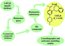
A bioisosteric strategy was successfully implemented with a screening protocol for new, potent 5-HT6R ligands. Initially, 2-[5-(4-methylpiperazin-1-yl)-2-nitrophenyl]-1,2,3,4-tetrahydroisoquinoline (9) was found in commercial databases using a bioisosteric query (screening 5-HT6R Ki = 128 nM). Then, the hit compound was bioisosterically modified (ring alteration) leading to a novel, high affinity (Ki = 6 nM) 5-HT6R ligand (10). Extensive docking studies followed by structural interaction fingerprint analysis supported by single-crystal X-ray structures of the investigated ligands suggest different binding modes with 5-HT6R models for compounds with varying activity. An alternative anchoring point for protonated amine (D7.36) that has not been previously reported was identified.


Introduction

Isosteric or, in the case of biologically active compounds, bioisosteric substitution is one of the most commonly used methods for developing new compounds with required characteristics, e.g., drugs with improved pharmacodynamics and/or pharmacokinetic properties.1 The first known bioisosteric replacement that revolutionized drug development and medicine was the discovery of Prontosil.2 Prontosil was the first antibiotic of the sulfonamide family. Its active metabolite, sulfanilamide, is an actual bioisostere of p-aminobenzoic acid, an intermediate in the bacterial synthesis of folate. Since then, this strategy, usually in the context of structure–activity relationship studies, has been widely used in the search for novel active compounds for different biological targets, among others for the modification of serotonin receptor ligands.

One of the first studies concerning bioisosterism in serotonin receptor ligands was published in 1987 and utilized indazoles as indole substitutes in 5-HT3R antagonists.3 Another report from 1992 described the bioisosteric replacement of indole with benzofuran in a series of 5-HT2R agonists.4 Twelve years later, a 5-HT1fR agonist with an indole–furopyridine ring substitution was reported.5 The most recent paper, published in 2010, reported the replacement of a benzene ring in haloperidol with [2,2]-paracyclophane.6 In spite of the large number of reports of bioisosterism, none discuss explicitly the application of nonclassical bioisosteric strategy to 5-HT6 receptor ligands.7

The 5-HT6 receptor, a member of the GPCR superfamily, shows significant therapeutic potential and is a promising target for novel psychotropic drugs.8 This receptor is responsible primarily for motor control, memory and learning, and its antagonists have several applications, e.g., improving cognitive function and memory in cognitive impairments.9–11 These antagonists are also potential antiobesity drugs.12,13 The 5-HT6R and some other members of serotonergic receptor family have been investigated in our laboratories for a long time, and this research has included the synthesis of novel ligands, in vitro testing, homology modeling, pharmacophore filtering and multistep virtual screening.14–18

In this study, we present an implementation of bioisosteric query to the virtual screening (VS) of potential 5-HT6R ligands in a 5.4 million database of commercial compounds and subsequent bioisosteric modification of the VS hit. All alterations of the hit structure were ring modifications which resulted in the discovery of a new, potent 5-HT6 receptor ligand. To explain differences in 5-HT6R ligands affinity, the single crystal X-ray structure analysis and docking studies were performed.

Initial screening – selection of a hit compound

The hit compound was found from a multistep ligand-based virtual screening cascade performed on compound collections from five vendors with a query based on bioisosteres of 5-HT6R ligands (Fig. 1). The ChEMBL database version 13, extended by data from ca. 30 patents, was used as a source of compounds with known 5-HT6 affinity.19 For each of the 4298 compounds from the final set, all possible bioisosteres were generated in Pipeline Pilot.20 After removal of duplicates, a collection of 137[thin space (1/6-em)]951 unique compounds was obtained. Next, from the database of 5.4 million commercially available structures (Chembridge, Chemdiv, UORSY, Vitas M and Enamine), those with Tanimoto similarity coefficients (Tc) higher than or equal to 0.9 to any previously generated bioisostere were fetched, and the resulting set of 1718 analogues was subjected to further filtering steps.
image file: c5ra00054h-f1.tif
Fig. 1 Workflow for the virtual screening protocol with bioisostere query for the identification of a hit compound.

The applied protocol followed the hierarchical scheme reported previously and consisted of four filters: physicochemical, ADME, 3D pharmacophore and visual inspection.21 First, the Lipinski rules of 5, the Veber rules and the criteria of the strongest basic pKa > 5 were applied (Calculator Plugins, JChem).22 After generation of 3D structures (Ligprep), the ADME descriptors (QikProp) were calculated, and only compounds fulfilling all ADME requirements (see experimental) were considered.23,24 In the next phase, a linear combination of four different pharmacophore models, developed using four of the most populated chemical scaffolds of 5-HT6R antagonists (indoles, indole-like, monocyclic arylpiperazines and polycyclic arylpiperazines), were applied.17 Finally, 129 compounds, each matching at least one of the pharmacophore models, were evaluated by team members to select structures with the most diverse chemotypes for biological investigation. As a result, a set of 11 compounds (ESI) was acquired from Vitas M, Chembridge and Enamine and tested for 5-HT6 receptor activity at two concentrations (see Experimental). Of the tested compounds, (2-[5-(4-methylpiperazin-1-yl)-2-nitrophenyl]-1,2,3,4-tetrahydroisoquinoline), STK148877 (9), was the most potent (screening 5-HT6R Ki = 128 nM) and was selected as a hit compound for further modification.

Chemistry

The parent of the hit compound, N-benzyl-5-(4-methylpiperazin-1-yl)-2-nitroaniline (4), was previously reported by Tasler et al.25 A modified route of its resynthesis, where instead of coupling of 2-bromo-4-chloronitrobenzene with Boc-protected benzylamine, a coupling of 5-chloro-2-nitroaniline with benzylbromide with subsequent Boc protection was employed (Scheme 1).26
image file: c5ra00054h-s1.tif
Scheme 1 Synthesis of N-benzyl-5-(4-methylpiperazin-1-yl)-2-nitroaniline (4). Reagents and conditions: (a) H2O, reflux, 1, 5 h; (b) KHMDS, Boc2O, THF, 0 °C; (c) Pd(OAc)2, JohnPhos, K3PO4·H2O, N–Me–piperazine, DME, 100 °C, 20 h; (d) MeOH, conc. HCl, reflux, 3 h.

Syntheses of the hit compound 9 and its two isosteres (2-[5-(4-methylpiperazin-1-yl)-2-nitrophenyl]-2,3-dihydro-1H-isoindole 8 and 2-[5-(4-methylpiperazin-1-yl)-2-nitrophenyl]-2,3,4,5-tetrahydro-1H-2-benzazepine) 10 were performed according to the synthetic route shown in Scheme 2. The general procedure utilized coupling of 4-chloro-2-fluoronitrobenzene with amines at the 2-position and subsequent addition of N–Me–piperazine through the Buchwald–Hartwig reaction.


image file: c5ra00054h-s2.tif
Scheme 2 Synthesis of 4-methylpiperazin-1-yl-2-nitroanilines (8, 9, 10). Reagents and conditions: (a) DMSO, K2CO3, 120 °C, 2 h; (b) Pd(OAc)2, JohnPhos, K3PO4·H2O, N–Me–piperazine, DME, 100 °C, 20 h.

Pharmacology

In vitro evaluation

Radioligand binding assays were employed to determine the affinity and selectivity profiles of the synthesized compounds for human serotonin 5-HT1AR, 5-HT6R, 5-HT7bR and D2LR, which were stably expressed in HEK293 cells. This was accomplished by displacement of [3H]-8-OH-DPAT (187 Ci mmol−1) for 5-HT1AR; [3H]-LSD (85.2 Ci mmol−1) for 5-HT6R; [3H]-5-CT (39.2 Ci mmol−1) for 5-HT7bR and [3H]-Raclopride (74.4 Ci mmol−1) for D2LR. Each compound was tested in triplicate at 7 to 8 different concentrations (10−11 to 10−4 M). The inhibition constants (Ki) were calculated from the Cheng–Prusoff equation.27 The results are expressed as the mean of at least two separate experiments (Table 1).
Table 1 Affinities of the synthesized compounds
  R Cmpd Ki [nM] Function at 5-HT6e KBe [nM]
5-HT1Aa 5-HT6b 5-HT7c D2d
a Buspirone as a reference Ki = 20 nM.b Olanzapine as a reference Ki = 7 nM.c Clozapine as a reference Ki = 18 nM.d Olanzapine as a reference Ki = 7 nM.e Methiotepine as a reference KB = 3.1 nM.f Resynthesized STK148877.
image file: c5ra00054h-u1.tif image file: c5ra00054h-u2.tif 4 1278 22 2783 557 Antagonist 79
image file: c5ra00054h-u3.tif 8 2292 245 5117 1396 Antagonist 1000
image file: c5ra00054h-u4.tif 9f 3740 62 4148 227 Antagonist 340
image file: c5ra00054h-u5.tif 10 1015 6 5181 358 Antagonist 29


The most potent and selective 5-HT6R ligand, 2-[5-(4-methylpiperazin-1-yl)-2-nitrophenyl]-2,3,4,5-tetrahydro-1H-2-benzazepine 10, was further investigated for other activities by CEREP (Table 2). It is noteworthy that compound 10 was found to be 60-fold selective, versus only 25-fold selectivity for compound 4, against D2R. Antagonist effect at 5-HT6R was determined by CEREP (Table 1). The parent compound 4, as well as compounds 8–10, exhibit antagonistic properties of different potency, proportional to 5-HT6 affinity values. Tasler et al. reported the function of analogue of parent compound 4 with unmethylated piperazine to be full agonist.25 The function of methylated parent compound 4 was not reported but it was shown that similar compound with 2-furane moiety instead of phenyl ring possessed partial agonistic properties.

Table 2 % inhibition of specific binding at a compound 10 concentration of 1 μM
Receptor Serotonin 5-HT2C Serotonin 5-HT3 Dopaminergic D3 Adrenergic α1 Adrenergic α2C Histamine H1 Muscarinic M1
% Inhibition 72 25 89 12 12 85 3


Crystal structure analysis of ligands

To obtain the protonated form of the synthesized compounds, several salts were considered. However, crystallization of the salts were unsuccessful. Good quality single crystals of free bases were obtained instead.

The crystal structures of two of the most potent 5-HT6R ligands (4 and 10) revealed a similar hairpin-like conformation of the molecules, which differs from the bent conformation of the less active bioisosteres (8 and 9). The distinct orientation of the p-nitroaryl fragment and the second aromatic ring of the molecules is represented by the torsion angle C(2)–N(2)–C(11)–C(12) (Table 3). The hairpin conformation of 4 is a result of intramolecular hydrogen bond formation N(2)–H(2)⋯O(1) (Fig. 2 and Table 4), while in the case of 10 it is related to the conformation of the introduced seven-membered ring.

Table 3 Selected geometrical values for the presented crystal structures [Å and °]
Compound 4 8 9 10
N(1)–C(1) 1.429(2) 1.443(1) 1.439(2) 1.444(1)
N(2)–C(2) 1.352(2) 1.380(1) 1.388(2) 1.380(1)
N(3)–C(4) 1.398(2) 1.385(1) 1.372(2) 1.385(1)
C(3)–C(4)–C(5) 118.5(1) 118.4(1) 116.6(1) 117.98(8)
C(2)–C(1)–C(6) 120.1(1) 121.0(1) 119.9(1) 121.00(8)
C(2)–N(2)–C(11)–C(12) −69.1(2) −173.34(9) −165.0(1) 90.7(1)
O(1)–N(1)–C(1)–C(2) 4.0(2) −36.9(2) −22.6(2) −33.5(1)
O(2)–N(1)–C(1)–C(6) 3.4(2) −39.7(1) −27.5(2) −37.5(1)
C(3)–C(4)–N(3)–C(7) 30.76(2) −1.9(2) −6.5(2) −30.01(1)
C(5)–C(4)–N(3)–C(9) −8.5(2) 35.5(2) 21.3(2) 3.7(1)


image file: c5ra00054h-f2.tif
Fig. 2 Molecular geometry and common geometrical features (distance in Å and torsion angle value in °) that determine arrangement of molecules in the crystal environment. For 4, an intramolecular hydrogen bond is highlighted. The displacement ellipsoids of the non-hydrogen atoms are drawn at the 30% probability level. H atoms are presented as small spheres with an arbitrary radius.
Table 4 Geometry of strong and weak hydrogen bonds in the crystal structures [Å and °]a
Cmpd D–H⋯A d(D–H) d(H⋯A) d(D⋯A) <(DHA)
a Symmetry transformations used to generate equivalent atoms: _1: −x, −y, −z; _2: −x, y + 1/2, −z + 1/2; _3: −x, 1 − y, −z; _4: x − 1, y, z; _5: x, −y + 3/2, z + 1/2 _6;−x, y − 1/2, −z + 1/2; _7: −x + 1, −y + 1, −z + 1.
4 N(2)–H(2)⋯O(1) 0.88(2) 1.95(2) 2.639(2) 133(2)
N(2)–H(2)⋯O(1)_1 0.88(2) 2.31(2) 2.994(2) 134(1)
C(10)–H(10A)⋯O(2)_2 0.99 2.52 3.419(2) 151
C(13)–H(13)⋯O(2)_3 0.95 2.56 3.505(2) 175
8 C(11)–H(11A)⋯O(1) 0.99 2.12 2.870(1) 130
C(11)–H(11A)⋯N(1) 0.99 2.52 3.051(2) 113
C(7)–H(7A)⋯O(1)_4 0.99 2.57 3.301(1) 130
C(9)–H(9B)⋯O(2)_5 0.99 2.59 3.537(2) 161
C(5)–H(5)⋯O(2)_5 0.95 2.61 3.549(1) 171
9 C(11)–H(11B)⋯N(4)_6 0.99 2.61 3.293(2) 126
C(18)–H(18B)⋯O(1) 0.99 2.55 3.164(2) 120
C(19)–H(19A)⋯N(1) 0.99 2.47 3.124(2) 123
10 C(11)–H(11B)⋯O(1)_7 0.99 2.49 3.437(1) 161
C(18)–H(18B)⋯O(1)_7 0.99 2.48 3.419(1) 159
C(20)–H(20B)⋯N(1) 0.99 2.35 3.004(1) 123


The hairpin conformation of the two most active bioisosteres allows the two aromatic fragments to be closer, which is reflected by centroids distance (Cg1–Cg2) of 5.1–5.5 Å. This conformation also results in a shorter distance between the basic nitrogen atom N(4) and the centroid (Cg2) of the aromatic ring C(12)–C(17), which varies between 6.8 to 7.7 Å for the most active ligands, whereas it is approximately 10 Å for the two less active compounds (Fig. 2). Additionally, the close proximity of the C(12)–C(17) π-electron system influences the mutual orientation of both rings of the arylpiperazine moiety, which can be defined by the torsion angle C(3)–C(4)–N(3)–C(7). This angle for both active compounds is approximately ±30°. The opposite sign of this value is related to the opposite orientation of the aromatic C(12)–C(17) ring with respect to the piperazine ring. This distinct orientation is a result of the packing in the crystal structure. It is potentially based upon the formation of weak interaction C(13)–H(13)⋯O(2) in the crystal structure of 4, which is not observed in 10. For the latter structure, similar interactions occur between the two methylene groups of the seven-membered ring and the oxygen atom O(1) as a bifurcated acceptor of weak hydrogen bond (see Table 4).

Within the crystal structure of the presented compounds, weak hydrogen bonds C–H⋯O and C–H⋯N are primarily observed. The only exception is the crystal structure of 4, in which the presence of the strong donor N(2) allowed centrosymmetric dimer formation through hydrogen bonding system (Table 3). The intramolecular interaction N(2)–H(2)⋯O(1) mentioned above results in a co-planar arrangement of the nitro group with respect to the aromatic ring C(1)–C(6). For compounds 8, 9 and 10, this co-planarity is not observed. The p-NO2 substituent demonstrated different inductive effects in these molecules. The nitro group exhibits an electron withdrawing effect in 8 and 10 and a slightly electron donating effect in 9. The electron withdrawing effect is shown by the elongation of the N(1)–C(1) bond and the increased C(2)–C(1)–C(6) angle (Table 3). The differing properties of the nitro group are related to the number of intermolecular interactions in which the oxygen atoms are involved.

Docking studies

Compounds 4, 8, 9 and 10 were ionized and docked without any restraints to 200 homology models of 5-HT6R (aligned on an β2-adrenergic template) developed in our laboratory employing a previously utilized approach (see Experimental).14,28,29 For each compound, only the best docking pose per receptor was considered, and the 100 best scoring complexes were transformed into bitstrings by applying SIFt formalism to statistically describe the interactions between ligand and receptor.30,31 The most potent compounds 4 and 10 (Fig. 3A and D) were classically docked near TM3 and TM5. Crucial charge-reinforced hydrogen bonds of protonated nitrogen were formed with D3.32, and hydrophobic interactions were created with W3.28, T3.29 and C3.36. The methyl group of the N-methyl-arylpiperazine also interacted with TM7 (Y7.43). The central nitrophenyl group directed toward TM6 participated in edge-to-face contacts with F6.51 and F6.52. In case of compound 4, the benzyl moiety was near EL2 and participated in a hydrophobic interaction with L164. On the other hand, benzazepine moiety of 10 was situated deep into the subpocket near TM5 and interacted with F5.38, A5.42, S5.43 (statistically, the most important TM5 residue) and T5.46. Surprisingly, both of the less potent compounds (8 and 9) formed hydrogen bonds between the positive, ionized nitrogen atom and D7.36 (instead of D3.32). Thus, they were located shallowly in the binding pocket, close to TM6 and TM7; consequently, these compounds had almost no interaction with TM3 (Fig. 3B and C). The central nitrophenyl group did not contact F6.52, but participated in face-to-edge stacking with F6.51 and strongly interacted with EL2 (especially L164). Despite the fact that the side phenyl moiety was positioned toward TM5 deeper into the receptor, it did not interact with S5.43 and T5.46.
image file: c5ra00054h-f3.tif
Fig. 3 Computational models of the best scoring complexes between ligand 4 (A), 8 (B), 9 (C), 10 (D) and 5-HT6R. The important residues that are statistically crucial for the more potent ligands are marked in cyan. Residues (D7.36 and L164) that interact with the less active compounds are indicated in orange. Hydrogen bonds with D3.32 or D7.36 are shown in red. Both 4 and 10 are situated deeper into the receptor cavity and strongly interact with TM3, TM5 and TM6, whereas the less potent 8 and 9 are located shallowly and interact with TM7 and EL2.

Discussion

Common VS campaigns use the structures of active compounds as direct queries to search databases; however, here, we introduced an additional step of bioisostere generation to cover more biologically relevant chemical space. Indeed, the similarity between the 11 VS hits and the 5-HT6R ligand database ranged from Tc = 0.57–0.83. Particularly, Tc equaled 0.79 for the hit compound (9), which means that this compound would not have been selected using a standard VS procedure with the criteria of Tc ≥ 0.8. Further bioisosteric modification of 9 based on ring alteration produced two additional compounds with significantly different affinities for 5-HT6R (Ki = 245 and 6 nM for 8 and 10, respectively). The parent compound N-benzyl-5-(4-methylpiperazin-1-yl)-2-nitroaniline 4 was previously reported by Tasler et al. in a series of 5-HT6R ligands developed by modifying virtual screening hits.25 The most potent compound of a group of obtained nitrophenylpiperazines displayed affinity Ki = 8 nM towards 5-HT6R.25

Crystallographic studies of synthesized bioisosteres revealed that both compounds with higher binding affinities (4 and 10) adopt a similar molecular arrangement. The mutual orientation of both aromatic moieties, and consequently, the shorter distance between the C(12)–C(17) ring and the basic nitrogen atom N(4) seems to play an important role. Interestingly, the binding poses of the docked ligands resemble crystalline structures. Distances between the aromatic rings are 1 Å greater, on average, in less potent compounds than in compounds 4 and 10, similar to the intervals between the N(4) atoms of the N-methylpiperazine moiety and the terminal aromatic system. These substructures are relatively distant for compounds 8 and 9, whereas in the structures 4 and 10, the distance values are decreased by 2–3 Å. Similar orientations of the rings in the arylpiperazine fragment for both of the more active compounds may be critical for ligand–receptor recognition and may play an important role in selective ligand–receptor binding. The semi-rigid molecule 10 represents optimal spatial orientation of the crucial molecular fragments, thus allowing more precise definition of the pharmacophore. The same conformation is also possible for the most flexible molecule 4, although this was not observed in the crystalline form because of the additional interactions formed in the crystal lattice. This favorable conformation cannot be attained by 8 and 9 because of their increased rigidity, which explains the lower binding affinity of these compounds for the 5-HT6 receptor.

The statistical analysis of multiple ligand–receptor complexes (Table 5) emphasizes the importance of L163, F6.51 and F7.35 in ligand binding. In addition, the more potent compounds, 4 and 10, interacted classically with D3.32, S5.43, T5.46 and F6.52, whereas the binding modes of the rigid, less potent compounds 8 and 9 indicated atypical interactions with D7.36 (instead of D3.32). Interestingly, of the entire serotonin GPCR family of 13 receptors, the D7.36 residue is present only in 5-HT1B and 5-HT1D receptors. As far as we know, this alternative side interaction of a protonated nitrogen with D7.36 has not been previously reported. This information can be used as a potential starting point for the design of novel, potent, selective serotonin receptor ligands.

Table 5 Averaged SIFt profiles calculated for the 100 best scoring complexes. Only values higher than 0.50 were selected as significant
Residue Compound
4 8 9 10
W3.28 0.79 0.74
T3.29 0.61 0.56 0.64
D3.32 0.99 0.98
V3.33 0.85 0.79 0.84 0.96
C3.36 0.88 0.90
L4.61 0.51
R162 0.57
L163 0.98 0.96 0.98 0.99
L164 0.59 0.60 0.64
A165 0.87 0.83 0.86 0.98
F5.38 0.63 0.89 0.82 0.99
V5.39 1.00 0.93 0.82 0.99
A5.42 0.65 0.76 0.68 0.95
S5.43 0.95 0.72
T5.46 0.63 0.85
F6.51 1.00 0.96 0.95 1.00
F6.52 0.98 0.56 0.99
N6.55 0.99 0.86 0.86 1.00
V6.58 0.67 0.58 0.50
P7.32 0.50
F7.35 0.99 0.99 1.00 1.00
D7.36 0.58 0.77
T7.39 1.00 0.74 0.75 1.00
Y7.43 0.94 0.54 0.93


Conclusion

In conclusion, bioisosteric exchange based on ring modification was successfully applied to the design of potent 5-HT6R ligands with a modified selectivity profile. Whereas a rigid conformation between two aromatic moieties in compounds 8 and 9 resulted in decreased affinity, the introduction of a seven-membered ring in compound 10 conserved the active spatial orientation of the important pharmacophore features. These observations were confirmed and explained by crystallographic and computational studies. Compared to the previously reported compound 4, the most potent compound 10 exhibited greater selectivity towards 5-HT1A (three-fold), 5-HT7 (seven-fold) and D2 (two-fold). These differences in biological activity suggest that 5-HT6 receptors are sensitive to subtle changes in the orientation of the aromatic moieties. Additionally, analysis of the ligand–receptor complexes indicated the possibility of the formation of an unusual interaction with D7.36, which has not been previously reported.

Experimental protocols

Synthesis

Melting points were determined on an Electrothermal 9100. 1H NMR spectra were recorded on Bruker Avance III HD 400 NMR spectrometer. Mass spectra were recorded using a TQD Waters LC/MS spectrometer with electrospray ionization. Substrates and solvents were purchased from Sigma-Aldrich and Apollo Scientific and were used without further purification.

N-Benzyl-5-chloro-2-nitroaniline (1)

5-Chloro-2-nitroaniline (1.0 g) was added to water (12.0 ml), benzyl bromide (1.19 g) was added, and the reaction was heated to reflux for 1.5 h. After cooling to RT, a saturated sodium bicarbonate solution (10.0 ml) was added to the reaction mixture, and the product was extracted 3 times with ethyl acetate (3 × 20 ml). The combined extracts were washed with brine, dried over anhydrous MgSO4 and evaporated under reduced pressure. Purification by column chromatography on silica eluting with chloroform/hexane 1[thin space (1/6-em)]:[thin space (1/6-em)]1 afforded a yellow solid (0.71 g). Yield 45%.

LCMS [M + 1] = 263.09 m/z (262.05 calcd). 1H NMR (CDCl3, 400.17 MHz) δ (ppm): 4.54 (d, J = 5.5 Hz, 2H); 6.66 (dd, J = 9.1, 2.1 Hz, 1H); 6.86 (d, J = 2.1 Hz, 1H); 7.48–7.27 (m, 5H); 8.22–8.12 (m, 1H); 8.47 (d, J = 6.1 Hz, 1H).

13C NMR (CDCl3, 400.17 MHz) δ (ppm): 47.27; 113.60; 116.32; 127.17; 127.99; 128.27; 129.07; 130.89; 136.9; 142.86; 145.62.

tert-Butyl N-benzyl-N-(5-chloro-2-nitrophenyl)carbamate (2)

N-Benzyl-5-chloro-2-nitroaniline (1) (0.66 g) was dissolved in anhydrous THF (10 ml), and the solution was cooled in an ice bath. Then, a 0.5 M solution (5.82 ml) of KHMDS in toluene was added dropwise. After standing for 30 min, a solution of Boc2O (0.64 g) in anhydrous THF (10 ml) was added dropwise. The reaction mixture was allowed to warm to RT and was stirred until completion (as indicated by TLC). The reaction was quenched by the addition of distilled water (50 ml), and the product was extracted with chloroform. Purification by column chromatography on silica eluting with chloroform afforded a red solid (0.35 g). Yield 40%. The substance could not be ionized under ESI conditions. 1H NMR (CDCl3, 400.17 MHz) δ (ppm): 1.56 (s, 9H); 4.54 (d, J = 5.5 Hz, 2H); 6.66 (dd, J = 9.1, 2.1 Hz, 1H); 6.86 (d, J = 2.1 Hz, 1H); 7.48–7.29 (m, 5H); 8.17 (dd, J = 9.1, 0.3 Hz, 1H).

13C NMR (CDCl3, 400.17 MHz) δ (ppm): 27.43; 47.27; 85.17; 113.60; 116.32; 127.16; 127.98; 128.27; 129.07; 136.58; 142.85; 145.62; 146.75.

General procedure for the coupling of 4-chloro-2-fluoro-1-nitrobenzene with heterocyclic secondary amines

4-Chloro-2-fluoro-1-nitrobenzene (5.7 mmol) and the appropriate amine (5.7 mmol) were dissolved in DMSO (10 ml), and anhydrous potassium carbonate (14.2 mmol) was added. The reaction mixture was heated to 120 °C for 2 h. After cooling to RT, the reaction mixture was poured into distilled water (100 ml) and extracted with ethyl acetate (3 × 30 ml). The combined extracts were washed with distilled water (2 × 100 ml) and brine (20 ml), dried over anhydrous MgSO4 and evaporated under reduced pressure. Purification was performed by flash chromatography on silica gel eluting with chloroform.

2-(5-Chloro-2-nitrophenyl)-2,3-dihydro-1H-isoindole (5)

A red solid was obtained (1.2 g). Yield 76%. LCMS [M + 1] = 275.05 m/z (274.05 calcd). 1H NMR (CDCl3, 400.17 MHz) δ (ppm): 4.68 (s, 4H); 6.77 (dd, J = 8.7, 2.0 Hz); 7.03 (d, J = 2.0 Hz); 7.40–7.25 (m, 4H); 7.67 (d, J = 8.7 Hz).

13C NMR (CDCl3, 400.17 MHz) δ (ppm): 55.67, 116.00, 116.38, 122.25; 127.72; 127.84; 135.95; 136.36; 139.02, 142.07.

2-(5-Chloro-2-nitrophenyl)-1,2,3,4-tetrahydroisoquinoline (6)

A red solid was obtained (1.34 g). Yield 82%. LCMS [M + 1] = 289.06 m/z (288.06 calcd). 1H NMR (CDCl3, 400.17 MHz) δ (ppm): 3.05 (t, 2H); 3.43 (t, J = 6.2, 5.4 Hz, 2H); 4.32 (s, 2H); 6.92 (dd, J = 8.7, 2.1 Hz, 1H); 7.15–7.08 (m, 1H); 7.30–7.15 (m, 4H); 7.82 (d, J = 8.7 Hz, 1H). 13C NMR (CDCl3, 400.17 MHz) δ (ppm): 28.72; 49.78; 52.12; 119.12; 119.58; 126.30; 126.32; 126.87; 128.07; 128.71; 133.17; 134.37; 138.89; 139.78; 146.34.

2-(5-Chloro-2-nitrophenyl)-2,3,4,5-tetrahydro-1H-2-benzazepine (7)

A red solid was obtained (0.32 g). Yield 18%. LCMS [M + 1] = 303.08 m/z (302.08 calcd). 1H NMR (CDCl3, 400.17 MHz) δ (ppm): 2.09–1.98 (m, 2H); 2.99–2.92 (m, 2H); 3.45–3.37 (m, 2H); 4.50 (s, 2H); 6.76 (dd, J = 8.7, 2.1 Hz, 1H); 7.03 (d, J = 2.1 Hz, 1H); 7.31–7.12 (m, 4H); 7.67 (d, J = 8.7 Hz, 1H).

13C NMR (CDCl3, 400.17 MHz) δ (ppm): 27.53; 33.72; 55.49; 56.65; 118.37; 119.45; 126.68; 127.78; 127.82; 128.22; 129.83, 137.36; 137.97; 139.18; 140.85; 146.21.

General procedure for the coupling of N–Me–piperazine with aryl chloride

An oven-dried round-bottom flask was charged with the aryl chloride (3.6 mmol), palladium acetate (0.1 eq.), biphenyl tert-butyl phosphine (JohnPhos) (0.2 eq.) and potassium phosphate monohydrate (1.4 eq.). The flask was evacuated under vacuum and filled with argon three times. Then, DME (2 ml/1 mmol) and N-methylpiperazine (1.3 eq.) were added via syringe. The reaction mixture was heated at 100 °C for 20 h. After completion, the reaction mixture was filtered through celite, and the solvent was evaporated. The product was purified by column chromatography on silica eluting with CHCl3[thin space (1/6-em)]:[thin space (1/6-em)]MeOH (19[thin space (1/6-em)]:[thin space (1/6-em)]1).

N-Benzyl-5-(4-methylpiperazin-1-yl)-2-nitroaniline (4)

A red oil was obtained (0.21 g). Yield 51%. LCMS [M + 1] = 327.24 m/z (426.23 calcd); the compound undergoes deprotection during ionization. tert-Butyl N-benzyl-N-[5-(4-methylpiperazin-1-yl)-2-nitrophenyl]carbamate (3) (0.20 g) was dissolved in MeOH (10 ml) with a few drops of concentrated hydrochloric acid. The reaction was heated to reflux for 3 h, and distilled water (50 ml) and 15% NaOH solution (5 ml) were added. The products were extracted with chloroform (3 × 20 ml), and the extracts were dried over anhydrous MgSO4 and evaporated under reduced pressure. The obtained substance was dissolved in diethyl ether and converted to the hydrochloride salt by addition of a 20% solution of HCl in diethyl ether. The collected solids were recrystallized from ethanol to afford an orange solid (0.094 g). Yield 55%.

For the purpose of crystallographic studies, the free base was crystallized from hexane–acetone.

Hydrochloride mp. 206–208 °C. LCMS [M + 1] = 327.17 m/z (326.17 calcd). 1H NMR (CDCl3, 400.17 MHz) δ (ppm): 2.31 (s, 3H), 2.47–2.42 (m, 4H), 3.32–3.27 (m, 4H), 4.50 (d, J = 5.4 Hz, 2H), 5.84 (d, J = 2.6 Hz, 1H), 6.22 (dd, J = 9.7, 2.6 Hz, 1H), 7.33–7.24 (m, 1H), 7.39–7.30 (m, 4H), 8.08 (d, J = 9.7 Hz, 1H), 8.79 (t, J = 5.5 Hz, 1H).

13C NMR (CDCl3, 400.17 MHz) δ (ppm): 46.02; 46.80; 47.08; 47.20; 54.48; 94.42; 104.48; 124.46; 127.12; 127.59; 128.88; 128.94; 137.62; 147.44; 155.73.

2-[5-(4-Methylpiperazin-1-yl)-2-nitrophenyl]-2,3-dihydro-1H-isoindole (8)

An orange solid was obtained (0.5 g). Hydrochloride mp. 208–210 °C. LCMS [M + 1] = 339.17 m/z (338.17 calcd). 1H NMR (CDCl3, 400.17 MHz) δ (ppm): 2.39 (s, 3H); 2.66–2.55 (m, 4H); 3.47–3.37 (m, 4H); 4.68 (s, 4H); 6.26 (d, J = 2.5 Hz, 1H); 6.36 (dd, J = 9.3, 2.4 Hz, 1H); 7.30 (s, 4H); 7.83 (d, J = 9.3 Hz, 1H).

13C NMR (CDCl3, 400.17 MHz) δ (ppm): 45.92; 47.37; 54.56; 55.90; 99.52; 104.47; 122.14; 127.40; 129.39; 130.17; 136.62; 144.46; 154.51.

2-[5-(4-Methylpiperazin-1-yl)-2-nitrophenyl]-1,2,3,4-tetrahydroisoquinoline (9)

An orange solid was obtained (0.7 g). Hydrochloride mp. 157–159 °C. LCMS [M+1] = 353.17 m/z (352.19 calcd). 1H NMR (CDCl3, 400.17 MHz) δ (ppm): 2.39 (s, 3H); 2.62–2.55 (m, 4H); 3.10 (t, J = 5.8 Hz, 2H); 3.41 (ddd, J = 7.7, 5.5, 4.5 Hz, 6H); 4.34 (d, J = 1.0 Hz, 2H); 6.50–6.40 (m, 2H); 7.26–7.07 (m, 4H); 8.10–8.00 (m, 1H).

13C NMR (CDCl3, 400.17 MHz) δ (ppm): 28.92; 46.04; 47.15; 50.89; 52.56; 54.60; 102.64; 106.33; 125.96; 126.27; 126.56; 128.83; 129.91; 131.54; 133.92; 134.75; 148.97; 154.92.

2-[5-(4-Methylpiperazin-1-yl)-2-nitrophenyl]-2,3,4,5-tetrahydro-1H-2-benzazepine (10)

An orange solid was obtained (0.8 g). Hydrochloride mp. 136–138 °C. LCMS [M + 1] = 367.22 m/z (366.20 calcd). 1H NMR (CDCl3, 400.17 MHz) δ (ppm): 2.05–1.94 (m, 2H); 2.33 (s, 3H); 2.51–2.42 (m, 4H); 2.99–2.89 (m, 2H); 3.23–3.16 (m, 4H); 3.45–3.36 (m, 2H); 4.57 (s, 2H); 6.35–6.24 (m, 2H); 7.32–7.09 (m, 4H); 7.87 (dd, J = 9.0, 0.5 Hz, 1H).

13C NMR (CDCl3, 400.17 MHz) δ (ppm): 28.00; 34.06; 45.94; 47.03; 47.17; 54.44; 54.52; 56.59; 56.68; 103.08; 105.43; 126.26; 126.30; 127.41; 128.01; 129.49; 129.99; 130.39; 139.12; 141.23; 148.76; 154.43.

X-ray structure determination

Single crystals of all compounds were obtained from a 1[thin space (1/6-em)]:[thin space (1/6-em)]1 acetone[thin space (1/6-em)]:[thin space (1/6-em)]hexane mixture by slow evaporation of the solvent under ambient conditions. X-ray diffraction data for crystals of 4, 8 and 10 were collected with MoKα radiation (λ = 0.71073 Å) using a Nonius Brucker KappaCCD diffractometer with the software COLLECT.32 Data were processed with HKL SCALEPACK and HKL DENZO.33 For the selected crystal 9, intensities of the diffracted X-ray beam were collected with CuKα radiation (λ = 1.54184 Å) using SuperNova diffractometer. Data were processed with CrysAlisPro.34 Experiments were performed at 100(2) K for all compounds except 9. For 9, the temperature was set at 110(2) K. The phase problem was solved by direct methods with SHELXS-97 program. The model parameters were refined by full-matrix least-squares on F2 using SHELXL-97.35 All non-hydrogen atoms were refined anisotropically. In the structure of 4, the hydrogen atom attached to N2 was localized on the difference Fourier map and refined without restraints. For all compounds, the positions of the hydrogen atoms attached to C atoms were constrained with C–H = 0.95 Å for aromatic protons, C–H = 0.99 Å for methylene protons, and C–H = 0.98 Å for methyl groups and were refined using the riding model with the isotropic displacement parameter Uiso = 1.2Ueq and Uiso = 1.5Ueq (methyl groups only) of the parent atom.

All crystallographic data for the presented structures are shown in Table 6.

Table 6 Crystal data and structure refinement
Identification code 4 8 9 10
Molecular formula C18H22N4O2 C19H22N4O2 C20H24N4O2 C21H26N4O2
Formula weight 326.39 338.40 352.43 366.46
Wavelength 0.71073 Å 0.71073 Å 1.54184 Å 0.71073 Å
Crystal system Monoclinic Monoclinic Monoclinic Triclinic
Space group P21/c P21/c P21/c P[1 with combining macron]
Unit cell dimensions a = 14.8320(4) Å a = 9.2516(3) Å a = 14.6428(3) Å a = 9.5413(2) Å
b = 4.8941(2) Å b = 18.4177(5) Å b = 7.5669(1) Å b = 10.1771(2) Å
c = 23.9340(7) Å c = 12.6426(3) Å c = 16.1976(3) Å c = 11.0759(2) Å
β = 108.015(2)° β = 128.998(2)° β = 100.828(2)° α = 105.049(2)°
      β = 92.969 (2)°
      γ = 114.710 (1)°
Volume [Å3] 1652.15(10) 1674.18(9) 1762.75(6) 927.62(3)
Z 4 4 4 2
Dcalc [g cm−3] 1.312 1.343 1.328 1.312
Absorption coefficient [mm−1] 0.088 0.090 0.707 0.086
F(000) 696 720 752 392
Crystal size [mm3] 0.50 × 0.30 × 0.05 0.50 × 0.40 × 0.20 0.35 × 0.25 × 0.05 0.50 × 0.50 × 0.30
θ range [°] 2.62–28.68 2.35–27.50 3.07–71.24 2.79–27.45
Index ranges −19 ≤ h ≤ 19, −11 ≤ h ≤ 12, −17 ≤ h ≤ 17, −12 ≤ h ≤ 11,
−6 ≤ k ≤ 4, −21 ≤ k ≤ 23, −9 ≤ k ≤ 9, −12 ≤ k ≤ 13,
−32 ≤ l ≤ 32 −15 ≤ l ≤ 16 −19 ≤ l ≤ 18 −14 ≤ l ≤ 14
Reflections collected 10 299 9948 23 634 7289
Independent reflections 4234 3819 3397 4191
[R(int) = 0.0329] [R(int) = 0.0185] [R(int) = 0.0483] [R(int) = 0.0107]
Completeness % 99.6 (to θ = 25.24°) 99.5 (to θ = 25.24°) 100.0 (to θ = 67.68°) 99.4 (to θ = 25.24°)
Data/restraints/parameters 4234/0/221 3819/0/226 3397/2/235 4191/0/244
Goodness-of-fit on F2 1.016 1.038 1.021 1.027
Final R indices [I > 2σ(I)] R1 = 0.0469, R1 = 0.0378, R1 = 0.0385, R1 = 0.0352,
wR2 = 0.1076 wR2 = 0.0949 wR2 = 0.0977 wR2 = 0.0938
R indices (all data) R1 = 0.0736, R1 = 0.0444, R1 = 0.0465, R1 = 0.0376,
wR2 = 0.1199 wR2 = 0.0993 wR2 = 0.1051 wR2 = 0.0961
Δρmaxρmin [e Å−3] 0.243/−0.292 0.321/−0.268 0.200/−0.211 0.298/−0.245


Unit cell layout is presented in ESI Figures.

WinGX software was used to prepare the materials for publication.36 The figures showing the asymmetric units of the structures were obtained using Mercury CSD 3.3.37

In vitro pharmacology

Cell culture and preparation of cell membranes. HEK293 cells with stable expression of human serotonin 5-HT1AR, 5-HT6, 5-HT7bR or dopamine D2LR (prepared with the use of Lipofectamine 2000) were maintained at 37 °C in a humidified atmosphere with 5% CO2 and were grown in Dulbecco's Modifier Eagle Medium containing 10% dialyzed fetal bovine serum and 500 μg ml−1 G418 sulfate. For membrane preparations, cells were subcultured in 10 cm diameter dishes, grown to 90% confluence, washed twice with phosphate buffered saline (PBS), pre-warmed to 37 °C and pelleted by centrifugation (200 g) in PBS containing 0.1 mM EDTA and 1 mM dithiothreitol. Prior to membrane preparations, the pellets were stored at −80 °C.
Radioligand binding assays. Cell pellets were thawed and homogenized in 20 volumes of assay buffer using an Ultra Turrax tissue homogenizer and centrifuged twice at 35[thin space (1/6-em)]000 g for 20 min at 4 °C, with incubation for 15 min at 37 °C in between rounds of centrifugation. The composition of the assay buffers was as follows: for 5-HT1AR: 50 mM Tris–HCl, 0.1 mM EDTA, 4 mM MgCl2, 10 μM pargyline and, 0.1% ascorbate; for 5-HT6R: 50 mM Tris–HCl, 0.5 mM EDTA and 4 mM MgCl2, for 5-HT7bR: 50 mM Tris–HCl, 4 mM MgCl2, 10 μM pargyline and 0.1% ascorbate; for dopamine D2LR: 50 mM Tris–HCl, 1 mM EDTA, 4 mM MgCl2, 120 mM NaCl, 5 mM KCl, 1.5 mM CaCl2 and 0.1% ascorbate.

All assays were incubated in a total volume of 200 μl in 96-well microliter plates for 1 h at 37 °C, except for 5-HT1AR, which was incubated at room temperature for 1 h. The process of equilibration was terminated by rapid filtration through unifilter plates with a 96-well cell harvester, and the radioactivity retained on the filters was quantified on a Microbeta plate reader.

For displacement studies, the assay samples contained the following as radioligands: 1.5 nM [3H]-8-OH-DPAT (187 Ci mmol−1) for 5-HT1AR; 2 nM [3H]-LSD (85.2 Ci mmol−1) for 5-HT6R; 0.6 nM [3H]-5-CT (39.2 Ci mmol−1) for 5-HT7bR or [3H]-Raclopride (74.4 Ci mmol−1).

Non-specific binding was defined with 10 μM of 5-HT in 5-HT1AR and 5-HT7bR binding experiments, whereas 10 μM methiothepine and 1 μM of (+)butaclamol were used in the 5-HT6R and D2L assays, respectively. Each compound was tested in triplicate at 7 to 8 different concentrations (10−11 to 10−4 M). The inhibition constants (Ki) were calculated from the Cheng–Prusoff equation.27 The results are expressed as the means of at least two separate experiments.

Initial screening experiments were performed using the same conditions with two compound concentrations: 10−6 and 10−7 M.

Molecular modeling.
Preparation of ChEMBL database. Due to a large diversity of activity measures, only the compounds with defined Ki (IC50 – assumed as 2 × Ki, pKi or pIC50 were converted to Ki), as assayed on human cloned receptors or on rat cloned or native receptors, were considered. In case there were multiple data for one ligand, the Ki and human receptors were given preference; a median value was used if there were many biological results.
Compounds preparation. The appropriate ionization states at pH = 7.4 for all structures used in screening and docking procedures were assigned using Epik.28 3D structures were generated in Ligprep under default settings (force field used OPLS2005, retention of specified chiralities and generation of only one low energy ring conformation per ligand).
ADME filters. The following criteria were used to filter compounds with unfavorable profiles: the number of reactive functional groups (desirable range of values: 0–2), logarithm of calculated aqueous solubility (−6.5–0.5 mol l−1), gut-blood barrier (>500 nm s−1) and blood–brain barrier penetration coefficient (−3.0–1.2).
Docking protocol. All receptors were centered at D3.32 with the grid box size set to 25 × 25 × 25 Å. Docking runs were performed in Glide software at the SP level under default settings (sampling nitrogen inversion, sampling ring conformations with energy window equal to 2.5 kcal mol−1, penalizing nonplanar conformation of amides up to 100 steps during energy minimization and performing post-docking optimization).

Acknowledgements

This research was supported by the following projects: Interdisciplinary PhD Studies “Molecular sciences for medicine” (co-financed by the European Social Fund within the Human Capital Operational Programme) and UDA-POIG.01.03.01-12-063/09-00 “Antagonists of 5-HT6 receptor as advanced antipsychotics with pro-cognitive properties” co-financed by European Union from the European Fund of Regional Development (EFRD). The crystallographic research was carried out with equipment purchased using financial support of the European Regional Development Fund in the framework of the Polish Innovative Economy Operational Program (contract no. POIG.02.01.00-12-023/08).

Notes and references

  1. L. M. Lima and E. J. Barreiro, Curr. Med. Chem., 2005, 12, 23–49 CrossRef CAS PubMed.
  2. G. Domagk, DMW, Dtsch. Med. Wochenschr., 1935, 61, 250–253 CrossRef CAS.
  3. P. Fludzinski, D. A. Evrard, W. E. Bloomquist, W. B. Lacefield, W. Pfeifer, N. D. Jones, J. B. Deeter and M. L. Cohen, J. Med. Chem., 1987, 30, 1535–1537 CrossRef CAS PubMed.
  4. Z. Tomaszewski, M. P. Johnson, X. Huang and D. E. Nichols, J. Med. Chem., 1992, 35, 2061–2064 CrossRef CAS PubMed.
  5. B. M. Mathes, K. J. Hudziak, J. M. Schaus, Y.-C. Xu, D. L. Nelson, D. B. Wainscott, S. E. Nutter, W. H. Gough, T. A. Branchek, J. M. Zgombick and S. A. Filla, Bioorg. Med. Chem. Lett., 2004, 14, 167–170 CrossRef CAS PubMed.
  6. M. Skultety, H. Hübner, S. Löber and P. Gmeiner, J. Med. Chem., 2010, 53, 7219–7228 CrossRef CAS PubMed.
  7. A. Burger, Medicinal Chemistry, Wiley, NY, EUA, 3rd edn, 1970 Search PubMed.
  8. A. V. Ivachtchenko, Y. A. Ivanenkov and A. V Skorenko, Expert Opin. Ther. Pat., 2012, 22, 1123–1168 CrossRef CAS PubMed.
  9. G. Rossé and H. Schaffhauser, Curr. Top. Med. Chem., 2010, 10, 207–221 CrossRef.
  10. W. J. Geldenhuys and C. J. Van der Schyf, Expert Rev. Neurother., 2009, 9, 1073–1085 CrossRef CAS PubMed.
  11. A. Quiedeville, M. Boulouard, V. Da Silva Costa-Aze, F. Dauphin, V. Bouet and T. Freret, Rev. Neurosci., 2014, 25, 417–427 CAS.
  12. D. Heal, J. Gosden and S. Smith, Int. Rev. Neurobiol., 2011, 96, 73–109 CrossRef CAS PubMed.
  13. A. S. Garfield, L. K. Burke, J. Shaw, M. L. Evans and L. K. Heisler, Behav. Brain Res., 2014, 266, 1–6 CrossRef PubMed.
  14. K. Rataj, J. Witek, S. Mordalski, T. Kosciolek and A. J. Bojarski, J. Chem. Inf. Model., 2014, 54, 1661–1668 CrossRef CAS PubMed.
  15. R. Kurczab, M. Nowak, Z. Chilmonczyk, I. Sylte and A. J. Bojarski, Bioorg. Med. Chem. Lett., 2010, 20, 2465–2468 CrossRef CAS PubMed.
  16. M. Kołaczkowski, M. Nowak, M. Pawłowski and A. J. Bojarski, J. Med. Chem., 2006, 49, 6732–6741 CrossRef PubMed.
  17. D. Warszycki, S. Mordalski, K. Kristiansen, R. Kafel, I. Sylte, Z. Chilmonczyk and A. J. Bojarski, PLoS One, 2013, 8, e84510 Search PubMed.
  18. R. Kurczab and A. J. Bojarski, J. Chem. Inf. Model., 2013, 53, 3233–3243 CrossRef CAS PubMed.
  19. A. Gaulton, L. J. Bellis, A. P. Bento, J. Chambers, M. Davies, A. Hersey, Y. Light, S. McGlinchey, D. Michalovich, B. Al-Lazikani and J. P. Overington, Nucleic Acids Res., 2012, 40, D1100–D1107 CrossRef CAS PubMed.
  20. W. A. Warr, J. Comput.-Aided Mol. Des., 2012, 26, 801–804 CrossRef CAS PubMed.
  21. P. Zajdel, R. Kurczab, K. Grychowska, G. Satała, M. Pawłowski and A. J. Bojarski, Eur. J. Med. Chem., 2012, 56, 348–360 CrossRef CAS PubMed.
  22. Calculator Plugins Were Used for Structure Property Prediction and Calculation, Marvin 6.2.2, 2014, ChemAxon, http://www.chemaxon.com.
  23. Schrödinger Release 2014-1, LigPrep, Version 2.9, Schrödinger, LLC, New York, NY, 2014 Search PubMed.
  24. Small-Molecule Drug Discovery Suite 2014-1, QikProp, Version 3.9, Schrödinger, LLC, New York, NY, 2014 Search PubMed.
  25. S. Tasler, J. Mies and M. Lang, Adv. Synth. Catal., 2007, 349, 2286–2300 CrossRef CAS.
  26. Marvin Was Used for Drawing, Displaying and Characterizing Chemical Structures, Substructures and Reactions, Marvin 6.2.2, 2014, ChemAxon, http://www.chemaxon.com.
  27. C. Yung-Chi and W. H. Prusoff, Biochem. Pharmacol., 1973, 22, 3099–3108 CrossRef.
  28. Epik, Version 2.6, Schrödinger, LLC, New York, NY, 2013 Search PubMed.
  29. M. Nowak, M. Kołaczkowski, M. Pawłowski and A. J. Bojarski, J. Med. Chem., 2006, 49, 205–214 CrossRef CAS PubMed.
  30. Z. Deng, C. Chuaqui and J. Singh, J. Med. Chem., 2004, 47, 337–344 CrossRef CAS PubMed.
  31. J. Witek, K. Rataj, S. Mordalski, S. Smusz, T. Kosciolek and A. J. Bojarski, J. Cheminf., 2013, 5, P28 Search PubMed.
  32. Nonius, COLLECT, Nonius BV, Delft, The Netherlands, 1998.
  33. Z. Otwinowski and W. Minor, Macromol. Crystallogr. A, 1997, 276, 307–326 CAS.
  34. Agilent, CrysAlisPro, Agilent Technologies UK Ltd., Yarnton, England, 2011 Search PubMed.
  35. G. M. Sheldrick, Acta Crystallogr., Sect. A: Found. Crystallogr., 2008, 64, 112–122 CrossRef CAS PubMed.
  36. L. J. Farrugia, J. Appl. Crystallogr., 1999, 32, 837–838 CrossRef CAS.
  37. C. F. Macrae, I. J. Bruno, J. A. Chisholm, P. R. Edgington, P. McCabe, E. Pidcock, L. Rodriguez-Monge, R. Taylor, J. van de Streek and P. A. Wood, J. Appl. Crystallogr., 2008, 41, 466–470 CrossRef CAS.

Footnote

Electronic supplementary information (ESI) available: Table showing structure of 11 selected and purchased compounds from virtual screening procedure and NMR spectra for synthesized compounds. CCDC 995671 (4), 995672 (8), 995673 (9) and 995674 (10). For ESI and crystallographic data in CIF or other electronic format see DOI: 10.1039/c5ra00054h

This journal is © The Royal Society of Chemistry 2015
Click here to see how this site uses Cookies. View our privacy policy here.