DOI:
10.1039/C5RA00052A
(Paper)
RSC Adv., 2015,
5, 26051-26060
Scaffold-hopping and hybridization based design and building block strategic synthesis of pyridine-annulated purines: discovery of novel apoptotic anticancer agents†
Received
2nd January 2015
, Accepted 3rd March 2015
First published on 4th March 2015
Abstract
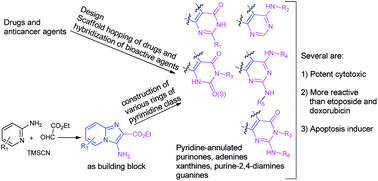
A set of novel pyridine-annulated analogs of purinones, adenines and their oxo/thio congeners, xanthines, guanines, and purine-2,4-diamines as potential anticancer agents was considered based on the scaffold-hopping and hybridization of known anticancer agents/drugs, purine derivatives and our recently developed imidazo-pyridine derivatives. Towards the synthesis of these compounds, a new approach involving a convenient preparation of 3-amino-2-carboxyethyl substituted imidazo[1,2-a]-pyridine and its use as a building block for the construction of fused rings was developed. The approach enabled the preparation of a number of compounds with relevant substitutions for each class. Several of pyridine-annulated adenine and its oxo/thio analogs, xanthine and purine-2,4-diamine were found to possess significant anticancer activities in kidney cancer cells and relatively less cytotoxicity to normal cells. They were relatively more active than the anticancer drugs etoposide and doxorubicin. A representative pyridine-annulated adenine derivative compound(22) was found to exert significant apoptosis.
Introduction
Cancer is the second major cause of death worldwide after cardiovascular diseases.1 Towards the development of new chemical entities (NCEs), various approaches are followed in medicinal chemistry research. Among them, scaffold/template hopping2 has been recognized as a valuable approach. Scaffold-hopping offers an opportunity to explore new chemotypes that can possess potentially similar biological activities and are outside the coverage of existing patents. Several marketed drugs and clinical trial agents have been discovered using this strategy. For example, the drug vardenafil, a PDE5-inhibitor, was discovered from the heterocyclic scaffold-hopping of another drug called sildenafil, likewise, the cyclooxygenase-inhibiting anti-inflammatory drugs etoricoxib and valdecoxib were discovered from celecoxib. A selective DPP-4 inhibitor called imigliptin was developed by a scaffold hopping-based design on alogliptin and a structure–activity guided optimization is currently under clinical trial.3 Another important approach is the molecular hybridization of chemotypes of known drugs and therapeutic agents.4 A well-known example is azatoxin, which was derived by the structural hybridization of the topoisomerase II-targeting drugs etoposide and ellipticine.5 Azatoxin with relevant substitutions showed better topoisomerase II inhibitory activity than etoposide or ellipticine. Therefore, an amalgamation of template-hopping and molecular hybridization on drugs and known bioactive agents can be an important design approach towards the discovery of new chemical entities (NCEs).
The purine class of compounds is known for its wide range of anticancer activities. These compounds interfere with various biochemical pathways such as the inhibition of CDK,6 PDE7 and Hsp90,8 and disruption9 of microtubule dynamics. Various marketed drugs10 also possess a purine nucleus (Fig. 1). QAP 1, an adenine derivative that shows ATP-competitive catalytic inhibition of topoisomerase II, is on clinical trial.11 In 2014, the US FDA approved a drug called idelalisib, an adenine derivative, for the treatment of various leukemia and lymphoma.12 For some specific pharmacological activities, purine derivatives have been found to be less potent, while their analogs with an annulated ring have been found to possess enhanced pharmacological activities and better pharmacokinetic profiles.13 These features incited medicinal and organic chemists to generate a biologically important class of compounds: heterocyclic-condensed purines. These compounds have been shown to possess various bioactivities14 such as, antihypertension,15 anti-inflammatory,13a human A3 adenosine receptor antagonism,13c and inhibition of PDE1/5 (ref. 16) and tyrosine kinase EphB4.17
 |
| Fig. 1 Pharmacological activities of purine-based compounds and drugs. | |
Recently, we developed imidazo-pyridines, designed based on the scaffold-hopping of existing drugs and known agents, as novel anticancer agents that induced apoptosis in the G1/S phase.18a As part of our program aimed at anticancer drug discovery,18 employing the amalgamation of scaffold-hopping and molecular hybridization design approaches on these structural classes of anticancer agents/drugs, purines (Fig. 1), annulated purines and imidazo-pyridines, we considered a series of novel pyridine-annulated purine compounds as potential anticancer agents (Fig. 2).
 |
| Fig. 2 Design of target compounds for potential anticancer activity. | |
Accordingly, studies for the establishment of a diversity-feasible building block approach for the synthesis of pyridine-annulated analogs of purinone, adenine and its oxo/thio congener, xanthine, purine-2,4-diamine and guanine derivatives, preparation of relevant substituted compounds, and evaluation of their anticancer activities were undertaken. Several of these new compounds were found to show significant anticancer activities, lower cytotoxicities to normal cells, and apoptosis in cancer cells.
Results and discussion
Chemistry
The known methods for the preparation of heterocyclic-annulated purine derivatives involve mostly the construction of new annulated rings on purines.13a,c,d,15,19 Following these methods, the preparation of pyridine-condensed purinones, adenines, xanthines, and purine-2,4-diamines with a variety of relevant substitutions would require, for each compound, a separate reaction route involving the construction of the annulated pyridine ring on the purine motif. In addition, the construction of the annulated pyridine ring is difficult. We considered a building block synthetic strategy via a suitably functionalized compound that could enable the construction of a fused pyrimidine ring and follow up derivatization towards the preparation of the whole series of designed compounds (Scheme 1a and b and 2). In this direction, we recently developed a method for the preparation of 3-amino-2-carboxyethyl substituted imidazo[1,2-a]-pyridine (I) in one step and explored its use as a building block in the synthesis of pyridine-fused purinones and adenines (Scheme 1a and b).20 In the present work, the synthesis of pyridine-annulated xanthines, guanines and purine-2,4-diamines was explored using the building block strategy. Some relevant pyridine-fused adenines (ten compounds) were prepared.
 |
| Scheme 1 (a) Synthesis of pyridine-condensed purinones (1–7).20 (b) Synthesis of pyridine-condensed adenines and their oxo/thio analogs (9–32).20 | |
 |
| Scheme 2 Synthesis of pyridine-condensed xanthines (33–41), thioxanthine (42), purin-2,4-diamines (43–46) and guanines (47–49). | |
The development of a method towards the preparation of pyridine-annulated xanthines was initially investigated. The preparation of a fused pyrimidine-dione ring on 2-aminobenzenecarboxamide by its reaction with urea at 190 °C is known.21 Following this method, the reaction of the amide derivative (II) of the building block (I) with urea was performed. However, the reaction did not proceed. Variation of the conditions including the addition of acid/base promoters also did not help. These attempts reveal the poor amine-nucleophilicity of the C2-carboxyethyl substituted imidazopyridinyl-3-amine towards urea. A set of reactions on the amide derivative (II) was then performed using various reactants such as di-ethyl/methyl carbonate, ethyl chloroformate, and Boc-anhydride, which could promote the domino conversions of the C3-amine to carbamate and the follow up trans-amidation. However, none of these reactions provided the construction of the fused pyrimidine-dione. In each case, either the reaction did not proceed or a mixture of non-isolable products was obtained on prolonging the reaction or enhancing the reaction temperature. The mass spectroscopic studies of the crude mixture obtained under higher temperature indicated the formation of the desired product as well as the unwanted product generated from the intermolecular trans-amidation of the derivative II. Therefore, this domino reaction is associated with a chemoselectivity issue also. The construction of the fused pyrimidine-dione ring directly from the building block (I) by reaction with urea, ethyl carbamate or isocyanate was then investigated. Gratifyingly, the reaction of the building block with isocyanate was found to be promising. In the reaction, bases were found to be more efficient as promoters than acids. In the evaluation of various bases, to our delight, the NaOEt-mediated reaction under microwave irradiation was found to be the most effective and afforded the pyridine-annulated xanthine in good yield (Scheme 2). This developed method enabled the preparation of various pyridine-annulated xanthines (33–41). The protocol was also suitable for the construction of thioxanthine (42) by reaction of the building block with isothiocyanate.
Towards the synthesis of pyridine-condensed guanines starting from the building block, condensed thioxanthine (42) was considered as a suitable substrate. The thio-methylation and SNAr with amines produced pyridine-enlarged guanines (47–49).
Purine-2,6-diamines are well known for their inhibitory activities against various kinases.22 This incited us to also investigate the use of the building block in the synthesis of pyridine-enlarged purines with 2,4-differential amine substitutions. In this aspect, we were required to obtain the chemoselective differential amination of the dichloro-derivative of the purine-annulated xanthine (VI), prepared by deoxychlorination of the pyridine-condensed xanthine (V). Compared to the 2-Cl group, the 4-Cl functionality was found to be more reactive for SNAr and underwent rapid reaction at room temperature, whereas the reaction of the 2-Cl with amine required microwave irradiation at a higher temperature and neat conditions. This differential reactivity of the 4-Cl vs. 2-Cl group enabled the preparation of the desired annulated purine-2,6-diamines (43–46) in moderate to good yields (Scheme 2). Using the developed building block synthetic strategy, we prepared new pyridine ring-condensed analogs of all known classes of purine scaffolds, purinone, adenine and its oxo/thio-congeners, xanthine, guanine, and purine-2,6-diamine. This approach is convenient, concise, and flexible for the incorporation of various substituents/functionalities, including those that are therapeutically important, into the products (Fig. 3). This approach bears potential application in the synthesis of various annulated purines.
 |
| Fig. 3 Synthesized pyridine-condensed purinones, adenines and their oxo/thio analogs, xanthines, guanines, and purine-2,4-diamines as potential anticancer agents. | |
Biological studies
Cytotoxicity assay. The cytotoxic activities of the synthesized compounds (1–49, Fig. 3) were measured by a well-known colorimetric-based MTT assay. A representative cancer cell, kidney cancer cell line (HEK 293T) and its corresponding normal cell (Vero) were considered for testing the cytotoxic activities. Doxorubicin (DOX) and etoposide (clinically used anticancer drugs) were used as positive controls. The cells were treated with doxorubicin, etoposide and the test compounds for 48 h, each with increasing concentrations, following a protocol described in the experimental section. The percent viability was measured (Table 1). The IC50 (concentration of compound to effect 50% cell growth inhibition in culture) values of 22, 23, 30, 32, 34, 37 and 46 were found to be relatively more active and showed remarkably low IC50 values (2.5, 3, 2.5, 3, 4, 3 and 2.5 μM, respectively) in HEK 293T as compared to doxorubicin (20 μM) or etoposide (22 μM). For the Vero cell line these compounds exhibited relatively higher IC50 (58, 54, 50, 60, 56.5, 45 and 40 μM, respectively) (Fig. 4). To further confirm the cytotoxicity, a clonogenic assay was performed according to the protocol described in the experimental section. Table 2 demonstrates the LC50 (concentration of compound to cause 50% death of cells) values. For compounds 22, 23, 30, 32, 34, 37 and 46, the LC50 values were found to be 1.4, 1.1, 1.7, 1.2, 3.4, 1.4 and 2.2 μM respectively. A dose dependent decrease in colony formation was observed (Fig. 5).
 |
| Fig. 4 Cell survivals in the MTT assay for the selected compounds 22, 23, 30, 32, 34, 37 and 46. Cells were treated with the investigated compounds and doxorubicin of various concentrations for 48 h. Symbols, , , , represent HEK 293T/compound, Vero/compound, HEK 293T/DOX and Vero/DOX, respectively. Data presented is the mean ± SD of three independent sets of experiments. | |
Table 1 IC50 (μM) values for the cytotoxicities in HEK 293T and Vero cells
Compound |
IC50 (μM) HEK 293T |
IC50 (μM) Vero |
Compound |
IC50 (μM) HEK 293T |
IC50 (μM) Vero |
1 |
49 ± 2.0 |
60 ± 1.9 |
28 |
8 ± 1.7 |
40 ± 1.7 |
2 |
60 ± 1.5 |
50 ± 1.0 |
29 |
2.5 ± 1.1 |
44 ± 2.0 |
3 |
60 ± 1.8 |
50 ± 1.1 |
30 |
2.5 ± 0.5 |
50 ± 1.5 |
4 |
29 ± 3.0 |
55 ± 2.1 |
31 |
3 ± 1.6 |
38 ± 2.2 |
5 |
25 ± 2.0 |
49 ± 2.0 |
32 |
3 ± 1.5 |
60 ± 1.7 |
6 |
30 ± 1.5 |
55 ± 1.8 |
33 |
10 ± 2.0 |
50 ± 2.5 |
7 |
28 ± 1.7 |
58 ± 2.5 |
34 |
4 ± 0.7 |
56.5 ± 1.0 |
8 |
12 ± 1.8 |
48 ± 1.8 |
35 |
4 ± 1.8 |
35 ± 1.5 |
9 |
5 ± 2.0 |
49 ± 1.1 |
36 |
12 ± 1.3 |
42 ± 2.0 |
10 |
8 ± 1.6 |
30 ± 1.2 |
37 |
3 ± 0.5 |
45 ± 2.2 |
11 |
5.2 ± 3.1 |
28 ± 1.0 |
38 |
4 ± 2.0 |
55 ± 1.1 |
12 |
7 ± 3.0 |
29 ± 2.1 |
39 |
18 ± 2.5 |
60 ± 2.5 |
13 |
6 ± 2.5 |
25.5 ± 1.0 |
40 |
12 ± 1.8 |
58 ± 1.2 |
14 |
6.2 ± 2.2 |
27 ± 1.5 |
41 |
4 ± 2.1 |
38 ± 0.7 |
15 |
3.0 ± 2.1 |
29 ± 2.1 |
42 |
5 ± 2.0 |
36 ± 0.5 |
16 |
6.0 ± 2.0 |
24.2 ± 0.5 |
43 |
4 ± 2.2 |
24 ± 0.2 |
17 |
1.5 ± 1.8 |
29.5 ± 0.8 |
44 |
4 ± 1.8 |
18 ± 0.5 |
18 |
4 ± 2.0 |
30 ± 1.1 |
45 |
6 ± 1.0 |
24 ± 1.1 |
19 |
8 ± 1.9 |
22 ± 1.2 |
46 |
2.5 ± 1.5 |
40 ± 2.0 |
20 |
4 ± 2.2 |
60 ± 1.0 |
47 |
2.5 ± 0.5 |
8 ± 2.1 |
21 |
5 ± 2.5 |
38 ± 0.8 |
48 |
8.5 ± 1.1 |
14 ± 2.5 |
22 |
2.5 ± 1.0 |
58 ± 0.5 |
49 |
6 ± 1.0 |
10 ± 2.0 |
23 |
3 ± 1.1 |
54 ± 1.1 |
IV |
8 ± 1.9 |
44 ± 2.0 |
24 |
4 ± 1.7 |
50 ± 1.2 |
V |
10 ± 2.1 |
60 ± 2.0 |
25 |
4 ± 2.0 |
52 ± 1.0 |
VI |
8 ± 1.0 |
42 ± 2.1 |
26 |
5 ± 2.2 |
25 ± 0.5 |
VII |
6 ± 1.1 |
50 ± 1.1 |
27 |
5 ± 1.5 |
44 ± 0.2 |
VIII |
6.5 ± 1.5 |
60 ± 2.0 |
Doxorubicin |
20 ± 1.1 |
60 ± 1.1 |
Etoposide |
22 ± 1.5 |
50 ± 1.5 |
Table 2 LC50 (μM) values for the clonogenic assay using HEK 293T cells
Compound |
LC50 (μM) HEK 293T |
Compound |
LC50 (μM) HEK 293T |
22 |
1.4 ± 0.5 |
34 |
3.4 ± 1.5 |
23 |
1.1 ± 0.8 |
37 |
1.4 ± 0.5 |
30 |
1.7 ± 1.1 |
46 |
2.2 ± 1.0 |
32 |
1.2 ± 0.2 |
|
|
 |
| Fig. 5 Clonogenic cell survival in HEK 293T cells. , , , , , , symbols represent compounds 22, 23, 30, 32, 34, 37 and 46, respectively. Data presented is the mean ± SD of three individual sets of experiments. | |
Measurement of apoptosis in HEK 293T cells by Annexin V/FITC assay. The apoptosis measurement was performed by Annexin-V/FITC staining followed by FACS after treatment with compound 22 for 48 h. Fig. 6 demonstrates the distribution of cells in various phases of apoptosis. Approximately 46.8 percent apoptotic cells (Q3) were observed for cells treated with 2.5 μM of the compound and more than 54 percent necrotic cells (Q4) were observed for cells treated with 4 μM of the compound. Thus, the result indicated that cells were moving towards the necrotic phase followed by a true apoptotic phase with increasing concentration of compound 22 (Fig. 6).
 |
| Fig. 6 Measurement of apoptosis by the Annexin-V/FITC method. Cells were treated with compound 22 of various concentrations for 48 h and the experiment was carried out as described in the experimental section. Data given here is one of three separate experiments. | |
Expressions of CASPASE 3. Increased expression of CASPASE 3, an important marker, is an indication of apoptosis. To check whether exposure to compound 22 caused the apoptosis in the HEK 293T cells, we measured the CASPASE 3 expression by immunostaining CASPASE 3 after exposure to compound 22. HEK 293T cells were treated with compound 22 of increasing concentrations and probed with the CASPASE-3 antibody (cat #9662 from Cell Signaling, MA, USA). They were re-probed with secondary anti-rabbit conjugated to TRITC (tetramethylrhodamine, a red fluorescent dye) followed by counterstaining with DAPI and visualized under a fluorescence microscope. Increasing expression of CASPASE-3 (red fluorescence) was noted with an increasing dose of the compound (Fig. 7).
 |
| Fig. 7 Compound 22 increased the CASPASE 3 expression in HEK 293T cells. The left and middle panels represent the cells stained with DAPI and CASPASE 3/TRITC (tetramethylrhodamine, a red fluorescent dye) and the right panel represents the MERGED image of both. Images were taken under a fluorescence microscope at 40× magnification. Images presented here are the best of three individual sets of experiments (scale bar 10 μm). | |
Measurement of apoptosis using DAPI stain. To further confirm the apoptotic effect of compound 22, a nuclear staining experiment using HEK 293T cells was performed. The cells were treated with compound 22 of increasing concentrations and the fluorescence was observed after staining with DAPI. Significantly higher chromatin condensation and nuclear fragmentation in treated cells compared to untreated cells were observed and these were found to enhance with increasing concentrations of the compound (Fig. 8A and B). Approximately 4, 13 and 15 fold increases in apoptotic nuclei in cells treated with the compound at 1, 2.5 and 4 μM concentrations compared to untreated cells were observed.
 |
| Fig. 8 (A) DAPI nuclear staining: images were taken under a fluorescence microscope at 40× magnification (scale bar 10 μm). (B) Bar-diagram presentation is the number of apoptotic and non-apoptotic nuclei taken from (A). | |
Experimental section
Chemistry
General considerations. The starting materials and solvents were used as received from commercial sources without further purification. The 1H and 13C NMR spectra were recorded in CDCl3/DMSO-d6/CD3OD solvents on 400 MHz spectrometer using TMS as the internal standard. HRMS was measured using a TOF analyzer. The melting points determined are uncorrected. Microwave assisted reactions were carried out in a sealed reaction vessel using a Biotage Initiator.Ethyl 3-aminoimidazo[1,2-a]pyridine-2-carboxylate (I), 3-aminoimidazo[1,2-a]pyridine-2-carboxamide (II), N-benzoylimidazo[1,2-a]pyridine-3-amine-2-carboxamide (III), pyrido[1,2-e]purin-4(3H)-one (1–7), pyrido[1,2-e]purin-4(3H)-one (8), 4-chloropyrido[1,2-e]purine (IV), pyrido[1,2-e]purin-4-amines (9, 11, 12, 14, 17, 19, 20, 22, 25, 27, 28, 29, 31 and 32) were prepared by our reported procedures.20 Some more relevant pyridine-fused adenines (10, 13, 15, 16, 18, 21, 23, 24, 26 and 30) were prepared by following our reported procedures.20
Representative experimental procedure for the synthesis of 3-benzylpyrido[1,2-e]purine-2,4(1H,3H)-dione (33). To a solution of ethyl 3-aminoimidazo[1,2-a]pyridine-2-carboxylate (I, 205 mg, 1 mmol) in anhyd. ethanol (2 mL) in a vial, were added subsequently benzyl isocyanate (200 mg, 1.5 mmol) and sodium ethoxide (68 mg, 1 mmol) under a flow of N2. The reaction mixture was heated at 120 °C under microwave irradiation. After completion of the reaction (monitored by TLC, 20 min), the solvent was evaporated under a vacuum. The column chromatographic purification of the crude mass on neutral alumina provided 3-benzylpyrido[1,2-e]purine-2,4(1H,3H)-dione (33, 181 mg, 62%).The other compounds (34–42, Scheme 2) were prepared following this procedure.
Experimental procedure for the synthesis of 3-benzyl-2-(methylthio)pyrido[1,2-e]purin-4(3H)-one (VIII, Scheme 2). 3-Benzyl-2-thiopyrido[1,2-e]purin-4(1H)-one (42) was taken in an ethanol–water mixture (1
:
1, 2 mL) in a round bottom flask. Sodium hydroxide (40 mg, 1 mmol) and methyl iodide (142 mg, 1 mmol) were subsequently added to it. The reaction mixture was heated at 60 °C in a closed system. After completion of the reaction (monitored by TLC, 1 h) the solvent mixture was evaporated under a vacuum. The column chromatographic purification of the crude mass on neutral alumina provided 3-benzyl-2-(methylthio)pyrido[1,2-e]purin-4(3H)-one (VIII, 209 mg, 65%).
Representative experimental procedure for the synthesis of 3-benzyl-2-(hexylamino)pyrido[1,2-e]purin-4(3H)-one (47, Scheme 2). 3-Benzyl-2-(methylthio)pyrido[1,2-e]purin-4(3H)-one (VIII, 322 mg, 1 mmol) was taken in a sealed tube and hexylamine (2 mL) was added to it and the tube was closed. The reaction mixture was heated at 170 °C. After maximum conversion in the reaction (monitored by TLC, 8 h), the column chromatographic purification of the crude mass on neutral alumina provided 3-benzyl-2-(hexylamino)pyrido[1,2-e]purin-4(3H)-one (47, 199 mg, 53%).The products 48 and 49 were prepared following this procedure.
Experimental procedure for the synthesis of pyrido[1,2-e]purine-2,4(1H,3H)-dione (V). To a solution of ethyl 3-aminoimidazo[1,2-a]pyridine-2-carboxylate (I, 205 mg, 1 mmol) in phenol (2 mL) in a round bottom flask, urea (315 mg, 5 mmol) was added. The reaction mixture was heated at 150 °C. After the completion of the reaction (monitored by TLC, 24 h), the column chromatographic purification of the crude mass on neutral alumina provided pyrido[1,2-e]purine-2,4(1H,3H)-dione (V, 102 mg, 50%).
Experimental procedure for the synthesis of 2,4-dichloropyrido[1,2-e]purine (VI). Pyrido[1,2-e]purine-2,4(1H,3H)-dione (V, 203 mg, 1 mmol) was taken in a round bottom flask. N,N-Diethylaniline (149 mg, 1 mmol) and POCl3 (2 mL) were added subsequently to it under a flow of N2. The reaction mixture was heated at reflux. After reaction completion (monitored by TLC, 4 h), the reaction mixture was poured into ice cold water (30 mL). 20% aq. NaOH solution was added dropwise till pH 8 was achieved. Then the mixture was extracted with EtOAc (2 × 40 mL). The combined organic layers were washed with water, dried with anhyd. Na2SO4, and concentrated under a vacuum. The column chromatographic purification of the crude mass on silica gel provided 2,4-dichloropyrido[1,2-e]purine (VI, 127 mg, 53%).
Representative experimental procedure for the synthesis of 2-morpholino-N-phenethylpyrido[1,2-e]purin-4-amine (43, Scheme 2). To a solution of 2,4-dichloropyrido[1,2-e]purine (VI) in THF (1 mL) in a round bottom flask, phenethylamine (121 mg, 1 mmol) and triethylamine (101 mg, 1 mmol) were added. The reaction mixture was stirred at room temperature (25–27 °C). After the completion of the reaction (monitored by TLC, 10 min), the solvent was evaporated under a vacuum. To this crude reaction mixture morpholine (1 mL) was added and the mixture was further heated at 120 °C under microwave irradiation. After the completion of the reaction (monitored by TLC, 10 min), the column chromatographic purification of the crude mass on neutral alumina provided 2-morpholino-N-phenethylpyrido[1,2-e]purin-4-amine (43, 307 mg, 82%).The other products (44–46, Scheme 2) were prepared following this procedure. 2-Chloro-N-phenethylpyrido[1,2-e]purin-4-amine (VII) was obtained only by 4-amination of compound VI. The column chromatographic purification of the crude mass on neutral alumina provided VII in 90% isolated yield.
Biology
Cell line and culture conditions. HEK 293T (cat #CRL-11268), a kidney cancer cell line, and Vero, normal kidney cells (cat #CCL-81), were grown in a DMEM medium supplemented with 10% FBS, 1% antibiotic (100 units of penicillin and 10 mg streptomycin per mL in 0.9% normal saline) and maintained in 5% carbon dioxide in humidified conditions at 37 °C. All cell culture reagents were purchased from Hi Media, Mumbai, India.
MTT assay. The cell proliferation ability of the investigated compounds (1–49) was measured by performing a colorimetric based assay using MTT reagent. Approximately 10
000–12
000 cells per well were seeded in a 96 well tissue culture plate and were incubated for 24 h. After 24 h of incubation the cells were exposed to different concentrations of the investigated compounds for 48 h. Then the cells were washed with 1× PBS followed by the addition of 100 μL of 0.05% MTT reagent to each well. The plate was incubated overnight at 37 °C. Purple formazan crystals were formed which were dissolved using 10% NP-40 with 4 mM HCl. The plate was incubated for 1 h at 37 °C and then the intensity of the developed color was measured at 570 nm using a microplate reader (Barthold, Germany). The data were calculated and a graph representing the percent viability was plotted in comparison to the control. The data presented are the mean ± SD of at least three experiments.
Clonogenic assay. The colony forming abilities of the cells after treatment with the investigated compounds were tested by performing a clonogenic assay. This assay was carried out to check the anti-proliferating or colony forming ability of the selected investigated compounds. Approximately 500 cells per well of HEK 293T, a kidney cancer cell line, were seeded in 12 well plates. The plates were incubated for 24 h. The cells were treated with various concentrations of the selected compounds for 48 h. Then the media was aspirated and replaced with fresh media. The plates were incubated further to allow the cells to grow for 5–6 doublings by changing the media every 72 h. Then the media was removed and the cells were washed with 1× PBS followed by the addition of 0.2% crystal violet stain to each well of the plates. It was kept for 1 h and the excess stain was removed by washing with 1× PBS. The plates were air dried and the colonies were counted under a gel documentation system (UVP, Germany). The colonies formed in the treated wells were counted against the untreated well that served as the control (100% survival) and a graph was plotted representing the percent survival of the cells. Data presented here are the mean ± SD of three different experiments.
Annexin-V/FITC staining. Annexin-V/FITC staining is used to detect the distribution of cells in early, apoptotic and late apoptotic (necrosis) phases. For this assay, the cells were seeded in a 6 well cell culture plate at a density of 1 × 10⁵ cells per well. 70–80 percent confluent grown cells were treated with varying concentrations of compound 22 for 48 h. Then, the cells were harvested, washed and stained with Annexin-V/FITC and PI according to manufacturer’s protocol (Sigma). Finally, the stained cells were sorted by FACS using the PE-A (to capture the PI stained cells) and FITC-A (to capture the Annexin-V stained cells) channels. According to principle, Annexin-V dye stains living and apoptotic cells, whereas PI stains all cells including necrotic cells. The percentage of cells population in Q1, Q2, Q3 and Q4 represent the normal, early apoptotic, apoptotic and necrotic cells, respectively.
Immunofluorescence of CASPASE 3. Immunofluorescence of CASPASE 3 was performed to check the expression of CASPASE 3 after the treatment of the cells with compound 22. HEK 293T cells (1 × 10⁴) were seeded on sterile cover slips and allowed to attain 50–60% confluency. The cells were treated with various concentrations of the drug for 48 h. The drug treated media were removed and the cells were washed with 1× PBS. They were then fixed with acetone–methanol (1
:
1) for 15 min at −20 °C. The fixative solution was removed and the cells were blocked for 30 min at 37 °C with 2% BSA in 0.02% Triton X 100 in 1× PBS. The cells were stained with CASPASE 3 antibody (cat #9662 from Cell Signaling, MA, USA) and incubated for 3 h. The cells were washed with 1× PBS followed by immunostaining with secondary antibody (anti-rabbit) conjugated with TRITC and incubated for 1 h at 37 °C. After completion of the incubation time, the cells were washed with 1× PBS and stained with DAPI. The cover slips containing cells were washed properly with 1× PBS, air dried and mounted on slides. Images were taken using a fluorescence microscope.
DAPI nuclear staining assay. This assay helps in the detection of apoptosis and nuclear fragmentation. HEK 293T cells were seeded in a 96 well tissue culture plate (1 × 10⁴ per well) and incubated for 24 h. The cells were treated with various concentrations of compound 22 and incubated for another 48 h. Then, the plates were washed with 1× PBS and fixed with acetone–methanol (1
:
1) and kept at −20 °C for 15 min. After removing the fixative, the cells were washed with 1× PBS and stained with 4,6-diamidino-2-phenylindole (DAPI). They were again incubated for another 30 min in the dark at 37 °C. After the end of the incubation time, the slides were washed once with 1× PBS and then visualized under a fluorescence microscope. The data provided here are from one of three independent experiments.
Conclusions
A set of novel pyridine annulated purinones, adenines and their oxo/thio analogs, xanthines, guanines, and purine-2,4-diamines were considered as potential anticancer agents based on scaffold-hopping and hybridization of known drugs and bioactive agents. They, with relevant substitutions, were synthesized conveniently via a building block strategy with 3-amino-2-carboxyethyl substituted imidazo[1,2-a]-pyridine as the building block. Their MTT-cytotoxicity and clonogenic cell survival studies revealed several of the pyridine-annulated purines were significantly active in terms of anticancer properties and relatively less cytotoxic to normal cells. They were found to be relatively more potent than the anticancer drugs etoposide and doxorubicin. A representative compound (22) exerted significant apoptosis. The present work has potential application in the synthesis of the annulated purine class of compounds via a building block strategy and will incite further investigation towards the exploration of bioactive heterocyclic-annulated purines. Further optimization of the new anticancer agents is underway.
Acknowledgements
We gratefully acknowledge financial support from DST, New Delhi for this investigation. VC is thankful to CSIR, New Delhi for his fellowship.
Notes and references
- A. Jemal, F. Bray, M. M. Center, J. Ferlay, E. Ward and D. Forman, Ca-Cancer J. Clin., 2011, 61, 69 CrossRef PubMed.
- S. Hongmao, G. Tawa and A. Wallqvist, Drug Discovery Today, 2012, 17, 310 CrossRef PubMed.
- C. Shu, H. Ge, M. Song, J.-h. Chen, H. Zhou, Q. Qi, F. Wang, X. Ma, X. Yang, G. Zhang, Y. Ding, D. Zhou, P. Peng, C.-k. Shih, J. Xu and F. Wu, ACS Med. Chem. Lett., 2014, 5, 921 CrossRef CAS PubMed.
-
(a) B. Meunier, Acc. Chem. Res., 2008, 41, 69 CrossRef CAS PubMed;
(b) A. Müller-Schiffmann, J. März-Berberich, A. Andreyeva, R. Rönicke, D. Bartnik, O. Brener, J. Kutzsche, A. H. C. Horn, M. Hellmert, J. Polkowska, K. Gottmann, K. G. Reymann, S. A. Funke, L. Nagel-Steger, C. Moriscot, G. Schoehn, H. Sticht, D. Willbold, T. Schrader and C. Korth, Angew. Chem., Int. Ed., 2010, 49, 8743 CrossRef PubMed;
(c) J. A. Lenhart, X. Ling, R. Gandhi, T. L. Guo, P. M. Gerk, D. H. Brunzell and S. Zhang, J. Med. Chem., 2010, 53, 6198 CrossRef CAS PubMed.
-
(a) S. D. Cline, T. L. Macdonald and N. Osheroff, Biochemistry, 1997, 36, 13095 CrossRef CAS PubMed;
(b) E. Solary, F. Leteurtre, K. D. Paull, D. Scudiero, E. Hamel and Y. Pommier, Biochem. Pharmacol., 1993, 45, 2449 CrossRef CAS PubMed;
(c) J. S. Madalengoitia, J. J. Tepe, K. A. Werbovetz, E. K. Lehnert and T. L. Macdonald, Bioorg. Med. Chem., 1997, 5, 1807 CrossRef CAS PubMed.
-
(a) L. Havlicek, J. Hanus, J. Vesley, S. Leclerc, L. Meijer, G. Shaw and M. Strnad, J. Med. Chem., 1997, 40, 408 CrossRef CAS PubMed;
(b) R. Jorda, L. Havlicek, I. W. McNae, M. D. Walkinshaw, J. Voller, A. Sturc, J. Navratilova, M. Kuzma, M. Mistrik, J. Bartek, M. Strnad and V. Krystof, J. Med. Chem., 2011, 54, 2980 CrossRef CAS PubMed.
- P. Raboisson, C. Lugnier, C. E. Muller, J.-M. Reimund, D. Schultz, G. Pinna, A. Le Bec, H. Basaran, L. Desaubry, F. Gaudiot, M. Selouma and J.-J. Bourguignon, Eur. J. Med. Chem., 2003, 38, 199 CrossRef CAS PubMed.
- L. Llauger, H. He, J. Kim, J. Aguirre, N. Rosen, U. Peters, P. Davies and G. Chiosis, J. Med. Chem., 2005, 48, 2892 CrossRef CAS PubMed.
- Y.-T. Chang, S. M. Wignall, G. R. Rosania, N. S. Gray, S. R. Hanson, A. I. Su, J. Merlie Jr, H.-S. Moon, S. B. Sangankar, O. Perez, R. Heald and P. G. Schultz, J. Med. Chem., 2001, 44, 4497 CrossRef CAS PubMed.
- M. Hoffmann, M. Chrzanowska, T. Hermann and J. Rychlewski, J. Med. Chem., 2005, 48, 4482 CrossRef CAS PubMed.
- P. Chène, J. Rudloff, J. Schoepfer, P. Furet, P. Meier, Z. Qian, J.-M. Schlaeppi, R. Schmitz and T. Radimerski, BMC Chem. Biol., 2009, 9, 1 CrossRef PubMed.
- R. R. Furman, J. P. Sharman, S. E. Coutre, B. D. Cheson, J. M. Pagel, P. Hillmen, J. C. Barrientos, A. D. Zelenetz, T. J. Kipps, I. Flinn, P. Ghia, H. Eradat, T. Ervin, N. Lamanna, B. Coiffier, A. R. Pettitt, S. Ma, S. Stilgenbauer, P. Cramer, M. Aiello, D. M. Johnson, L. L. Miller, D. Li, T. M. Jahn, R. D. Dansey, M. Hallek and S. M. O’Brien, N. Engl. J. Med., 2014, 370, 997 CrossRef CAS PubMed.
-
(a) D. J. Blythin, J. J. Kaminski, M. S. Domalski, J. Spitler, D. M. Solomon, D. J. Conn, S. C. Wong, L. L. Verbiar and L. A. Bober, J. Med. Chem., 1986, 29, 1099 CrossRef CAS PubMed;
(b) E.-M. Priego, J. F. D. Kuenzel, A. P. IJzerman, M.-J. Camarasa and M.-J. Pérez-Pérez, J. Med. Chem., 2002, 45, 3337 CrossRef CAS PubMed;
(c) C. E. Müller, M. Thorand, R. Qurishi, M. Diekmann, K. A. Jacobson, W. L. Padgett and J. W. Daly, J. Med. Chem., 2002, 45, 3440 CrossRef;
(d) P. G. Baraldi, D. Preti, M. A. Tabrizi, F. Fruttarolo, R. Romagnoli, N. A. Zaid, A. R. Moorman, S. Merighi, K. Varani and P. A. Borea, J. Med. Chem., 2005, 48, 4697 CrossRef CAS PubMed.
- H. Suzuki, M. Yamamoto, S. Shimura, K. Miyamoto, K. Yamamoto and S. Sawanishi, Chem. Pharm. Bull., 2002, 50, 1163 CrossRef CAS PubMed.
- H.-S. Ahn, A. Bercovici, G. Boykow, A. Bronnenkant, S. Chackalamannil, J. Chow, R. Cleven, J. Cook, M. Czarniecki, C. Domalski, A. Fawzi, M. Green, A. Gündes, G. Ho, M. Laudicina, N. Lindo, K. Ma, M. Manna, B. McKittrick, B. Mirzai, T. Nechuta, B. Neustadt, C. Puchalski, K. Pula, L. Silverman, E. Smith, A. Stamford, R. P. Tedesco, H. Tsai, D. Tulshian, H. Vaccaro, R. W. Watkins, X. Weng, J. T. Witkowski, Y. Xia and H. Zhang, J. Med. Chem., 1997, 40, 2196 CrossRef CAS PubMed.
-
(a) C. D. Boyle, R. Xu, T. Asberom, S. Chackalamannil, J. W. Clader, W. J. Greenlee, H. Guzik, Y. Hu, Z. Hu, C. M. Lankin, D. A. Pissarnitski, A. W. Stamford, Y. Wang, J. Skell, S. Kurowski, S. Vemulapalli, J. Palamanda, M. Chintala, P. Wu, J. Myers and P. Wang, Bioorg. Med. Chem. Lett., 2005, 15, 2365 CrossRef CAS PubMed;
(b) G. Xia, J. Li, A. Peng, S. Lai, S. Zhang, J. Shen, Z. Liu, X. Chen and R. Ji, Bioorg. Med. Chem. Lett., 2005, 15, 2790 CrossRef CAS PubMed.
- K. Lafleur, D. Huang, T. Zhou, A. Caflisch and C. Nevado, J. Med. Chem., 2009, 52, 6433 CrossRef CAS PubMed.
-
(a) A. T. Baviskar, C. Madaan, R. Preet, P. Mohapatra, V. Jain, A. Agarwal, S. K. Guchhait, C. N. Kundu, U. C. Banerjee and P. V. Bharatam, J. Med. Chem., 2011, 54, 5013 CrossRef CAS PubMed;
(b) S. Kandekar, R. Preet, M. Kashyap, R. Prasad, M. U. P. Mohapatra, D. Das, S. R. Satapathy, S. Siddharth, V. Jain, M. Choudhuri, C. N. Kundu, S. K. Guchhait and P. V. Bharatam, ChemMedChem, 2013, 8, 1873 CrossRef CAS PubMed;
(c) N. Sanghai, V. Jain, R. Preet, S. Kandekar, S. Das, N. Trivedi, P. Mohapatra, G. Priyadarshani, M. Kashyap, D. Das, S. R. Satapathy, S. Siddharth, S. K. Guchhait, C. N. Kundu and P. V. Bharatam, MedChemComm, 2014, 5, 764 RSC;
(d) M. Kashyap, D. Das, R. Preet, P. Mohapatra, S. R. Satapathy, S. Siddharth, C. N. Kundu and S. K. Guchhait, Bioorg. Med. Chem. Lett., 2012, 22, 2474 CrossRef CAS PubMed;
(e) M. Kashyap, S. Kandekar, A. T. Baviskar, D. Das, R. Preet, P. Mohapatra, S. R. Satapathy, S. Siddharth, S. K. Guchhait, C. N. Kundu and U. C. Banerjee, Bioorg. Med. Chem. Lett., 2013, 23, 934 CrossRef CAS PubMed.
-
(a) H. Sawanishi, H. Suzuki, S. Yamamoto, Y. Waki, S. Kasugai, K. Ohya, N. Suzuki, K.-I. Miyamoto and K. Takagi, J. Med. Chem., 1997, 40, 3248 CrossRef CAS PubMed;
(b) P. Virta, A. Koch, M. U. Roslund, P. Mattjus, E. Kleinpeter, L. Kronberg, R. Sjöholm and K. D. Klika, Org. Biomol. Chem., 2005, 3, 2924 RSC;
(c) S. K. Guchhait and C. Madaan, Tetrahedron Lett., 2011, 52, 56 CrossRef CAS;
(d) I. Cerna, R. Pohl, B. Klepetarova and M. Hocek, J. Org. Chem., 2010, 75, 2302 CrossRef CAS PubMed.
- S. K. Guchhait and V. Chaudhary, Org. Biomol. Chem., 2014, 12, 6695 Search PubMed.
- S. W. Schneller, A. C. Ibay, W. J. Christ and R. F. Burns, J. Med. Chem., 1989, 32, 2247 CrossRef CAS PubMed.
- N. S. Gray, L. Wodicka, A. M. W. H. Thunnissen, T. C. Norman, S. Kwon, F. H. Espinoza, D. O. Morgan, G. Barnes, S. LeClerc, L. Meijer, S. H. Kim, D. J. Lockhart and P. G. Schultz, Science, 1998, 281, 533 CrossRef CAS PubMed.
Footnote |
† Electronic supplementary information (ESI) available: General information, spectral data, and NMR spectra (1H and 13C) of compounds. See DOI: 10.1039/c5ra00052a |
|
This journal is © The Royal Society of Chemistry 2015 |
Click here to see how this site uses Cookies. View our privacy policy here.