
A facile label-free colorimetric aptasensor for ricin based on the peroxidase-like activity of gold nanoparticles†
Jingting Huab,
Pengjuan Niab,
Haichao Daiab,
Yujing Suna,
Yilin Wangab,
Shu Jiangab and
Zhuang Li*a
aState Key Laboratory of Electroanalytical Chemistry, Changchun Institute of Applied Chemistry, Chinese Academy of Sciences, Changchun, Jilin 130022, PR China. E-mail: zli@ciac.jl.cn; Fax: +86 431 85262057; Tel: +86 431 85262057
bUniversity of Chinese Academy of Sciences, Beijing, 100039, PR China
First published on 28th January 2015
Abstract
AuNPs possess peroxidase-like activity that could catalyze 3,3,5,5-tetramethylbenzidine (TMB) in the presence of H2O2, leading to color change of the solution. Herein we propose a simple and sensitive colorimetric aptasensor for the quantitative analysis of ricin by using AuNPs. It is shown that the peroxidase-like activity of AuNPs can be improved by surface activation with target-specific aptamers. However, with target molecules, the aptamer is desorbed from the AuNPs' surface, resulting in a decrease of the catalytic abilities of AuNPs. The color change of the solution was relevant to the target concentration, and this can be judged by the naked eye and monitored by UV-vis spectrometry. The linear range for the current analytical system was from 0.05 nM to 10 nM. The corresponding limit of detection (LOD) was 0.05 nM. Some other proteins such as thrombin (Th), glucose oxidase (GOx), and bovine albumin (BSA) all had a negligible effect on the determination of ricin. What is more, several practical samples spiked with ricin were analyzed using the proposed method with excellent recoveries. This colorimetric aptasensor is superior to the other conventional methods owing to its simplicity, low cost, and high sensitivity.
1. Introduction
Ricin is a highly toxic protein which is isolated from the seeds of the castor bean Ricinus communis.1 It is categorized as a type II ribosome inactivating protein, which contains a catalytic A chain with N-glycosidase activity and a lectin B chain assisting cellular uptake.2,3 The ricin A chain is toxic to cells which inhibits the protein synthesis, thus leading to cell death. The ricin B chain is essential for binding to galactosyl residues on the cell surface and it is responsible for delivering the ricin A chain into the cell via endocytosis.4,5 It is estimated that the LD50 for ricin in humans is approximately 5 to 10 μg kg−1 through inhalation and there is no known antidote for it.6Ricin has received significant attention since the infamous umbrella tip assassination of Georgi Markov demonstrated the extreme lethality of the toxin.7,8 Recently, several terrorist groups have experimented with ricin and caused several incidents of the poison's being mailed to U.S. politicians. On May 29, 2013, two anonymous letters sent to New York City Mayor Michael Bloomberg contained traces of ricin. Another letter containing ricin was also alleged to have been sent to American President Barack Obama at the same time. Due to its ease of extraction in large quantities from castor beans, which are processed worldwide on an industrial scale, there is a real threat of ricin being used as a biological warfare agent.9 Therefore, development of a rapid and sensitive detection method to monitor ricin has become more urgent.
Contemporary ricin detection methods are using such as Fourier transform near-infrared reflectance spectroscopy assay,10 capillary electrophoresis assay,11 surface-enhanced Raman spectroscopy12 and antibody-based immunoassays.13–19 Although, these assays are effective for ricin detection, most of these analytical methods rely on sophisticated instruments and skilled manpower, making such approaches impractical for on-site detection.
Of the alternatives to antibody-based sensing techniques, aptamer-based methods have become popular over the past decades due to their inherent selectivity, affinity, and multifarious advantages over the traditional recognition elements. As a new class of single-stranded DNA/RNA molecules, aptamers are selected in vitro by the systematic evolution of the ligand by the exponential enrichment (SELEX) process from random-sequence nucleic acid libraries. These molecular ligands have the ability to recognize their cognate targets with high affinity and specificity. This property makes aptamers an ideal tool to detect a variety of target molecules including proteins, drugs, small molecules, inorganic ions and even cells.20–25
Recently, increasing attention has been paid to the nanoscaled peroxidase mimetics and their potential applications in bioanalysis26–31 since the first Fe3O4 magnetic nanoparticles-based peroxidase was reported.32 A variety of nanomaterials have been also demonstrated to possess intrinsic peroxidase-like activity. Although gold is usually viewed as an inert metal, surprisingly it has been found that AuNPs possess intrinsic peroxidase-like activity.33,34 The findings showed that a catalytic AuNPs-based strategy has enormous potentials in detection and can be exploited to develop biosensors for a wide variety of target analytes.
Enlightened by the above facts, we propose a colorimetric aptasensor for ricin based on the peroxidase-like activity of AuNPs. Ricin binding aptamer (RBA) could be absorbed onto the surface of AuNPs and improve the ability of AuNPs to catalyze 3,3,5,5-tetramethylbenzidine (TMB) in the presence of H2O2 and lead to the significant enhancement of the absorbance. However, in the presence of ricin, the RBA undergoes target-responsive structural changes followed by desorption from the AuNPs surface to allow an aptamer-target binding event, resulting in a decrease in the catalytic abilities of AuNPs. The high selective and sensitive detection of ricin was possible using the naked eye or monitored by UV-vis spectrometer. Furthermore, the proposed method did not require any modification of the aptamer, making it time-saving and cost-effective.
2. Experimental
2.1. Reagent and chemicals
Ricin was purchased from Beijing Hapten and Protein Biomedical Institute (Beijing, China). The sequence of ricin binding aptamer (RBA) was 5′-ACACCCACCGCAGGCAGACGCAACGCCTCGGAGACTAGCC-3′.35 The ssDNA oligonucleotides were synthesized by Shanghai Sangon Biotechnology Co. Ltd. (Shanghai, China) and the lyophilized powder was dissolved in distilled water and before use it was stored at 4 °C. The concentration of the oligonucleotide was determined by measuring the UV absorbance at 260 nm. Chloroauric acid (HAuCl4·4H2O) was obtained from Shanghai Chemical Reagent Company (Shanghai, China). 3,3,5,5-Tetramethylbenzidine (TMB) and hexadecyl trimethyl ammonium bromide (CTAB) was purchased from Aladdin Reagent Company (Shanghai, China). Thrombin (Th), glucose oxidase (GOx), and bovine albumin (BSA) were purchased from Sigma-Aldrich Chemical Co (Milwaukee, WI, USA). C6H5Na3O7, H2O2, Na2HPO4·12H2O and NaH2PO4·2H2O were obtained from Beijing Chemical Reagent Company. All of the reagents were analytic grade and used as received. Ultrapure water obtained from a Millipore water purification system (≥18 MΩ, Milli-Q, Millipore) was used in all runs.2.2. Instrumentation
The ultraviolet-visible (UV-vis) absorption spectra were recorded on a Cary 50 Scan UV-vis spectrophotometer (Varian, USA) at 25 °C. The zeta potentials were measured on Malvern Zetasizer Nanoseries at 25 °C. Transmission electron microscopy (TEM) measurements were made on a Hitachi H-8100 transmission electron microscope operated at an accelerating voltage of 100 kV.2.3. Synthesis of the citrate-protected AuNPs
AuNPs were synthesized using the classical citrate reduction method.36 Briefly, colloidal AuNPs with an average diameter of 13 nm were prepared by rapidly injecting a sodium citrate solution (10 mL, 38.8 mM) into a boiling aqueous solution of HAuCl4·4H2O (100 mL, 1 mM) with vigorous stirring. After boiling for 30 min, the reaction flask was removed from the heat to allow the reaction solution to cool at room temperature. The concentration of the AuNPs was about 10 nM, which was determined according to Beer's law by using the extinction coefficient of 2.78 × 10⁸ M−1 cm−1 for 13 nm AuNPs in diameter at 520 nm.37 The AuNPs was stored at 4 °C before use.2.4. Detection of ricin using colorimetric biosensing method
A typical colorimetric analysis was realized as following procedure: first, 100 μL of 5 nM 13 nm AuNPs was mixed with 20 μL of 2.5 μM RBA. Second, 50 μL ricin with appropriate concentration in PBS (pH 7.4) was added to the AuNPs–RBA solution. The solutions were allowed to react for 5 min and then 15 μL of 6.7 mM TMB and 15 μL of 6.7 M H2O2 were added to produce color change. The solution was equilibrated for 10 min at 25 °C, and then was transferred to a quartz cuvette. The UV-vis absorption spectra were measured over the wavelength ranging from 400 nm to 800 nm.2.5. Treatment of raw milk and Pepsi Cola
Milk samples were prepared following a previous method with a minor modification.38 Briefly, 5.0 mL of raw milk was placed in a 7 mL centrifuge tube, and 1.5 mL of 2 M trichloroacetic acid was introduced. After ultrasonication for 10 min, the mixture was centrifuged at 10![[thin space (1/6-em)]](https://www.rsc.org/images/entities/char_2009.gif) 000 rpm for 10 min. The Pepsi Cola was diluted 25 fold with PBS before use.
000 rpm for 10 min. The Pepsi Cola was diluted 25 fold with PBS before use.
3. Results and discussion
3.1. The mechanism of the sensing system
Recently, it has been demonstrated that the peroxidase-like activity of nanoparticles varied with respect to electrostatic affinity between nanoparticles and substrates. Two substrates that would be helpful to evaluate this view.39,40 ABTS (negatively charged) possesses two sulfonic acid groups, exhibiting high affinity toward a positively charged nanoparticle surface. With ABTS as the substrate, cationic nanoparticles displayed a high affinity and subsequently a high peroxidase activity. Conversely, TMB (positively charged) carries two amine groups, exhibiting strong affinity toward a negatively charged nanoparticle surface. With TMB as a substrate, anionic nanoparticles had a high affinity and exhibited a high catalytic activity.As mentioned above, Fig. 1 outlines the concept of the catalysis-based colorimetric assay. According to previously reported,41,42 a random coil ssDNA (RBA herein) could uncoil sufficiently to expose its bases, which could be easily adsorbed onto the surface of AuNPs. Thus, DNA phosphate backbone with a large number of negative charges existed on the surface of AuNPs. With positively charged TMB as a substrate, RBA–AuNPs complex had a high affinity and exhibited a significant enhancement of catalytic activity. However, when ricin was added, RBA bound to ricin followed by desorption from surface of the AuNPs and the conformation of RBA was changed from random coil structure to G-quadruplex structure (rigid structure). The rigid structure prevented the exposure of the RBA bases to AuNPs and the negative charges existed on the surface of AuNPs became less, resulting in a decrease of the catalytic abilities of AuNPs.
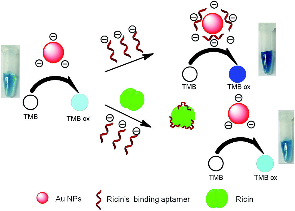 | ||
| Fig. 1 The mechanism of the colorimetric aptasensor for ricin. | ||
3.2. Spectral characteristics
To confirm the feasibility of our concept, the spectral responses of the colorimetric aptasensor were characterized under different conditions (Fig. 2A). The spectral value of AuNPs alone in solution at 650 nm was 0.98 absorbance units (a.u.) (1, Fig. 2A), explained by the oxidation of colorless TMB to blue TMB ox. In the presence of RBA, this value (2, Fig. 2A) increased to 1.5 a.u., indicating the enhancement of catalysis of AuNPs. When ricin molecules were present in the solution (3, Fig. 2A), RBA bound to ricin followed by desorption from the AuNPs, resulting in a decrease of the catalytic abilities of AuNPs. As expected, the addition of ricin alone did not cause the color change of the solution (4, Fig. 2A) compared with AuNPs alone (1, Fig. 2A). Further, the spectral value of AuNPs in the presence of CTAB (a kind of cationic surfactants) at 650 nm was 0.06 (5, Fig. 2A). These results confirmed that the negatively charged aptamer was the main cause of enhancement of AuNPs-dependent catalysis. Table 1 showed the zeta potentials of AuNPs at different conditions and the results were in accordance with our mechanism. | ||
| Fig. 2 (A) UV-vis spectra in the presence of TMB and H2O2 under different conditions of (1) 2.5 nM AuNPs, (2) 2.5 nM AuNPs + 0.25 μM RBA, (3) 2.5 nM AuNPs + 0.25 μM RBA + 0.5 nM ricin, (4) 2.5 nM AuNPs + 0.5 nM ricin, (5) 2.5 nM AuNPs + 0.5 g L−1 CTAB. Inset shows the corresponding digital images. (B) UV-vis spectra in the presence of TMB and H2O2 under different conditions of (1) 0.25 μM RBA, (2) 0.25 μM RBA + 0.5 nM ricin (3) 0.5 nM ricin. | ||
In order to get rid of the possible impact of oxidation of TMB caused by RBA, ricin, and the ricin–RBA complex, these three molecules were also employed to the oxidation of TMB (Fig. 2B). The results indicated that RBA, ricin, and the ricin–RBA complex did not possess the peroxidase-like activity.
Fig. S1† showed the morphology characteristics of AuNPs under different conditions. The AuNPs were monodisperse and spherical with the average size of 13 nm (ESI, Fig. S1†). The UV-visible spectra of AuNPs, AuNPs–RBA, AuNPs–ricin and AuNPs–RBA–ricin complex showed a typical AuNPs SPR peaks with maxima at 520 nm, confirming the stability of AuNPs on different conditions (ESI, Fig. S2†). The observation showed that there was no aggregation of AuNPs caused by RBA, ricin and RBA–ricin complex.
3.3. Optimization of the key parameters
The peroxidase-like activity of AuNPs depends on some key parameters, such as the concentration of RBA, TMB and H2O2, reaction time and temperature. Therefore, these key parameters were optimized prior to the application of our proposed method. We used the reduction of absorbance, that is, A0 − A(ΔA) as a criterion to optimize the detection conditions. A0 presents the absorbance at 650 nm in the absence of ricin. A presents the absorbance at 650 nm in the presence of 0.5 nM ricin.3.4. Colorimetric biosensing of ricin
Under the optimal detection conditions, the peroxidase-like of the AuNPs on TMB oxidation in the presence of various concentration of ricin was investigated. The ΔA at 650 nm in the presence of different amounts of ricin were shown in Fig. 3A. The digital images showed that with increasing ricin concentration, the color of the solution changed from dark blue to light blue, suggestive of the ricin concentration-dependent TMB catalysis. It could be evidently observed that the ΔA at 650 nm increase gradually with the increasing concentration of ricin. In another word, with the increasing concentration of ricin, the peroxidase-like activities of AuNPs decreased. This observation is accord with our hypothesis. As shown in Fig. 3B, ΔA exhibited a good linear relationship with logC (ricin, nM) in the concentration range from 0.05 nM to 10 nM (R2 = 0.9896). The detection limit can reach as low as 0.05 nM, which is lower than many previous reports. In addition, we compared the detection limit and detection time of the reported method. As shown in Table 2. The detection sensitivity of the proposed method is higher or comparable than the previous reports. Moreover, the detection time is much shorter than many previous reports. Most of the reported methods need tedious pretreatments while our method did not need them. Therefore, our method is simple and convenient.
 | ||
| Fig. 3 (A) ΔA at 650 nm in the presence of various concentration of ricin. (B) Typical calibration curve for ricin obtained using the colorimetric aptasensor. Inset shows the corresponding digital images. | ||
| Detection method | Detection limit | Detection time (including pre-treatment time) |
|---|---|---|
| Fluoroimmunoassay43 | 1000 ng mL−1 | 12 h |
| Aptamer arrays biosensor assay44 | 320 ng mL−1 | 6 h |
| SPR biosensor assay45 | 200 ng mL−1 | 20 min |
| Immunochromatographic assay18 | 50 ng mL−1 | 3 h |
| Aptamer-based SERS biosensor assay46 | 25 ng mL−1 | 21 h |
| Aptamer-based colorimetric biosensor assay47 | 20 ng mL−1 | 1 h |
| Microarray biosensor assay48 | 10 ng mL−1 | 4 h |
| Nanoparticle-based colorimetric biosensor assay19 | 4 ng mL−1 | 2 days |
| ELISA49 | 400 pg mL−1 | 20 h |
| Electrochemiluminescent assay49 | 50 pg mL−1 | 11 h |
| Nanoparticle-based bio-barcode assay50 | 1 fg mL−1 | 40 h |
| This work | 3.2 ng mL−1 | 1 h |
Moreover, in order to investigate the stability of the sensor, we compared with the UV-vis spectra of fresh prepared AuNPs and aged AuNPs (Fig. S8A†). The UV-visible spectra of the two samples both exhibited a typical AuNPs SPR peaks with maxima at 520 nm, and the absorbance made no obvious differences between two samples. The results showed that AuNPs was stable even one month later. Further, we compared the performance for colorimetric biosensing between fresh prepared AuNPs and aged AuNPs (Fig. S8B†). It was shown that there existed slight differences between the two samples. In view of the above results, the sensor had a high stability (ESI, Fig. S8†).
3.5. Selectivity
The selectivity of this colorimetric aptasensor to ricin was evaluated by measuring the ΔA at 650 nm to some other proteins such as thrombin (Th), glucose oxidase (GOx), and bovine albumin (BSA). Fig. 4A showed that ΔA at 650 nm in the presence of ricin was considerably larger than those of other proteins. All results indicated that our assay approach had a high specificity to ricin. | ||
| Fig. 4 The selectivity of the proposed system towards ricin detection. The concentration of ricin and other proteins are 7 nM. Inset shows the corresponding digital images. | ||
3.6. Application in practical samples
In order to evaluate the feasibility of the present method in practical applications, the detection of ricin in raw milk and Pepsi Cola was carried out. The practical samples were spiked with certain amounts of ricin. Table 3 shows that the recoveries of the samples are in the range 93.6% to 115.0%. The desirable recoveries demonstrate the reliability of the proposed method for detection of ricin in practical applications.4. Conclusions
In summary, we have successfully developed a sensitive, accurate and reliable colorimetric aptasensor for the determination of ricin based on peroxidase-like activity of gold nanoparticles without complicated modification and expensive instruments. The color change of dark-blue to light-blue was found to be easily observed by the naked eye or measured by UV-vis spectrometer. The linear range for the current analytical system was from 0.05 nM to 10 nM. The corresponding limit of detection (LOD) was 0.05 nM. More importantly, the proposed method was successfully applied to the detection of ricin in practical samples. Therefore, this study may offer a new approach for developing simple, low cost and high sensitive sensors for ricin detection.Acknowledgements
Financial support by the National Basic Research Program of China (973 program, no. 2010CB933600), the National Natural Science Foundation of China (21275135 and 21405146) is gratefully acknowledged.References
- L. M. Roberts and D. C. Smith, Toxicon, 2004, 44(5), 469–472 CrossRef CAS PubMed.
- J. M. Lord, L. M. Roberts and J. D. Robertus, FASEB J., 1994, 8(2), 201–208 CAS.
- J. Robertus, Semin. Cell Biol., 1991, 2(1), 23–30 CAS.
- Y. Endo, K. Mitsui, M. Motizuki and K. Tsurugi, J. Biol. Chem., 1987, 262, 5908 CAS.
- N. Sphyris, J. M. Lord, R. Wales and L. M. Roberts, J. Biol. Chem., 1995, 270, 20292 CrossRef CAS PubMed.
- L. L. He, B. Deen, T. Rodda, I. Ronningen, T. Blasius, C. Haynes, F. Diez-Gonzalez and T. P. Labuza, J. Food Sci., 2011, 76(5), N49–N53 CrossRef CAS PubMed.
- S. Olsnes, Toxicon, 2004, 44(4), 361–370 CrossRef CAS PubMed.
- M. Papaloucas, C. Papaloucas and A. Stergioulas, Pak. J. Biol. Sci., 2008, 11(19), 2370–2371 CrossRef CAS.
- K. Jasheway, J. Pruet, E. V. Anslyn and J. D. Robertus, Toxicon, 2011, 3, 1233–1248 CAS.
- L. E. Rodriguez-Saona, F. S. Fry and E. M. Calvey, J. Agric. Food Chem., 2000, 48(11), 5169–5177 CrossRef CAS PubMed.
- W. S. B. Yeung, G. A. Luo, Q. G. Wang and J. P. Ou, J. Chromatogr. B: Anal. Technol. Biomed. Life Sci., 2003, 797(1–2), 217–228 CrossRef CAS.
- L. L. He, B. Deen, T. Rodda, I. Ronningen, T. Blasius, C. Haynes, F. Diez-Gonzalez and T. P. Labuza, J. Food Sci., 2011, 76(5), N49–N53 CrossRef CAS PubMed.
- C. Lubelli, A. Chatgilialoglu, A. Bolognesi, P. Strocchi, M. Colombatti and F. Stirpe, Anal. Biochem., 2006, 355(1), 102–109 CrossRef CAS PubMed.
- E. A. E. Garber and T. W. O'Brien, J. AOAC Int., 2008, 91(2), 376–382 CAS.
- N. Koja, T. Shibata and K. Mochida, Toxicon, 1980, 18(5–6), 611–618 CrossRef CAS.
- M. A. Poli, V. R. Rivera, J. F. Hewetson and G. A. Merrill, Toxicon, 1994, 32(11), 1371–1377 CrossRef CAS.
- U. Narang, G. P. Anderson, F. S. Ligler and J. Burans, Biosens. Bioelectron., 1997, 12(9–10), 937–945 CrossRef CAS.
- R. H. Shyu, H. F. Shyu, H. W. Liu and S. S. Tang, Toxicon, 2002, 40(3), 255–258 CrossRef CAS.
- H. Z. Liu, J. J. Tang, X. X. Ma, L. Guo, J. W. Xie and Y. X. Wang, Anal. Sci., 2011, 27(1), 19–24 CrossRef.
- A. D. Ellington and J. W. Szostak, Nature, 1990, 346, 818–822 CrossRef CAS PubMed.
- T. Kunii, S. Ogura, M. Mie and E. Kobatake, Analyst, 2011, 136, 1310–1312 RSC.
- J. Lee, M. Jo, T. H. Kim, J. Y. Ahn, D. K. Lee, S. Kim and S. Hong, Lab Chip, 2011, 11, 52–56 RSC.
- R. L. Srinivas, S. C. Chapin and P. S. Doyle, Anal. Chem., 2011, 83, 9138–9145 CrossRef CAS PubMed.
- C. Tuerk and L. Gold, Science, 1990, 249, 505 CAS.
- W. Yuanboonlim, W. Siripornnoppakhun, N. Niamnont, P. Rashatasakhon, T. Vilaivan and M. Sukwattanasinitt, Biosens. Bioelectron., 2012, 33, 17–22 CrossRef CAS PubMed.
- Y. Jv, B. Li and R. Cao, Chem. Commun., 2010, 46(42), 8017–8019 RSC.
- S. Wang, W. Chen, A. Liu, L. Hong, H. Deng and X. Lin, ChemPhysChem, 2012, 13(5), 1199–1204 CrossRef CAS PubMed.
- Z. Gao, M. Xu, L. Hou, G. Chen and D. Tang, Anal. Chim. Acta, 2013, 776, 79–86 CrossRef CAS PubMed.
- H. Jiang, Z. Chen, H. Cao and Y. Huang, Analyst, 2012, 137(23), 5560–5564 RSC.
- X. Wang, Q. Wu, Z. Shan and Q. Huang, Biosens. Bioelectron., 2011, 26(8), 3614–3619 CrossRef CAS PubMed.
- J. Tian, Q. Liu, A. Asiri, A. Qusti, A. Al-Youbi and X. Sun, Nanoscale, 2013, 5(23), 11604–11609 RSC.
- L. Gao, J. Gao, L. Nie, J. Zhang, Y. Zhang, N. Zhang, T. Wang, J. Wang, D. Wang, S. Perrett and X. Yan, Nat. Nanotechnol., 2007, 2(9), 577–583 CrossRef CAS PubMed.
- M. Comotti, C. Della Pina, R. Matarrese and M. Rossi, Angew. Chem., Int. Ed., 2004, 43, 5812 CrossRef CAS PubMed.
- Y. Lin, Z. Li, Z. Chen, J. Ren and X. Qu, Biomaterials, 2013, 34, 2600 CrossRef CAS PubMed.
- J. J. Tang, J. W. Xie, N. S. Shao and Y. Yan, Electrophoresis, 2006, 27, 1303–1311 CrossRef CAS PubMed.
- G. N. Mayer, Nucleic Acid and Peptide Aptamers: Method and Protocols, Humana, New York, NY, 2009 Search PubMed.
- M. M. Maye, L. Han, N. N. Kariuki, N. K. Ly, W. B. Chan, J. Luo and C.-J. Zhong, Anal. Chim. Acta, 2003, 496, 17–27 CrossRef CAS.
- X. F. Li, J. Li, H. Y. Kuang, L. Feng, S. J. Yi, X. D. Xia, H. W. Huang, Y. Chen, C. R. Tang and Y. L. Zeng, Anal. Chim. Acta, 2013, 802, 82–88 CrossRef CAS PubMed.
- F. Yu, Y. Huang, A. J. Cole and V. C. Yang, Biomaterials, 2009, 30, 4716–4722 CrossRef CAS PubMed.
- Y. P. Liu and F. Q. Yu, Nanotechnology, 2011, 22, 145704 CrossRef PubMed.
- H. X. Li and L. J. Rothberg, Anal. Chem., 2004, 76(18), 5414–5417 CrossRef CAS PubMed.
- H. X. Li and L. J. Rothberg, J. Am. Chem. Soc., 2004, 126(35), 10958–10961 CrossRef CAS PubMed.
- G. P. Anderson and N. L. Nerurkar, J. Immunol. Methods, 2002, 271, 17–24 CrossRef CAS.
- R. Kirby, E. J. Cho, B. Gehrke, T. Bayer, Y. S. Park, D. P. Neikirk, J. T. McDevitt and A. D. Ellington, Anal. Chem., 2004, 76, 4066–4075 CrossRef CAS PubMed.
- B. N. Feltis, B. A. Sexton, F. L. Glenn, M. J. Best, M. Wilkins and T. J. Davis, Biosens. Bioelectron., 2008, 23, 1131–1136 CrossRef CAS PubMed.
- E. A. Lamont, L. He, K. Warriner, T. P. Labuza and S. Sreevatsan, Analyst, 2011, 136, 3884–3895 RSC.
- J. T. Hu, H. C. Dai, Y. J. Sun, P. J. Ni, Y. L. Wang, S. Jiang and Z. Li, RSC Adv., 2014, 4, 43998–44003 RSC.
- J. B. Delehanty and F. S. Ligler, Anal. Chem., 2002, 74, 5681–5687 CrossRef CAS.
- V. Guglielmo-Viret and P. Thullier, J. Immunol. Methods, 2007, 328, 70–78 CrossRef CAS PubMed.
- H. Q. Yin, M. X. Jia, S. Yang, S. Q. Wang and J. G. Zhang, Toxicon, 2012, 59, 12–16 CrossRef CAS PubMed.
Footnote |
| † Electronic supplementary information (ESI) available. See DOI: 10.1039/c4ra17327a |
| This journal is © The Royal Society of Chemistry 2015 |