The role of hydroxyl channel in defining selected physicochemical peculiarities exhibited by hydroxyapatite

Vuk Uskoković *
Advanced Materials and Nanobiotechnology Laboratory, Department of Bioengineering, University of Illinois, Chicago, IL 60607-7052, USA. E-mail: vuk21@yahoo.com

Received 28th December 2014 , Accepted 27th March 2015

First published on 27th March 2015


Abstract

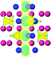
Mysteries surrounding the most important mineral for vertebrate biology, hydroxyapatite, are many. Perhaps the Greek root of its name, απαταo, meaning ‘to deceive’ and given to its mineral form by the early gem collectors who confused it with more precious stones, is still applicable today, though in a different connotation, descriptive of a number of physicochemical peculiarities exhibited by it. Comparable to water as the epitome of peculiarities in the realm of liquids, hydroxyapatite can serve as a paradigm of peculiarities in the world of solids. Ten of the peculiar properties of hydroxyapatite are sketched in this study, ranging from (i) the crystal lattice flexibility to (ii) notorious surface layer instability, (iii) finite piezoelectricity, pyroelectricity and conductivity to protons, (iv) accelerated growth and improved osteoconductivity in electromagnetic fields, (v) a high nucleation rate at low supersaturation and low crystal growth rate at high supersaturation, (vi) higher bioactivity and resorbability of biological apatite compared to those of the synthetic ones, and beyond. An attempt has been made to explain this array of curious characteristics by referring to a particular element of the crystal structure of hydroxyapatite: the hydroxyl ion channel extending in the direction of the c-axis, through a crystallographic column created by the overlapping calcium ion triangles.


image file: c4ra17180b-p1.tif

Vuk Uskoković

Vuk Uskoković is Director of the Advanced Materials and Nanobiotechnology Laboratory and Assistant Professor in the Department of Bioengineering at the University of Illinois at Chicago. His primary research interests include nanostructured biomaterials for controlled drug delivery and tissue engineering applications. Bridging the gap between materials science and life science, his lab specializes in the synthesis, characterization and biological testing of materials for the next generation of bionic implants. His main goal at the moment is the design and fabrication of smart, theranostic platforms for finely localized and targeted in situ remediation of various medical conditions, ranging from bone infection to brain tumors.


1. Introduction

Figuration of structure–property relationships is engrained in the heart of materials science, but it has suffered immensely in the recent past. Demands for practicality and applicability in natural sciences have taken their toll. No longer is the mainstream materials scientist interested in paralleling practical findings with fundamental insights. Rather, the former is given priority over the latter. As a result, methods for the synthesis of materials with exciting properties multiply, though only rarely followed up by their developers’ eagerness to comprehend the physicochemical bases of the structural forms adopted by the materials, and to link those with the characteristic types of behavior displayed by them. In this work, a major turnaround has been made with the attempt to look back at one of the key structural features of a material that is of particular interest to bone biology, and to correlate it to some of the experimentally observed oddities displayed by this material and collected over the past century.

The compound that is the subject of this review is hydroxyapatite (HAp): Ca10(PO4)6(OH)2. It is a hydroxylated form of apatite, Ca10(PO4)6(F,Cl,OH)2, the most abundant phosphorus-bearing mineral on Earth, findable in igneous, sedimentary and metamorphic rocks alike.1 In its calcium-deficient and partially carbonated form, representable as (Ca,Z)10(PO4,Y)6(OH,X)2 [Z = Na+, Mg2+, K+, Sr2+, etc., Y = CO32−, HPO42−, and X = Cl, F], it acts as the sole mineral component of bone. Its role in this complex and multifunctional organ is to impart solid compressive properties to elastic but weak collagen fibers and thus participate in a synergy thanks to which deficiencies of both the soft and the hard components of bone alone become effectively cancelled out.

HAp, interestingly, is a compound that appeals to the curiosity of chemists owing to its broad range of peculiar properties, just about as much as water can. Like water, exhibiting so many extraordinary properties in the realm of fluids from (i) its highest surface tension among all liquids except mercury to (ii) its density being highest in the liquid, not the solid state, at 4 °C and 1 bar, (iii) its extremely low vapor pressure and an incredibly high boiling point compared to the neighboring hydrides in the Periodic Table, (iv) its being one of the rare carbonless liquids at room temperature, and (v) its being an almost universal solvent, HAp abounds with unexpected features that reveal themselves to the studious and inquisitive eye of the researcher. Perhaps due to Nature's finding utility in peculiarity, both of these compounds, water and HAp, have been chosen as central ingredients of the molecular machinery of life.

However, like most ceramics, HAp is difficult to work with. It cannot be easily processed into any particle size or shape, as metals can, nor can it be easily functionalized with therapeutic ligands, as polymers, silica or gold can. It is also very unattractive to a casual observer – pale, fragile, not very conductive to electricity or vibration as well as inexpensive, without really displaying many appealing characteristics at the first sight. Although in its mineral form it can be easily confused with more precious stones, ranging from beryl to amethyst to olivine, a drop of lime juice is enough to start dissolving it, the reason for which A. G. Werner named it in 1786 using the Greek word απαταo, meaning ‘to deceive’ or ‘to mislead’.2 On top of this, it is highly defective in its most natural and functional, bioactive and osteoconductive state. Yet, in spite of all this, it is the material selected by evolution to build the basis of our biophysical structures, yielding yet another glimpse into the wonders of natural creation whereby perfection is found almost exclusively in imperfection.

The central aim of this study, as elaborated in section 3, is to elucidate the idea that a particular structural feature of HAp, known as the hydroxyl ion channel, running straight through the center of the basal plane of its hexagonal lattice and parallel to the c-axis, is partially responsible for an array of its extraordinary characteristics. The preceding section 2, which follows next, presents a compilation of unusual properties exhibited by this strange member of the biomineral family: unappealing at first, but to those who spend enough time examining it, it may reveal semantic signs that extend across the world of atoms and molecules and into the spheres of our psyches, our social lives and beyond.

2. Peculiarities of HAp

2.1. Crystal lattice flexibility

HAp has the capacity to undergo enormous lattice distortion as it accommodates cations and anions that differ in size from Ca2+ and PO43−, respectively. Approximately one-fifth of the Ca2+ ions in biological HAp is substituted with cationic impurities and, for example, substitution of Ca2+ with Mg2+ reduces the lattice constant c by 0.33% and increases the lattice constant a by 0.1%,3 which itself is enough to produce significant lattice disturbances and increase the solubility of the compound.4,5 The more frequent incorporation of CO32− ions in the hexagonal channel of the apatite structure (A-type HAp) leads to an increase in lattice parameter a and a markedly lesser decrease in parameter c in comparison with those of the stoichiometric HAp.6 In contrast, the parameter a decreases while c increases with a partial substitution of PO43− with the smaller CO32− ions (B-type HAp).7

These changes in the crystal lattice parameters often induce changes in crystallinity, thermal stability, morphology, solubility and other physicochemical and biological properties of the material.8 The charge neutrality principle also implies the formation of structural defects in order to accommodate foreign ions. The extraordinary stoichiometric flexibility of the compound manifests itself in accommodating such defects, as the Ca/P molar ratio, equaling 1.67 for pure HAp, can drop down to 1.3 without breaking of the crystallographic symmetry: P63/m. The crystal structure of stoichiometric HAp is, in fact, monoclinic (P21/b),9 but due to foreign ion inclusion, vacancy formation and Ca2+ depletion it becomes hexagonal (P63/m)10 for more disordered biological HAp. The same transition to a crystallographic state of lower symmetry is observed following annealing at temperatures higher than 207 °C. This polymorphic transition is reversible and the hexagonal symmetry transforms back to the monoclinic one at temperatures lower than 204 °C in the cooling regime.11

Though the capacity to incorporate foreign ions may not be as large as that typifying silicate glasses, the lattice flexibility of HAp is large enough to accommodate one half of all the elements of the periodic table,12 in almost any valence state, from SiO44− to As5+. It is thanks to this effect of lattice flexibility that bone can act as the mineral reservoir of the body and the storage for toxic elements, thus fulfilling two of its essential physiological roles. Despite this flexibility, the driving force for crystallization is still sufficiently high to drive out most of the organic molecules present at the moment of precipitation and limit the loading capacity of the compound mostly to the amount that could be adsorbed on the surface. This surface layer of a drug bound by weak forces to a particle predisposes HAp to exhibit burst release, and prevents it from being used as a sustained release platform in its dispersed form. Powder compaction and drug capturing within the pores is an approach that overcomes this deficiency of HAp,13–16 but limits the use of such composites to their application as solid blocks. Although the entrapment of fluorophores has been reported for glassy silicate calcium phosphates prepared in an amorphous form in Igepal-based reverse micelles and stabilized with citric acid,17 the compound naturally present in bone coating HAp crystals at 0.5 molecules per nm2 and preventing their coalescence in the collagen matrix,18 it appears that loading HAp with organics in any amount greater than that present in nacre or tooth enamel (<3 wt%) via intercalation is not possible. The intra-crystalline loading also appears to be theoretically possible but only for sufficiently small molecules such as glycine, the smallest amino acid, with amounts ranging from 1–3 wt%, depending on the concentration of the paired cationic and OH vacancies in the material.19

2.2. Surface layer instability

Surface layer instability, another one of the key properties of HAp, is of vital importance in ensuring the timely resorption of this compound during in vivo remodeling in spite of its sparsely soluble nature. Dissolution of biological HAp, for example, yields twice higher Ca2+ concentrations in the supernatant compared to that of the theoretically more soluble, commercial β-tricalcium phosphate (β-TCP),20 and this effect is predominantly due to the highly mobile surface layer whose composition and structure are constantly fluctuating in response to the fluctuant physicochemical conditions of the fluid at the interface. As a result, the surface of HAp, particularly in dynamic environments, such as biological ones, can be imagined to be in a state of constant phase transition.

Although the exact mechanism of phase transformations in calcium phosphates has not been fully elucidated yet, it has been traditionally held that the solution-mediated dissolution/reprecipitation mechanism constitutes the dominant one.21,22 This was elegantly proven by the noticed acceleration of phase transformations by stirring and their slowing down by substituting water with nonpolar solvents,23 and later supported by the ability of organic adsorbates, such as alendronate sodium, to extend the phase transformation timescale from minutes to days.24 The complementary mechanism, on the other hand, which is similar in nature to the martensitic/austenitic transition involving bulk lattice rearrangements, is still a subject of debate. Namely, although there are documented cases of transitions that took place solely in the interiors of compact calcium phosphate blocks, it is usually the surface that changes the phase composition to fit the chemical and energy demands of the local environment. Thus, for example, the core of dicalcium phosphate monohydrate (DCPM, a.k.a. brushite) implants transformed to HAp even while the surface retained the DCPM composition,25 though such reports are a rarity in the literature and a far more prevalent scenario involves the surface transformation in the direction of the most stable phase under the given chemical conditions, which most often serves to protect the bulk from premature dissolution. DCP blocks implanted as bone substitutes would thus undergo restructuring of the surface, until the most stable phase under physiological conditions, HAp, is present on it. For this reason, it has been held that the solubility of calcium phosphates does not conform to the solubility product, just as much as experimentally determined solubilities cannot be readily converted to bioresorption rates.26

Interestingly, this constant restructuring involving periodic dissolution and reprecipitation of surface ions could provide a counterforce against the low surface charge that facilitates aggregation. Namely, the ξ-potential of HAp is almost always below ±15 mV, which is the lower threshold for colloidal instability.27 On the other hand, the observed surface charge fluctuations during the aging of HAp precipitates in solution,28 along with frequent swings from the positive to the negative values and vice versa, have been suggestive of constant restructuring of the surface, which may have the capacity to partially compensate for the low surface charge. This has enabled HAp particles in suspension to be comparatively stable in phosphate buffer saline ([HxPO4x−3] = 11.8 mM, overall salt concentration ∼150 mM), undergoing virtually no ‘salting out’ effect.29 The central practical downside of this constant restructuring of the surface, however, is the impossibility of chemical functionalization of the surface since the continuously dissolving and reprecipitating surface ions would ensure the rapid release of the ligand to the environment. HAp, otherwise, does have plenty of attributes that justify its use as a drug delivery vehicle, with the central one of them being the capability of launching the endosomal escape of drugs, owing to its dissolution in the moderately acidic interior of the late endosome (pH 4.5–5.5)30 before this intracellular compartment transitions into the enzymatically permeated lysosome which would degrade macromolecular drugs had they been retained by the particle until then.31 This has been linked to the great effectiveness of this compound in gene delivery,32,33 second to none in the realm of non-viral transfection agents.34 Despite all of these pros, however, the instability of the surface, along with the resistance to drug loading via trans-crystalline entrapment, has greatly limited the use of HAp in targeted and sustained drug delivery.

2.3. Multiphasic particle composition

So, we see that despite its sparse solubility in water, equaling only 0.3 mg dm−3 at 25 °C for Ksp of 117.3, HAp has a very unstable surface which undergoes constant restructuring and prevents stable chemical functionalization, achievable for some other ceramics, predominantly covalent ones such as alumina,35 titania36 or zirconia.37 On the other hand, a constant engagement in the exchange of ionic species across the interface with the solution has allowed HAp to be used as an efficient industrial sorbent.38,39 This surface instability, along with the relatively high specific surface area of the compound, has also contributed to the facileness with which HAp accommodates foreign ions and acts as a mineral reservoir for the body.

In spite of the long-time use of HAp as an adsorbent in waste removal industry40–42 and in chromatographic columns for the electrophoretic separation of a variety of biological entities, including proteins,43,44 nucleic acids45 and even microorganisms,46 all thanks to the surface alternation of highly charged divalent calcium and trivalent phosphate ions capable of interacting with both positively and negatively charged functional groups in biomolecules and binding them in zwitterionic forms,47 constant dissolution and reprecipitation of surface ions stands as an obstacle to their binding ligands stably. Such swift surface rearrangements are, on one hand, responsible for the fact that no grain boundary segregation of ionic species has ever been observed in HAp,48 suggesting that, perhaps with the help of structural defects, whose concentration is directly proportional to the diffusivity of ions through the lattice, foreign ions comparatively easily diffuse across the particle–solution interface and into the bulk. On the other hand, however, the constant exchange of ions with the solution creates conditions for the formation of surface calcium phosphate phases different in composition and structure from those comprising the core of the particle. As a result, a HAp particle in contact with a pH 6 solution might form a protective DCP layer on its surface that would considerably slow down its dissolution and the reverse effect might happen to a DCP particle in contact with a pH 7 solution. A single-phase particle could thus undergo a partial internal phase transition upon immersion into solution, yielding multiphasic and possibly even surface-gradient particle compositions. This effect is the direct consequence of the unstable surface layers that undergo constant rearrangement in contact with the solution or body fluids to find the most energetically favorable composition for the given chemical conditions of the local environment.

It is worth mentioning here that HAp is only one of a dozen existing calcium orthophosphate phases, which together cover a broad range of solubility values, as illustrated in Table 1. Combinations with more soluble phases and the possibility of achieving time-controlled solidification of the as-precipitated gels enables the use of HAp as a component of self-setting pastes with an immense therapeutic potential as bone grafts.49 In such a form, despite their ceramic nature, calcium phosphates are typified by excellent viscous flow properties, for which reason they are added to polymers to facilitate their injectability50,51 rather than vice versa, as common sense would expect, in analogy with the emulsifying properties of calcium phosphate nanoparticles naturally present in milk52 and utilized in Pickering emulsions.53

Table 1 Main calcium phosphate phases, along with their chemical formulas, space group symmetries, solubility product values (Ksp) and solubilities at 25 °C and pH 7.4 (ref. 54–58)
Phase Chemical formula Space group pKsp Solubility (mg dm−3)
a MCPA = monocalcium phosphate anhydrous; MCPM = monocalcium phosphate monohydrate; DCPD = dicalcium phosphate dihydrate, a.k.a. brushite; DCPA = dicalcium phosphate anhydrous, a.k.a. monetite; CPP = calcium pyrophosphate; ACP = amorphous calcium phosphate (data pertain to the phase obtainable at pH 9–11); TCP = tricalcium phosphate; TTCP = tetracalcium phosphate; OAp = oxyapatite; CDHAp = calcium-deficient hydroxyapatite; OCP = octacalcium phosphate; HAp = hydroxyapatite; FAp = fluoroapatite.
MCPAa Ca(H2PO4)2 Triclinic P[1 with combining macron] 1.14 17 × 103
MCPMa Ca(H2PO4)2·H2O Triclinic P[1 with combining macron] 1.14 18 × 103
DCPDa CaHPO4·2H2O Monoclinic Ia 6.6 88
DCPAa CaHPO4 Triclinic P[1 with combining macron] 7.0 48
β-CPPa Ca2P2O7 Tetragonal P41 18.5 7.6
ACPa Ca3(PO4)2·nH2O 25 0.8
α-TCPa Ca3(PO4)2 Monoclinic P21/a 25.5 2.5
β-TCPa Ca3(PO4)2 Rhombohedral R3cH 29.5 0.5
TTCPa Ca4(PO4)2O Monoclinic P21 37.5 0.7
OApa Ca10(PO4)6O Hexagonal P63/m 69 8.7
CDHApa Ca10−x(HPO4)x(PO4)6−x(OH)2−x (0 < x < 1) Hexagonal P63/m 85 9.4
OCPa Ca8H2(PO4)6·5H2O Triclinic P[1 with combining macron] 97.4 8.1
HApa Ca10(PO4)6(OH)2 Hexagonal P63/m 117.3 0.3
FApa Ca10(PO4)6F2 Hexagonal P63/m 120 0.2


2.4. Finite piezoelectricity, pyroelectricity and conductivity to protons

Both of the main components of bone, collagen and HAp, exhibit piezoelectricity.59 In the case of collagen, this is presumably due to charge separation under shear, the process in which HAp plays a facilitating role by restricting the access to water for collagen and distributing load across large spatial scales so as to enable a large number of collagen molecules to deform locally.60 In the case of HAp, piezoelectricity is due to the polarization of OH groups confined to the channel formed by the overlapping hexagonal calcium atoms (Ca2). It appears to be a property solely of the inherently disordered, nonstoichiometric P63/m HAp and not of its pure, monoclinic, P21/b counterpart.61 The order of magnitude of the piezoelectric response of HAp, with the piezoelectric coefficient D = 16 pC N−1,62 is lower than that exhibited by lead zirconate titanate (PZT), but higher than that of aluminum nitride and in about the same range as those of polyvinylidene fluoride and zinc oxide.63,64 In addition, HAp thin films exhibit the ferroelectric nature of polarization, with the remnant polarization being approximately one-fourth of the value of PZT films.65 Although nanocrystalline HAp is predominantly polar, its polycrystalline nature in bone and mostly elsewhere is the reason why this piezoelectric effect becomes averaged over three dimensions, resulting in minor values along its favored direction, perpendicular to the basal plane and parallel to the OH channel. HAp is also a pyroelectric material, being able to generate electrical charge as a function of the rate of temperature change and characterized by the pyroelectric coefficient pi = 12 μC m−2 K−1. It is also an efficient proton conductor with the potential to be used in energy conversion devices.66 Proton conduction is directly causative of spontaneous polarization observed in these materials.67

2.5. Accelerated growth and improved osteoconductivity in the electromagnetic field

The polarizability of HAp explains its responsiveness to electric and magnetic fields in the context of its application as a biomaterial for bone regeneration. For example, sintered and electrically polarized HAp with a total stored charge density of 3.9 μC cm−2 proved to be able to enhance the osteobonding and osteoconducting abilities when implanted in the cortical bone of the femoral diaphysis in white rabbits.68 A metacarpal fracture in goats healed much faster in the presence of static magnetic field stimulation (800 G cm−2) and the healing process was paralleled by an increase in preferential alignment and crystallinity of the HAp crystals in bone.69 Cobalt-doped HAp also facilitated the regeneration of mandibular osteoporotic bones in rats.70 Even outside the biomedical context, the effects of external fields on the habit of crystal growth of HAp have been observed. Thus, the growth of HAp was accelerated when it was grown on polarized HAp substrates and slowed down when grown on non-polarized HAp.71 To verify that the effect was due to the existence of lattice channels populated by OH ions, annealing of the substrates was paired with dehydration under low water vapor content, the result of which was tricalcium phosphate, not HAp, but the difference between the growth on polarized and non-polarized substrates was expectedly none. The presence of a static magnetic field of 0.1 T was also shown to increase the precipitation rate of HAp by accelerating phase transformations along the steps defined by the Ostwald–Lussac rule, predominantly DCP → OCP → HAp.72

2.6. The fastest growing face in enamel HAp grows considerably slower in bone HAp

Growing fast in the world of crystals does not pay off, given that either minimization of the surface eminence of the crystalline faces or their complete disappearance entails their overly fast growth. In contrast, the slowest growing faces tend to be the most prominent ones on the crystal surface.73 This is illustrated in Fig. 1, where two HAp crystals of geological origin are shown: one with the exposed (001) face by cutting and the other one with the diagonal (011) and (101) faces instead of (001), the result of the sole directional growth of (001). An example among apatites of biomineral origin comes from the tooth enamel, where HAp grows almost uniaxially in the [001] direction, leading to the comparatively minor presence of basal (001) faces in comparison with the prismatic (hk0) ones in the final crystal forms.74 The growth of HAp platelets (30 × 20 × 2 nm (ref. 75)) in bone, however, proceeds in a very different manner. Namely, their growth in the [001] direction is not considerably faster than that along the [010] direction, which is atypical for most hexagonal closely-packed symmetries, given that the growth along the central axis of the (001) plane is usually the most favorable. This difference can be explained by the fact that the crystals of HAp in enamel and bone grow under two vastly different conditions. Elongated HAp crystals in enamel grow without the direct involvement of cells, in a comparatively hydrophobic and gelatinous amelogenin matrix, and are therefore free to grow according to their elementary crystallographic propensities, that is, primarily along the c-axis.76 Still, that the enamel matrix proteins do not merely block the (hk0) faces so as to foster the uniaxial growth of the basal plane, as the dominant model of amelogenesis assumes,77 but directly participate in the growth of the (001) face, producing crystals with aspect ratios of up to 104 as a result, is suggested by the presence, not disappearance, of this face in HAp crystals comprising mature enamel. In contrast, HAp platelets in bone grow in a highly hydrophilic environment, with the direct involvement of bone cells that alternately deposit and resorb the solid material, so the crystals cannot effectively grow on average in equal measure along their most favorable axis of growth, the c-axis. This versatility in terms of crystal orientation and growth adds another type of crystallographic flexibility to the crystal lattice one mentioned earlier, i.e. symmetry preservation under considerable lattice compression or expansion caused by ionic substitutions and defects.
image file: c4ra17180b-f1.tif
Fig. 1 Two HAp crystals of mineralogical origin: one with the exposed (001) face (left) and the other one with the (001) face being overgrown by the (011) and (101) planes as a result of its preferred growth on the account of the stagnation of the adjacent planes (right). Reprinted with permission from ref. 78.

2.7. An ultrahigh nucleation rate even at extremely low supersaturation and an ultralow crystal growth rate even at extremely high supersaturation

Nanosize is the natural form of existence of HAp particles.79 In bone and in intertubular dentin, HAp particles are nanosized (<100 nm in length by convention) along all three dimensions, whereas in enamel they are nanosized in the two directions that define their diameter and microsized in their height. Most precipitation protocols performable in the lab likewise yield HAp nanoparticles. This means that, unlike what is the case for most other materials, an experimenter has to try hard not to obtain HAp in nanoparticle form. Which is precisely the consequence of an ultrahigh nucleation rate even at extremely low supersaturation. This phenomenon coincides with the complex precipitation mechanism starting with the formation of amorphous, ∼1 nm sized solid units colloquially called Posner’s clusters,80 which undergo aggregation and recrystallization over the early course of the ripening time.81 Depending on the extent of aggregation and compactness achieved in the process, the high nucleation rate need not be an obstacle for the formation of larger crystals, which will, however, always be composed of aggregates of distinct nanosized growth units in the absence of thermal treatment and extensive recrystallization that it brings about. HAp also has a relatively low interfacial energy in aqueous environments,82 especially when the surface of the material is terminated by OH groups,83 which additionally contributes to its propensity to remain stable in the form of nanoparticles and resist uncontrolled growth.

Another remarkable feature of this material, following in step with the high nucleation rate in its favoring the adoption of nanostructured forms, is a considerably low crystal growth rate even under relatively high supersaturation.84–86 For example, the step kinetic coefficient, a reciprocal measure of the resistance to crystallization at the surface step,87 was measured to be around 4 × 10⁻⁵ cm s−1 which is by 3–4 orders of magnitude lower than the values attributed to most inorganic crystals and in about the same range as those of protein crystals.88 The slow motion of elementary steps on the crystal surface can be both a puzzling and a logical observation in view of the rather high mobility of ionic species comprising the hydrated surface layers of HAp. For, while a high surface ion mobility can be translated to a high step propagation rate, under the conditions of intense hydration, as is the case with HAp, it can also provide conditions for the constant dissolution of the steps, which as such propagate back and forth by advancing and retreating in an alternating manner. There are other reasons too, involving most importantly the complex multistage growth mechanism with the initial precipitation of Posner’s clusters and their subsequent aggregation and structural rearrangement followed by an increase in size, compactness and crystallinity,81 all the while obeying the Ostwald–Lussac principle which dictates that, in the absence of kinetic factors, the least thermodynamically stable phases (i.e. the most soluble ones having the lowest interfacial energy) are the first to form before transforming sequentially to the most stable phase,89 which is HAp in alkaline (>pH 6.8 at 37 °C) and DCP in acidic solutions (<pH 6.8 at 37 °C). By bearing resemblance to the growth of protein and viral crystals through chirality selection and orientation fitting, the low crystal growth rate of HAp, involving not ions but amorphous nanoparticle precursors, is sometimes compared to that of these biologics.90 Despite the low crystal growth rate, the crystallinity of HAp precipitated under physiological conditions is remarkably low,91 the reason being the amorphous precursors that only partly crystallize as the growth proceeds. Slow crystal growth in ordinary crystals produces relatively smooth surfaces, but this is not so in HAp where both the leading edges and terraces in between the surface steps exhibit a highly irregular profile at the atomic scale.92 As a result, unlike DCP, which dissolves via a combined volume diffusion and surface reaction mechanism,93 the dissolution of HAp follows the polynucleation mechanism94 and is greatly hindered for atomically smooth, step- and pit-free HAp crystals.95 The crystallization process mediated by amorphous precursors is responsible for yet another peculiarity exhibited by HAp upon nucleation: the inversely proportional dependence of the nucleation rate on supersaturation, as indicated by the linear increase in nucleation lag time with pH in the range of 6.8–7.6.96 Finally, in the presence of kinetic factors which abound in biological milieus and a high content of impurities, this already low multistage growth rate can be prolonged indefinitely and take unexpected crystallographic turns, which is why it has been said that there is no direct correlation between maturity and crystallinity in biological HAp.97 This effect certainly contributes to the structural versatility of this compound, a versatility we have barely begun to translate into a functional one and harness as such.98

2.8. A higher bioactivity and resorbability of biological HAp compared to those of the synthetic one

As a result of the high carbonate content, calcium deficiency and topographic irregularities on the atomic scale, the bioactivity, osteoconductivity and resorbability of biological HAp are higher than those of its synthetic analog. The carbonate content of biological HAp, varying anywhere between 2 and 8 wt%, has a particularly large effect on the higher solubility and bioresorbability compared to those of the synthetic one. Interestingly, whereas synthetic carbonated HAp is usually of A-type (CO32− → OH) when prepared by annealing at high temperatures (owing to the high mobility of the OH groups), and B-type (CO32− → PO43−) when it is precipitated at the room temperature,99 biological HAp is of a mixed A and B type.100,101 This suggests a vastly different formation mechanism of the biomineralized HAp compared to the synthetic processes usually run in the lab. The surface energy and interaction potential of biological HAp were also measured to be 1.6 and 2.5 times higher, respectively, compared to those of pure synthetic HAp, indicating a considerably higher surface heterogeneity and a greater conduciveness to protein adhesion and cell binding.102 Multiple substitutions and defects in biological HAp are certainly the key to explaining its superior biological activity over that of its synthetic, pure and defect-free HAp counterpart.103,104

2.9. Ultrahigh sensitivity to variations in the environmental conditions

Precipitation of HAp, especially at low supersaturation when it bears resemblance to the biological formation pathway, is extraordinarily sensitive to the subtlest changes in the conditions. The reaction routes leading to HAp as the final phase in a long chain of transformations, frequently proceeding in concert with the Ostwald–Lussac rule,105–107 are subject to change depending on even the mildest modifications to the initial experimental conditions. For example, the formation pathways and properties of HAp are greatly sensitive to the amount and nature of impurities. Impurities create condensation centers for the primary particle formation during crystallization of synthetic HAp, and calcium phosphates in general appear to be sensitive to impurities with concentrations of less than 10−6% (0.01 ppm).108 The texture, porosity and chemical content of the reaction vessel also drastically affect the level of critical supersaturation.109 For example, nucleation of HAp will proceed at a considerably lower supersaturation level in borosilicate vessels than in plastic ones as a result of leaching of silicate ions and of more polar container walls which reduce the energy barrier for nucleation. Tadashi Kokubo, the inventor of the disputably thorough method for evaluating the bioactivity of a solid compound by immersing it in simulated body fluid (SBF) and looking for the signs of HAp deposition on its surface,110 consequently suggested discarding any plastic bottles with visible scratches and never reusing them for storing SBF,111 a metastable solution with a supersaturation per growth unit equaling 19.5 with respect to HAp. The kinetics of HAp dissolution are also strongly influenced by the presence of impurities released into the solution during the reaction.112 Solutions particularly prone to exhibit this sensitivity lie in the middle of the metastability zone, between the levels of saturation and critical supersaturation. In this supersaturation window, kinetic effects such as the presence of an organic matrix exert the finest level of control over the microstructure and hierarchical superstructure of HAp precipitates.113 As a result, there is a multitude of possible stoichiometries and phase combinations that could appear in the final products of the precipitation reaction depending on the subtlest changes in the synthesis conditions,114,115 and some of them such as the acidic and biosoluble transitory calcium phosphate phases, found exclusively in pathological calcification deposits, may be all but ideal components of HAp designed for specific biomedical applications. This versatility of reaction outcomes depending on variations in the experimental conditions becomes even more magnified when various preparation methods are introduced in the comparison. This is why different methods to synthesize HAp have led to materials that vastly differed in crystalline order and properties, with the most notable difference being found in the diffusivity across the hydroxyl ion channel, which spanned two orders of magnitude, ranging from ultralow for the ordered monoclinic samples prepared by solid state synthesis at 1100 °C, to ultrahigh for the disordered hexagonal samples prepared by room temperature precipitation.116 Tracing this journey of ions across the channel running through the center of the triple hexagons that define the crystal structure of HAp takes us to the elaboration of its properties, the subject of the next section.

3. The role of the hydroxyl ion channel

The question framing this opinion piece is this: what if there is a single structural detail that could explain all these peculiar phenomena demonstrated by the protean nature of HAp? Our imagination is limitless, we know, and, though unsupported by direct computational analyses, it has been given free rein in the following paragraphs. But let us look first at the crystal structure model of HAp.

3.1. The place for hydroxyl in the crystal lattice of HAp

Although its first chemical composition analyses of the mineral form date back to 1788 (ref. 117 and 118) and the first synthesis to 1851,119 HAp was first detected as a bone mineral phase in 1926.120 In 1930, four years later, in two simultaneously published independent studies, its crystal structure was determined.121,122 Pure HAp crystallizes in the monoclinic P21/b space group symmetry with 88 atoms per unit cell and Ca20(PO4)12(OH)4 stoichiometry, but presents a rarity in the lab, let alone in Nature, where it virtually never occurs in such a form. For, any precipitation under atmospheric conditions leads to the inclusion of carbonate ions which necessitate charge compensation and, thus, lead to vacancies, deformations of the lattice and adoption of the hexagonal P63/m space group symmetry with 44 atoms per unit cell and Ca10(PO4)6(OH)2 stoichiometry, whereas solid state processing at elevated temperatures, higher than 207 °C, produces the same transition to the hexagonal symmetry. When it comes to pure HAp, the structural difference between the two phases boils down to the ordered, head-to-tail arrangement of OH groups located in the center of every other Ca2 triangle in the monoclinic low-temperature symmetry, and the disordered arrangement of OH groups where the head-to-tail and tail-to-head arrangements alternate throughout the channel in the hexagonal high-temperature symmetry. The monoclinic-to-hexagonal transition at elevated temperatures is thus paralleled by a curious phenomenon: namely, thermal energy during annealing converts a more ordered OH channel structure to a more disordered one. As a result, the hexagonal phase is higher in energy than the monoclinic phase by a rather small amount of 22 meV per unit cell,123 and the transition between the two, typified by a relatively low transition enthalpy of 130 J mol−1,124 is initiated by the rotation and reorientation of the OH dipoles125,126 and may proceed by the subsequent progressive twinning of the monoclinic, pseudo-hexagonal lattice until the hexagonal symmetry is reached.127 Note that both the occupational and the orientational order of OH ions become perturbed by this transition from the state of the higher, P21/b space group symmetry to the state of the lower, P63/m one, leading to a greater diffusivity of OH species along the channel and a higher potential for restructuring of the material under the demands of the local biological environment. The hexagonal form is, thus, the one favored by biological systems, in part because it allows for an easier exchange of OH groups78 with ions such as F, Cl and CO32−, which brings about an increased level of structural disorder, usable during the incessantly ongoing process of bone remodeling. The symmetry elements present in the rather disordered biological HAp include a six-fold rotation axis parallel to the c-axis, a 1/2 translation along the c-axis and a mirror plane perpendicular to the screw axis and the c-axis. The theoretical lattice parameters are a = 9.418 Å and c = 6.884 Å for P63/m (b = 2a, α = 120° for P21/b). However, depending on the preparation method, particularly when wet room temperature syntheses are involved, the lattice parameters a and c could be found anywhere in the ranges of 9.41–9.44 Å and 6.84–6.94 Å, respectively, for the P63/m space group.

The unit cell formula of hexagonal HAp, Ca10(PO4)6(OH)2, can be more precisely given as Ca14Ca26(PO4)6(OH)2, where Ca1 denotes columnar calcium ions and Ca2 denotes the hexagonal ones.128 Columnar calcium ions are named so because they are arranged in columns that uninterruptedly traverse the lattice, while the hexagonal ones outline hexagons when projected on the basal plane. Such an etymology conceals a mild misnomer because the columnar Ca atoms also create a hexagon when viewed down the c-axis, just as the hexagonal Ca atoms do. The difference between the two hexagons projected on the basal plane is that the one created by columnar Ca atoms is wider and accommodates in its center the hexagon created by the hexagonal ones. As seen from Fig. 2, the outer hexagon created by the columnar Ca atoms outlines the edges of the P63/m unit cell, while the six hexagonal Ca atoms form a series of overlapping inner hexagons. While all six hexagonal Ca atoms occupy the interior of the P63/m unit cell, the columnar Ca atoms are positioned in the six corners of the two basal planes and the six corners of the equatorial mirror plane in between them, yielding eighteen Ca1 atoms within a unit cell but only four per unit cell (each of the six basal plane atoms is shared with five neighboring unit cells, while each of the six atoms on the mirror plane is shared with two adjacent cells). Each inner hexagon is formed by two overlapping Ca2 triangles positioned parallel to the basal plane at z = 1/4 and z = 3/4 and at a 60° angle with respect to each other (Fig. 3a). Each of the six PO43− tetrahedra populating the interior of the hexagonal unit cell is positioned between a pair of Ca2+ ions in the outer hexagon when viewed down the c-axis, with three of them lying on the Ca2 triangle plane at z = 1/4 and the other three lying on the Ca2 triangle plane at z = 3/4. Furthermore, one of the four oxygen atoms of each of the PO43− tetrahedra is found just outside the edge of the outer hexagon, which is compensated by the intrusion of an oxygen from the tetrahedra positioned on planes below and above (z = ±1/2). The PO43− tetrahedra, 2.6 Å in radius, are arranged so as to form hexagonal close packing (ABABA) and, given their size-wise dominance over the crystal structure, the Ca2+ ions, 1.06 Å in radius, could be visualized as merely filling the holes between them. These holes come in two sizes and both form long columns in the direction of the c-axis. While the smaller holes can accommodate ions ≤0.225r in radius (r being the radius of a PO43− tetrahedron), which is too small to fit Ca2+, the larger holes provide enough room for Ca2+, given that they can accommodate ions ≤0.41r in radius (∼1.1 Å), and are filled with Ca atoms.129 Each site at which a Ca1 atom is located lies in between two parallel PO43− sheets, A and B, and is coordinated by nine oxygen atoms from three PO43− tetrahedra on top and three more at the bottom. The Ca1 site is also called “octahedral” because the six PO43− groups it is coordinated to outline the corners of a regular octahedron together with Ca1. The Ca1 atoms are thus found in columns perpendicular to the PO43− sheets and parallel to the c-axis, with the Ca1/OH ratio of 2[thin space (1/6-em)]:[thin space (1/6-em)]1. The Ca2 atoms, on the other hand, lie in the same plane as the PO43− tetrahedra and reside inside the holes formed by the oxygen atoms of the tetrahedra that line the c-axis channel. The Ca2 ions are engaged in a seven-fold coordination with six oxygen ions belonging to five different PO43− tetrahedra and an OH group.130 Arranged in a series of triangles lying parallel to the basal plane, the Ca2 atoms outline the boundaries of the OH channel extending through the center of the crystal structure, perpendicularly to the basal plane.


image file: c4ra17180b-f2.tif
Fig. 2 View of a stoichiometric HAp crystal down the c [001] axis, showing an OH group (small green circle) in its center, surrounded by hexagonal (blue circles) and columnar (purple circles) Ca atoms and phosphate tetrahedra: each green pyramid is a P5+ ion and the four red circles in its corners are four O2− ions. Notice the misrepresentation though: the O2− ions are not the smallest ions in the lattice, as shown here, but, in fact, the largest, occupying most space in it. As parts of the PO4 sublattice, they determine the stacking order of the unit cell. All other ions could be imagined as merely occupying the interstices created by the oxygen atoms.

image file: c4ra17180b-f3.tif
Fig. 3 (a) View of the hexagonal unit cell of HAp when projected down the c [001] axis. Three parallel hexagons with columnar calcium atoms (Ca1) on their corners, positioned at the two basal planes (z = 0 and z = 1) and at the equatorial mirror plane in between them (z = 1/2), define the geometric form of the unit cell, while the hexagonal calcium atoms (Ca2) form a pair of inner triangles (z = 1/4 and z = 3/4) positioned at a 60° angle with respect to each other. Six PO4 tetrahedra occupying the interior of the unit cell are distributed in such a way so that three of them lie on the Ca2 triangle plane at z = 1/4 and three of them lie on the Ca2 triangle plane at z = 3/4, themselves forming triangles at a 60° angle with respect to the in-plane Ca2 triangles and with respect to one another. Reprinted with permission from ref. 144. (b) The effect of the phosphate-to-carbonate substitution on the creation of paired vacancies at adjacent Ca2 and hydroxyl group sites. The solid line represents the axis of the OH channel formed by a series of overlapping calcium triangles and running perpendicular to the basal plane of the hexagonal unit cell. Reprinted with permission from ref. 145.

The OH groups are thus lined up along the c-axis, being positioned inside the overlapping triangles formed by the hexagonal Ca2+ ions, with an average distance of 2.5 Å between them. The triangles are stacked on top of one another in the direction of the c-axis, with each neighboring triangle being rotated by 60° with respect to the triangles below and above it, and the line connecting all the OH groups in a stacked array of unit cells runs straight through their center, with both oxygen and hydrogen of the OH lying on the c-axis in the higher, monoclinic symmetry (Fig. 3b). However, being inherently disordered as part of the P63/m symmetry, the oxygen atoms of hydroxyl are equally likely to be found either slightly above or slightly below the plane of the calcium triangles, (Fig. 4a) and the same applies to their halide ion substitutes. Moreover, there is no energetic preference as to whether the hydrogen atom of the OH group is to point upwards or downwards with respect to the c-axis and, as a result, each OH ion has a 50% probability of being in one state or the other. Compared to other ionic groups in the lattice, particularly PO43−, the OH groups have a considerable freedom of movement along the Ca2 channel, which is circa 3 Å in diameter. To be more precise, the diameter of the channel varies across the unit cell, being widest in its center and in the basal plane (z = 0, z = 1/2, 2.85 Å) and narrowest at the two midpoints between the basal planes and the center of the unit cell, lying in plane with the Ca2 triangles (z = 1/4, z = 3/4, 2.73 Å).131 For this reason, the channel could be better visualized as a continuous array of ovoid cavities than as a hollow cylinder (Fig. 4b).


image file: c4ra17180b-f4.tif
Fig. 4 (a) Results of an ab initio calculation of the band gap deviation from the lowest energy configuration depending on different orientations of the OH groups with respect to the two Ca2 triplet planes (the two vertical lines in each image). Reprinted with permission from ref. 150. (b) Sketched view of the ordered channel structure, with all OH groups pointing in the same direction, as seen in the P21/b apatite space group.

As can be seen from Fig. 4b, there is not enough room in the lattice to accommodate two OH groups pointing towards each other (OH⋯ ⋯HO) in the adjacent calcium triangles without producing significant lattice distortion and the loss of the higher, monoclinic symmetry. For this reason, the OH groups in the channel must either be arranged in an ‘ordered column’ fashion, i.e. as OH OH OH OH… along the c-axis, or in a ‘disordered column’ fashion, implying the reversal of the direction from OH OH OH OH… to HO HO HO HO… and back at various places. While the former head-to-tail arrangement is seen in stoichiometrically pure, monoclinic HAp, the latter disordered arrangement is seen in biological, intrinsically impure, defective and hexagonal HAp. The reorientation of a single OH group from the ordered OH OH OH configuration to a disordered OH HO OH one seems also to be followed by the tilting of the flipped OH group by ∼20–40° from the [001] direction to decrease the unfavorably short distance between the two protons and the two oxygens of the adjacent OH groups.132 This entails widening of one edge of the Ca2 triangle accommodating the reoriented OH group and subsequent tilting of the PO43− tetrahedra adjacent to the channel by 2.4°, whereby the symmetric distribution of the electron density of the O2− atoms bound to the P5+ ions becomes restored.133 The disorder initiated in the channel thus extends to the neighboring regions of the lattice. Note, however, that even in the ordered channel structure there is no correlation between the orientations of the OH groups in neighboring channels in the absence of a polarization-inducing electric field.

So, we see that both the two different types of calcium atoms and the phosphate groups outline hexagons when viewed down the [001] axis, while the OH groups occupy a central place by creating a vertical line that spans across the interiors of all these three hexagons. The attempt to translate this structural centrality into a property-wise one presents the central aim of this discourse.

3.2. Effects on resorbability, surface composition variability and trends in the crystal growth/dissolution of HAp

The higher bioactivity and osteoclastic resorption rate of biological HAp compared to those of its synthetic analog are owing to the fact that less than 50% of the OH positions in it are filled. This is a direct consequence of the comparatively large amount of impurities accommodated by biological HAp. These impurities are the cause of structural defects, which are directly proportional to ion diffusivity and solubility of the compound in spite of its having a high melting point and allowing virtually no diffusion of Ca2+ and PO43− ions at room temperature and in the pure state.134 More specifically, as shown in Fig. 3b, substitution of trivalent phosphate with bivalent carbonate induces the formation of one Ca2 vacancy per PO43− → CO32− substitution as the result of charge compensation, which, on the other hand, entails the creation of a vacancy at the OH ion site. The content of OH in HAp isolated from the human cortical bone was estimated to an even lower level than 1/2 of the theoretical one: only 20% of that present in synthetic and stoichiometric HAp.135 The degree of hydroxylation, greatly dependent on age, weight and the overall health of the donor, was measured to be 45–65% of the stoichiometric one in chicken and mouse cortical tibia bone.136 The incomplete hydroxylation of the bone mineral is the reason why it was often insisted that it must be called apatite rather than HAp, even though apatite, technically, is a term reserved exclusively for the apatite group of minerals and not for any single member of this group.137 Nevertheless, the OH ion content in biological HAp is still sufficient to affect the properties of the material and, although bone apatite is often considered virtually dehydroxylated,138,139 the aqueous environment in which it forms necessitates the incorporation of this ion (note that even lunar apatite was found to contain up to 8% of the channel ion sites occupied by OH,140 which, if released in its entirety, would be enough to cover the surface of the Moon with a one-meter-deep ocean141). Studies that reported undetectable amounts of OH ions in the bone mineral, relying predominantly on solid state NMR and other dry methods, could have been done with dehydrated apatite samples and could be thus seen as an implicit evidence for the pronounced diffusivity of OH groups intrinsic to the structure of this material. Namely, the amorphous surface of biological apatite is known to comprise large amounts of trapped water,142 which has been hypothesized to even have a directional effect on the crystal growth of this compound.143 Its prompt dehydration outside of its natural, aqueous milieu might be envisaged to draw the threads of OH groups to the surface via osmosis if the ion channel is structurally loose enough to allow for this effect to take place.

Vacancies created by only partially filled OH ion sites in the central channel have, in fact, an essential role by allowing room for the direction-dependent mobility of OH groups, their mobile O2− substitutes and Ca2 ions, thus facilitating restructuring of the crystals during bone remodeling orchestrated by the mutually antagonistic bone-building cells, osteoblasts, and bone-resorbing cells, osteoclasts. Also, paired with vacancies at the adjacent Ca2 sites, they appear to be able to entrap modest amounts of small organic molecules, such as glycine,146 as mentioned in section 2.1, suggesting a wholly unexplored avenue in the utilization of the OH channel by replicating the molecular trapping functionality of zeolites or clathrates in highly defective, nonstoichiometric HAp. In addition, the missing OH stretch and libration bands suggested that the structural binding of OH groups in biological HAp must be weak or almost nonexistent,147 which is a result of the inherently disordered OH content with regard to both its location and orientation, obviously facilitating structural reorganization during bone remodeling due to a lower entropic cost. This inherent disorder may be responsible for the existence of a multitude of energetically acceptable ion channel configurations and might even be the key to explaining the weaker mechanical properties of HAp when compared to those of other apatites.148 Substituting OH with F reduces this disorder by restricting the number of possible channel ion configurations, but only to a certain extent, after which the rigidity of the channel structure starts to take hold, resulting in an increased proneness to cracking and an overall weakening of the material.149

The role of the OH ions in enhancing the solubility of the surface layer and the ease of its restructuring, having immense biological repercussions during bone remodeling, can be tentatively supported by the effects of their substitution with fluoride and other halide ions. The size, the electronegativity and the orientation of these ionic substitutes plays a vital role in determining the number and strength of primary bonds, which, in turn, shifts the ionic–covalent equilibrium extending through the entire crystal structure. Larger ions lying farther from the Ca2 triplet plane thus have a greater number of bonds whose strength is weaker on average. Such is the case with relatively large Br ions that lie in between the two parallel calcium triplet planes. Smaller Cl displays a minor offset from the calcium triplet plane, while even smaller F is positioned right on the plane, exhibiting consequently the strongest bonding. Because of its intrinsic polarity, OH has multiple ways of positioning, typically exhibiting a small offset from the plane, like Cl. Orientation of the OH ions in the channel can also have a profound effect on the bonding strength.150 This is illustrated in Fig. 4a, where the results of the theoretical band gap prediction based on an ab initio density-functional-theory-based computational model are shown: depending on the orientation of the two OH groups per unit cell with respect to the Ca2 triplet plane, the deviation from the lowest energy configuration can vary from 0–15% (i.e., 0–0.66795 eV for the band gap estimated at 4.71 eV). Another way in which halide substitutes, such as F and Cl, modify the structure and properties of HAp is by promoting hydrogen bonding between neighboring OH dipoles in the channel.151,152 These bonds have otherwise not been observed in pure HAp due to the large distance between adjacent OH groups,153,154 limiting hydrogen bonding only to that between OH groups and the nearest oxygen atoms belonging to the PO43− groups.155

Then, the OH group exchange is known to proceed much faster than the exchange of surface phosphate or calcium ions.156 This is even more facilitated as a result of the deformation of the Ca2 hexagon outlining the edges of the OH ion channel when it is being exposed on the surface157 (Fig. 5). The protonation of OH groups and their release into the solution is promptly followed by the release of Ca2 ions, which endows the surface with a negative net charge and increases the rate of binding of electrostatically attracted integrins and other adhesion proteins that precede bone cell attachment.158,159 The relatively light Ca2+ ions are, in fact, flexibly arranged within the lattice, resulting in the proneness to display Ca2+ vacancies, whereas the heavier PO43− groups are practically those that define the hexagonal structure of the crystal, given that the atomic arrangement of all calcium orthophosphates is built around the network of PO43− groups that provide stability to the structure.160 With the PO43− network being more resistant to restructuring and dissolution in the solid phase and with heavier HxPO3x−3 ions spending effectively more time on the surface than the lighter and more diffusible Ca2+ ions (the same effect explains the negative charge of neutral surfaces in water, as OH groups spend more time on them on average than the more mobile protons), the surface carries a negative charge, which, as it turns out (Fig. 6), facilitates the crystal growth and remodeling.161 For, unlike cations, which are generally more hydrated than anions and have a greater tendency to reside in the bulk aqueous medium, bigger anions tend to be specifically adsorbed, affecting the crystal habit to a greater degree than cations162 and leading to the most commonly negative charge of surfaces in water.163 At the same time, during dissolution events bulkier anions tend to be retained on the surface longer than cations, as exemplified by silver iodide, which preferentially releases smaller and more mobile Ag+ ions and retains heavier and less mobile I. Consequently, although the solubility product (aAg+aI) for AgI equals 10−16, the point of zero charge is found not at pAg 8, but at pAg 5.5 (pI 10.5).164 An NMR study of nanocrystalline HAp has indeed demonstrated the presence of a circa 1 nm thick, amorphous surface layer predominantly populated by hydrogen phosphate ions, contrasting in composition and structure the crystalline and electrically neutral HAp core.165 And because of the more extensive adsorption of the HxPO4x−3 species onto the growing particle surface and their greater retention during dissolution, the Ca/P molar ratios in the equilibrium solution were measured to be as high as 25 despite the stoichiometric composition of the precipitate.166 Low Ca/P molar ratios, naturally brought about by the binding of free Ca2+ by phosphorylated proteins in the bone matrix, have, for example, been shown to be critical for promoting the controlled growth of HAp on amelogenin-functionalized surfaces.167 This is in agreement with the fact that pre-nucleation clusters during the formation of HAp from supersaturated solutions are closest in composition to calcium triphosphate (Ca/P = 0.333).168 The transformation of these pre-critical nucleation clusters to stable HAp particles is accompanied by a gradual increase in the Ca/P molar ratio from 0.333 to 1–1.5 for the amorphous particles and for the short-lived OCP transient (if present) to 1.67 for stoichiometric HAp, indicating that the growth rate is highest when phosphate groups are dominant at the interface between solid phase and solution. Also, though this is highly dependent on the atomic scale roughness, surface irregularities and the exact groups that terminate the crystal faces, atomistic simulations of most (hk0) faces – e.g. preferentially exposed (010) – have displayed an excess of Ca2+ ions, which is supposed to endow them with a positive surface charge. In contrast, the prominence of OH and PO43− groups endows the (001) faces with a negative surface charge,169 which is supposed to additionally foster their growth along the [001] axis in light of low Ca/P molar ratios as the central condition for crystal growth. This preferential direction of growth is additionally reinforced by the presence of Ca2+-chelating agents, such as EDTA or citric acid, which constrict the growth along the a-axis and extend the crystals along their c-axis.170 The less pronounced growth of the bone mineral crystals along the latter direction than expected from these considerations might be explained by the lower surface energy of the (001) faces compared to those of the prismatic ones: 1 J m−2 for (001) vs. 1.7 J m−2 for (010)171 and twice as much for (100).172 Growth along the axes perpendicular to high energy planes, of course, minimizes their surface prominence and thus stabilizes the crystal by lowering its surface energy, resulting in the plate-shaped morphology of HAp crystals in bone. Literature reports have not, however, reached agreement over the surface energy difference between the crystal faces of HAp, primarily because of a variety of ways in which the surfaces could be terminated. Recent surface modeling studies have, thus, arrived at contradictory findings with respect to the idea that the (001) faces should be the least energetic: according to some of them, the OH-terminated (010) faces should, in fact, be the most stable ones on the HAp surface and the (001) faces the least.173 From the biological angle, the growth along the c-axis is also partially mitigated by the (001) faces being aligned along the collagen fibril axis and placed in direct contact with the fibrils,174 where they are constricted and partly shielded from the inflow of ionic growth units. Conversely, the fact that the turnover of HAp platelets via resorption by the lactic-acid-secreting osteoclasts is easiest on (hk0) planes abundant in alkaline Ca2+ ions and threads of easily dissolvable OH groups, explains the orientation of their c-axis in the direction of the long axis of the collagen fibers in the intrafibrillar region of bone and the exposure of the (hk0) faces to the surface of the fibers (Fig. 7). In turn, the (001) faces are the least prominent ones in enamel rods, but most exposed to the environment of the oral cavity, so as to minimize the dissolution of the mineral that does not get regenerated by cells during the lifetime of the organism and for which solubility is an undesired propensity. And indeed, the solubility of (001), the smallest habit face on hexagonal HAp crystals, was both theoretically predicted and experimentally determined to be less than that of (100),175 the dominant face in bone and enamel crystals alike, with the only difference that it is shielded from the environment in enamel and exposed to the cellular milieu in bone, along with other (hk0) faces. Therefore, it can be speculated that threads of OH groups extending along the c-axis of the unit cell, facilitating rapid OH ion exchange between the surface and the solution, are the key to endowing the material with comparative surface instability and ‘liveliness’ that their biological environment utilizes well. In support of this idea comes the observed increase in the thickness of the mobile, defective and intensely hydrated surface layer in more soluble forms of HAp, such as the Mg-substituted one.176 To that end, the concentration of OH groups emerging from the central c-axis channel to the (001) faces as a result of their migration between two hydrated c-planes (20% of bone is water, and the water sheath enfolding the collagen molecules provides an essential enthalpic contribution to its structural stability177) must be considerable and, when the entire exposed channels along the (hk0) faces are added to the picture, this yields a stiff and sturdy material, yet flexible enough to undergo constant remodeling over the course of the lifetime of bone, with the annual turnover rate averaging at 3% for cortical bone and 25% for the lighter, more porous and vascular cancellous bone.178


image file: c4ra17180b-f5.tif
Fig. 5 Results of an interatomic potential study showing the structural rearrangement of the (001) faces on the HAp surface (left and right of the diagram) as a result of free energy minimization requirements. Even though the unit cells adjacent to the surface appear to be perfectly hexagonal, while the channels closer to the surface assume a distorted hexagonal geometry, the simulation has suggested the deformation of the OH channel outlined by Ca2 atoms, extending together with a short-range interaction between the surfaces over the distance of several unit cells into the bulk of the material.

image file: c4ra17180b-f6.tif
Fig. 6 Surface step propagation velocity (vi/v0) as a measure of the crystal growth rate for HAp increasing exponentially when the concentration of free HxPO4x−3 ions dominates over the concentration of free Ca2+ ions.

image file: c4ra17180b-f7.tif
Fig. 7 Reigning model of the packing of polycrystalline HAp platelets inside the parallel arrays of collagen fibrils in bone:179 (a) HAp particles initially nucleate and grow within the gaps between the N- and C-termini of the collagen molecules in fibrils, but (b) either in the later stages of mineralization or in separate events they grow into the intermolecular spaces within the fibrils, so that effectively between 30 and 70% of HAp is found outside of the fibrils.180 Despite the polycrystalline nature of the particles, the c-axis is predominantly oriented in parallel with the long axis of the fibers.

The rapidly rearranging surface layer of HAp, which has been described in more detail in sections 2.2 and 2.3, is also heavily hydrated181 and is not to be confused with the Stern layer of transiently adsorbed counter-ionic solutes since it is, strictly speaking, a part of the crystal and not the solution, even though it belongs to an entropic transition zone between the two. This surface layer hydration must have a considerable effect on the migration of constituent OH groups in and out of the channel, to and from the surface, on the length scale of a couple of unit cells extending down the c-axis from a (001) face. Generically speaking, dissolution of HAp proceeds with the first step being the acidic neutralization of the channel OH groups and their release in form of water. As a result, HAp is sparsely soluble at physiological pH and its solubility increases approximately tenfold with every unit decrease in pH, equaling 6.8 mg dm−3 at pH 7, 4.8 g dm−3 at pH 4 and 88 g dm−3 at pH 3 (all in pure water at 20 °C),182 the point below which the compound becomes practically fully soluble. The release of OH groups induces charge instability in the calcium column and opening up of the channel. This can be the starting point for two different processes: continued dissolution via release of Ca2+ ions or the accommodation of foreign ions into the lattice, the process in which, as we see it, the OH channel plays an equally critical role.183 This role, holding the key to the extraordinary ionic substitution capacity of HAp, can also be evident from the fact that other calcium phosphate phases, such as DCP or OCP, do not tend to incorporate impurity ions despite containing water in their crystal lattices.184 After the release of OH ions, migration of Ca2 ions out of the channel and into the solution is the next step in the sequence of dissolution events,185,186 which leaves a highly negatively charged, PO43−-rich layer at the solid interface. This greater diffusivity of Ca2 ions compared to the Ca1 ones explains their being the primary sites for cationic substitution; only at higher weight contents of foreign ions do Ca1 ions begin to swap places with them.187 The PO43−-rich layer exposed by the release of Ca2 ions is the next to dissolve due to water penetration and destabilization of the cross-linked network of PO43− tetrahedra. For this reason, it is said that HAp undergoes incongruent dissolution,188 in a sense that the Ca/P ratio in the supernatant (Ca/P = 2) is higher than the stoichiometric one (Ca/P = 1.67)189 during the dissolution of HAp, whereas as a result of the acidic nature of DCP the trend is the opposite during the dissolution of this more soluble calcium phosphate phase (Ca/P < 0.5).190 The sequential dissolution of different ionic species also predisposes the supernatant to exhibit occasionally observed periodic fluctuations in the Ca/P ratio and pH over the course of dissolution or ripening of the precipitate.191 Additives and substitutions can alter the dissolution process, e.g., by binding the exposed Ca2 ions, as is the case with citrate ions,192 which inhibit the dissolution of HAp,193 or by restricting the primary ion channel, as is the case following OH → F substitution whereby fluoroapatite, the least soluble apatite, forms. Namely, F and OH ions are of similar size and identical charge, which facilitates easy substitution (ionic radii equal 110 pm for OH, 119 pm for F, 167 pm for Cl and 182 pm for Br). Fluoride forms a particularly strong ionic bond with the surrounding Ca2+ ions in part because it is positioned right on the Ca2 triplet plane and in part in analogy with CsF, a compound that forms the strongest ionic bond by involving the most electropositive nonradioactive atom of the periodic table, Cs (0.79 in dimensionless Pauling units), and the most electronegative one, F (3.98 in Pauling units). Of course, when it comes to CsF crystals, the discrepancy in size of the ions prevents their close packing and diminishes the bond strength, so that many other halides, such as NaF, KF and NaCl, outweigh them in the solid form in terms of bond strength. This, however, is not the case with fluoroapatite, where both the packing factor and the electronegativity factor favor the residence of F ions in the OH channel. As a result of the stronger bonding to the Ca2 ions, substitution of OH with F contracts the lattice spacing a (ref. 194) and thus limits the diameter of the channel, reducing the mobility of the OH ions, while it also expands the c-axis, increasing the length of the channel. Both of these effects, causing the lattice expansion in the [001] direction and contraction in the [hk0] ones, reduce the solubility of the compound through the hampered movement of OH groups along the channel. In contrast, substitution of Ca2+ with Mg2+, as mentioned in section 2.1, increases the channel diameter and contracts it in length, enabling easier escape of the OH ions and, as a result, increasing the solubility of the compound. A similar effect is achieved by the incorporation of Co2+ ions in place of Ca2+, resulting in the expansion of the HAp lattice in the [100] and [010] directions and contraction along the c-axis,195 as evidenced by the reduction of the aspect ratio and increase in sphericity of the HAp crystallites accompanying this substitution.196,197 Likewise, the introduction of divalent carbonate ions induces the formation of Ca2 vacancies, which additionally contributes to a decreased confinement of OH groups in the channel and fosters their migration to and from the surface, facilitating the dissolution and cell-guided restructuring processes.

3.3. Hydroxyl ion channel as the route to polarization, proton conduction and susceptibility to electromagnetic radiation

Spontaneous polarization of the OH ions in the c-axis channel is responsible for both the piezoelectric and the pyroelectric effect exhibited by HAp. As shown in Fig. 8, different arrangements of OH dipoles in the channel created by the overlapping Ca2 triangles are possible, resulting in either ferroelectric or anti-ferroelectric ordering and justifying the envisioning of a polarized HAp lattice as an array of parallel 1D ferroelectric nanowires embedded in a dielectric matrix composed of Ca2+ and PO43− groups.198 And If this polarization has been shown to accelerate the crystal growth, while the increased disorder in the OH channel increases solubility and facilitates particle restructuring, then there are also grounds to correlate the surface layer instability that typifies HAp and the difficulty of inducing the growth of singlets past the nanosize range with the structure of the OH channel. For example, it is conceivable that the rapid exchange of OH groups between the solution and the particle surface, along with the partial penetration of protons contributing to internal hydration and disordering of the channel structure, plays an important role in destabilizing the surface and disfavoring the rapid particle growth via a classical mechanism that involves the attachment of individual ions, not amorphous clusters, onto a growing particle. In turn, the rapidity of nucleation at low supersaturation may be due to a relatively low energy barrier standing in the way of the transition of a large concentration of pre-nucleated clusters to their spontaneous growth via coalescence into stable particles. The pivotal role that OH groups play in conducting the coalescence of Posner’s clusters into amorphous particles, later to be transformed into crystalline platelets, could be tentatively inferred from the fact that one Ca2+ ion and two OH groups constitute the difference between the stoichiometry of an anhydrous Posner’s cluster, Ca9(PO4)6, and that of a hexagonal HAp unit cell, Ca10(PO4)6(OH)2. With the external Ca2+ ions, all of which are thought to be the hexagonal Ca2 ones, initially acting as bridges between the organic surface and the anchored clusters, in analogy with Ca2+ immobilizing DNA by being sandwiched between two phosphate groups, one from a DNA molecule and another one from a zwitterionic derivative of phosphatidylcholine,199 and OH drawing a thread in between them that defines the central axis of the six-fold symmetry of the apatite hexagon and enables precise localization of adjacent clusters, the conditions for the unprecedented, virtually defect-free aggregative growth at ambient temperatures are obtained.
image file: c4ra17180b-f8.tif
Fig. 8 Energy levels corresponding to different arrangements of the OH groups inside the c-axis channel for the different lattice space groups: P63 ferroelectric, P21 ferroelectric and P21/b anti-ferroelectric. Reprinted with permission from ref. 65.

The brick-and-mortar model of growth, according to which the growth occurs through aggregation of small clusters whose structurally unstable, mortar-resembling surface swiftly rearranges itself when in contact with the growing crystal so as to enable a seamless blend with it, is thus favored overall, but with definite limitations. Namely, while the coalescence of clusters may proceed fast when the cluster size is sufficiently small, regardless of supersaturation (hence the high nucleation rate at low supersaturation), it may be hindered at larger sizes, which is when the surface instability brought about by the diffusion of OH groups across the particle/solution interface can induce the opposite effect, effectively slowing down the growth instead of facilitating it (hence the low crystal growth rate at high supersaturation). Note that the OH groups emerging on the particle surface from the c-axis channel are also causative of the intense hydration of the ionic growth units in the Stern layer, which presents an additional barrier that prevents the fast growth of HAp crystals even under high supersaturation, given that dehydration is most frequently the kinetic step with the largest energy barrier in the growth unit transfer from solution to the solid surface.200 An inverse effect is observed during the dissolution of HAp: namely, even when the initial crystals are coarse and polydisperse, the particle size and morphology often evolve towards the nanosize and monodispersity, with the high surface area unexpectedly stabilizing further dissolution,201 again presumably owing to the highly mobile and hydrated interfacial layer, being the outcome of the rapid exchange of OH ions between the channel and the solution. Dehydration effects involving the migration of the channel OH ions across the interface are also thought to be responsible for another peculiar effect exhibited by some HAp precipitates, which is the unexpected reduction in particle size following calcination at up to 750 °C, in spite of the evident increase in crystallinity.202,203 In any case, the exposure of OH groups necessitates major surface reconstructions in search of stability83 and their dynamic interaction with the aqueous environment makes the surface reconstruction processes perpetually fluctuant.

The ability to transmit protons across the OH channel also makes HAp a good proton conductor, with a potential to be used in H+-conducting fuel cells and chemical sensors.204 The proton diffusion path, proceeding straight through the OH channel in a sinusoidal fashion accompanied by the reorientation of the OH ions, is depicted in Fig. 9. Proton conduction is directly responsible for the polarization of the compound205 and for its responding to electrical and magnetic fields with accelerated growth and enhanced bone formation. The high surface ion mobility, originating predominantly from the diffusion of OH groups along the c-axis channel and the intense local hydration that it brings about, is also thought to be responsible for the higher electrical conductivity exhibited by HAp than the one theoretically predicted by assuming the transport of charges across a fixed lattice. The band gap associated with HAp at 4.51 eV is consequently lower than those of apatites with a more compact and constrained channel structure, including fluoroapatites (5.47 eV), bromoapatites (4.71 eV) and chloroapatites (5.27 eV).150 Interestingly, the diffusion path of protons through the HAp lattice bears resemblance to that taken by O2− diffusing along the c-axis of the hexagonal, apatitic ionic conductor La5(Si3−xMgx)O13 (x = 0–0.15).206 However, though the diffusion direction may be the same, the involvement of OH ions, whose reorientation accompanies the transfer of a proton from one OH group to another,133 makes the transport mechanism profoundly different in comparison with that present in La5(Si3−xMgx)O13. The same transport mechanism applies also to the conductivity of HAp for O2− ions, which becomes particularly pronounced in B-type HAp,207 in which larger PO43− groups are partially substituted by smaller CO32− ones, causing a moderate widening of the channel and increased diffusivity of ions through it. Finally, the frequently noted positive effect of external fields on the biological response to HAp implants in the literature is the sole result of reorientation of OH dipoles in these fields, an effect whose complexity needs to be explored in far more detail before it could be utilized for therapeutic purposes.


image file: c4ra17180b-f9.tif
Fig. 9 The sinusoidal proton diffusion path through the OH channel of the HAp lattice. Reprinted with permission from ref. 208.

3.4. Hydroxyl ion channel, one of many structural features that, together, define the physicochemical peculiarities of HAp

In the end, it would be overly simplistic to say that the OH ion channel is the only structural element responsible for the broad range of peculiarities exhibited by HAp. The complex interaction involving all the ionic groups must take an equal part in defining the properties for which Nature selected it through evolution as the indispensable ingredient of the vertebrate skeleton. The discourse provided herein should act as an incentive for more detailed explorations of the structure–property relationships with a particular emphasis on this central channel and ions residing in and diffusing through it. Future studies on this literal structural centerpiece of the HAp structure will benefit from in situ high-resolution spectroscopic, diffractometric and microscopic techniques, though with definite limitations: complementary techniques, such as small-angle X-ray scattering and Fourier-transform infrared spectroscopy, for example, oftentimes give incompatible information about the crystal size and structure parameters,209 alongside having remained largely inconclusive about the state of hydroxylation of the lattice, while HAp particles have disintegrated during transmission electron microscopic analyses into CaO, volatile P2O5 and water under high beam currents,29 whose optimization entails an inescapable trade-off in contrast and resolution. Also, since for half a century now X-ray diffraction and neutron scattering studies have not been able to reach an agreement over the precise location of OH groups in the channel210,211 nor distinguish between their different orientations, e.g., between the ordered monoclinic configuration and the disordered hexagonal one, molecular dynamics simulations will need to step forward to compensate for such critical experimental limitations.132 The rather limited spectroscopic, diffractometric and microscopic studies will also need to be coupled to traditional methods of analytical chemistry, which have also benefitted in the meantime from the availability of more precise instrumentation. In any case, combinatorial analytical techniques are hoped to derive new paths in our understanding of this exciting structural detail of the most essential mineral in vertebrate biology. Only the future can tell how many new insights relevant for the fundamental understanding of materials and for continuing to harness the untapped biomedical potentials existing within this unique biomineral will be derived by those who will proceed to investigate the hydroxyl ion channel that runs through the center of its crystal symmetry.

4. Conclusions and perspective

We became acquainted with a myriad of interesting features of HAp, the biomineral that constitutes the foundations of our biological make-up. The purpose of this review study was twofold: firstly, to compile physicochemical peculiarities exhibited by HAp, and, secondly, to engage in an effort to explain them by referring to the hydroxyl ion channel as an essential crystallographic element of this compound. We have seen that the spontaneous polarization of HAp, the increased solubility, resorption rate and bioactivity of biological apatite as well as the precise crystallographic orientation of apatite platelets in bone may all have their roots in the crystallographic column of hydroxyl ions extending along the 63 screw axis that runs through the center of a series of overlapping calcium ion triangles. Still, what this review has aimed at is to provide only a glimpse at a remarkable structural feature of a crystalline structure that is, inexplicably, evolutionarily favored over others, and assuring the readers that there are reasons for which it should be studied in more detail.

Ideally, therefore, the impact of this study is to be the stimulation of a greater interest in examining the relationships between the hydroxyl ion channel extending across the longest axis of the crystal structure of HAp and the properties of this compound in both simple chemical environments and more complex, biological niches. Unfortunately, as already insinuated in section 3.4, the level of sophistication of the current state-of-the-art microscopic and spectrometric techniques is not adequate for visualizing this channel and quantifying interatomic potentials in and around it, let alone solving the finest symmetry perturbations on its scale under dynamic conditions. We have just begun to visualize the ripening of nanoclusters during crystal growth in real time and there is a long way before the dynamic surface restructuring that distinguishes the ion channel from the rest of the lattice will be observable using atomic-resolution imaging techniques. Although combinatorial techniques – such as microbeam X-ray diffraction and small-angle scattering paired with electron microscopy212 – are capable of discerning the deposition of Posner’s clusters onto growing HAp crystals in real time, any ideas about the precise structural transformations taking place on the atomic scale upon the coalescence of amorphous precursors and their recrystallization, have so far emerged strictly from theorizing. To compensate for these definitive deficiencies of experimental methods, computational analyses have justifiably stepped in, yet their shortcomings, stemming from a range of approximations applied to enable mathematical solvability, are equally substantial. Continued examination of the effects of the hydroxyl ion channel on the properties of HAp will undoubtedly resemble the digging of a tunnel from two opposite sides, hoping that the hollows will eventually meet in the middle. And each time they do, a new line, like that running through the center of the crystal that was the subject of this study, will be drawn, perhaps to remind us, on a grander scale of things, of the infinitude of applicative and inspirational analogies latent in the structure of this magnificent material.

Acknowledgements

Writing of this review article was supported by the National Institutes of Health grant R00-DE021416: osteogenic calcium phosphate nanoparticles with designable drug release kinetics.

References

  1. N. H. de Leeuw, J. R. Bowe and J. A. L. Rabone, Faraday Discuss., 2007, 134, 195–214 RSC.
  2. A. G. Werner, Gerhards Grundr (as Apatit), 1786, 281.
  3. A. Farzadi, F. Bakhshi, M. Solati-Hashjin, M. Asadi-Eydivand and N. A. A. Osman, Ceram. Int., 2014, 40, 6021–6029 CrossRef CAS PubMed.
  4. M. Okazaki, Biomaterials, 1991, 12, 831–835 CrossRef CAS.
  5. E. Landi, G. Logroscino, L. Proietti, A. Tampieri, M. Sandri and S. Sprio, J. Mater. Sci.: Mater. Med., 2008, 19, 239–247 CrossRef CAS PubMed.
  6. S. Marković, L. Veselinović, M. Lukić, L. Karanović, I. Bračko, N. Ignjatović and D. Uskoković, Biomed. Mater., 2011, 6, 045005 CrossRef PubMed.
  7. F. Ren, X. Lu and Y. Leng, J. Mech. Behav. Biomed. Mater., 2013, 26, 59–67 CrossRef CAS PubMed.
  8. D. Shi, Introduction to Biomaterials, World Scientific Press, Singapore, 2006 Search PubMed.
  9. J. C. Elliot, P. E. Mackie and R. A. Young, Science, 1973, 180, 1055–1057 CrossRef PubMed.
  10. M. I. Kay, R. A. Young and A. S. Posner, Nature, 1964, 204, 1050–1052 CrossRef CAS.
  11. T. Ikoma, A. Yamazaki, S. Nakamura and M. Akao, Netsu Sokutei, 1998, 25, 141–149 CAS.
  12. B. Wopenka and J. D. Pasteris, Mater. Sci. Eng., C, 2005, 25, 131–143 CrossRef PubMed.
  13. V. Uskoković and T. A. Desai, J. Biomed. Mater. Res., Part A, 2013, 101, 1416–1426 CrossRef PubMed.
  14. M. Itokazu, W. Yang, T. Aoki, A. Ohara and N. Kato, Biomaterials, 1998, 19, 817–819 CrossRef CAS.
  15. C. Petrone, G. Hall, M. Langman and M. J. Filiaggi, Acta Biomater., 2008, 4, 403–413 CrossRef CAS PubMed.
  16. M. A. Rauschmann, T. A. Wichelhaus, V. Stirnal, E. Dingeldein, L. Zichner, R. Schnettler and V. Alt, Biomaterials, 2005, 26, 2677–2684 CrossRef CAS PubMed.
  17. B. M. Barth, E. Altinoğlu, S. S. Shanmugavelandy, J. M. Kaiser, D. Crespo-Gonzalez, N. A. DiVittore, C. McGovern, T. M. Goff, N. R. Keasey, J. H. Adair, T. P. Loughran Jr, D. F. Claxton and M. Kester, ACS Nano, 2011, 5, 5325–5337 CrossRef CAS PubMed.
  18. Y. Y. Hu, A. Rawal and K. Schmidt-Rohr, Proc. Natl. Acad. Sci. U. S. A., 2010, 107, 22425–22429 CrossRef CAS PubMed.
  19. C. Rey, J. C. Trombe and G. Montel, J. Chem. Res., Synop., 1978, 188, 2401 Search PubMed.
  20. R. Z. LeGeros, Biological and Synthetic Apatites, in Hydroxyapatite and Related Materials, ed. P. W. Brown and B. Constantz, CRC Press, Boca Raton, FL, 1994, pp. 3–28 Search PubMed.
  21. M. S.-A. Johnsson and G. H. Nancollas, Crit. Rev. Oral Biol. Med., 1992, 3, 61–82 CAS.
  22. R. Z. LeGeros, G. Daculsi and I. Orly, Scanning Electron Microsc., 1989, 3, 129–138 CAS.
  23. A. L. Boskey and A. S. Posner, J. Phys. Chem., 1973, 77, 2313–2317 CrossRef CAS.
  24. G. Zhang, R. Huang, Z. Li, X. Yang, X. Chen, W. Xia, X. Sun, G. Yang, C. Gao and Z. Gou, J. Inorg. Biochem., 2012, 113, 1–8 CrossRef CAS PubMed.
  25. M. Bohner, F. Theiss, D. Apelt, W. Hirsiger, R. Houriet, G. Rizzoli, E. Gnos, C. Frei, J. A. Auer and B. von Rechenberg, Biomaterials, 2003, 24, 3463–3474 CrossRef CAS.
  26. M. Bohner, Bioresorbable ceramics, in Degradation rate of bioresorbable materials, ed. F. Buchanan, Woodhead, Cambridge, 2008, pp. 95–114 Search PubMed.
  27. V. Uskoković, J. Dispersion Sci. Technol., 2012, 33, 1762–1786 CrossRef PubMed.
  28. V. Uskoković, R. Odsinada, S. Djordjevic and S. Habelitz, Arch. Oral Biol., 2011, 56, 521–532 CrossRef PubMed.
  29. T. T. Morgan, H. S. Muddana, E. Altınoğlu, S. M. Rouse, A. Tabaković, T. Tabouillot, T. J. Russin, S. S. Shanmugavelandy, P. J. Butler, P. C. Eklund, J. K. Yun, M. Kester and J. H. Adair, Nano Lett., 2008, 8, 4108–4115 CrossRef CAS PubMed.
  30. A. Sorkin and M. von Zastrow, Nat. Rev. Mol. Cell Biol., 2002, 3, 600–614 CrossRef CAS PubMed.
  31. I. Rodríguez-Ruiz, J. M. Delgado-López, M. A. Durán-Olivencia, M. Iafisco, A. Tampieri, D. Colangelo, M. Prat and J. Gómez-Morales, Langmuir, 2013, 29, 8213–8221 CrossRef PubMed.
  32. E. H. Chowdhury, M. Kunou, M. Nagaoka, A. K. Kundu, T. Hoshiba and T. Akaike, Gene, 2004, 341, 77–82 CrossRef CAS PubMed.
  33. M. S. Lee, J. E. Lee, E. Byun, N. W. Kim, K. Lee, H. Lee, S. J. Sim, D. S. Lee and J. H. Jeong, J. Controlled Release, 2014, 192, 122–130 CrossRef CAS PubMed.
  34. K. Khosravi-Darani, M. R. Mozafari, L. Rashidi and M. Mohammadi, Acta Med. Iran., 2010, 48, 133–141 CAS.
  35. T. Halfer, A. Rei, L. C. Ciacchi, L. Treccani and K. Rezwan, Biointerphases, 2014, 9, 031018 CrossRef PubMed.
  36. J. Veliscek-Carolan, K. A. Jolliffe and T. L. Hanley, ACS Appl. Mater. Interfaces, 2013, 5, 11984–11994 CAS.
  37. L. Treccani, T. Yvonne-Klein, F. Meder, K. Pardun and K. Rezwan, Acta Biomater., 2013, 9, 7115–71150 CrossRef CAS PubMed.
  38. I. Mobasherpour, E. Salahi and M. Pazouki, Desalination, 2011, 266, 142–248 CrossRef CAS PubMed.
  39. S. Bailliez, A. Nzihou, E. Beche and G. Flamant, Process Saf. Environ. Prot., 2004, 82, 175–180 CrossRef CAS.
  40. F. Monteil-Rivera and M. Fedoroff, Sorption of Inorganic Species on Apatites from Aqueous Solutions, Encyclopedia of Surface and Colloid Science, Marcel Dekker. Inc., New York, 2002, pp. 1–26 Search PubMed.
  41. I. Smičiklas, S. Dimović, I. Plećaš and M. Mitrić, Water Res., 2006, 40, 2267–2274 CrossRef PubMed.
  42. J. Reichert and J. G. P. Binner, J. Mat. Sci., 2004, 31, 1231–1241 CrossRef.
  43. T. Kawasaki, S. Takahashi and K. Ikeda, Eur. J. Biochem., 1985, 152, 361–371 CrossRef CAS PubMed.
  44. L. J. Cummings, M. A. Snyder and K. Brisack, Methods Enzymol., 2009, 463, 387–404 CAS.
  45. R. Giovannini and R. Freitag, Bioseparation, 2001, 9, 359–368 CrossRef.
  46. M. Saito, Y. Kurosawa and T. Okuyama, PLoS One, 2013, 8, e53893 CAS.
  47. A. Rimola, M. Corno, C. M. Zicovich-Wilson and P. Ugliengo, J. Am. Chem. Soc., 2008, 130, 16181–16183 CrossRef CAS PubMed.
  48. L. M. Gordon, L. Tran and D. Joester, ACS Nano, 2012, 6, 10667–10675 CAS.
  49. M. P. Ginebra, M. Espanol, E. B. Montufar, R. A. Perez and G. Mestres, Acta Biomater., 2010, 6, 2863–2873 CrossRef CAS PubMed.
  50. S. Aghyarian, L. C. Rodriguez, J. Chari, E. Bentley, V. Kosmopoulos, I. H. Lieberman and D. C. Rodrigues, J. Biomater. Appl., 2014, 29, 688–698 CrossRef CAS PubMed.
  51. R. Marefat-Seyedlar, A. Nodehi, M. Atai and M. Imani, Carbohydr. Polym., 2014, 99, 257–263 CrossRef CAS PubMed.
  52. C. Holt, J. A. Carver, H. Ecroyd and D. C. Thorn, J. Dairy Sci., 2013, 96, 6127–6146 CrossRef CAS PubMed.
  53. S. Fujii, M. Okada and T. Furuzono, J. Colloid Interface Sci., 2007, 315, 287–296 CrossRef CAS PubMed.
  54. C. Combes and C. Rey, Acta Biomater., 2010, 6, 3362–3378 CrossRef CAS PubMed.
  55. M. C. F. Magalhaes, P. A. A. P. Marques and R. N. Correia, Calcium and magnesium phosphates: normal and pathological mineralization, in Biomineralization – medical aspects of solubility, ed. E. Königsberger and L. C. Königsberger, John Wiley & Sons, Chichester, 2006, pp. 71–124 Search PubMed.
  56. N. C. Webb, Acta Crystallogr., 1966, 21, 942–948 CrossRef CAS.
  57. R. M. Bennett, J. R. Lehr and D. J. McCarty, J. Clin. Invest., 1975, 56, 1571–1579 CrossRef CAS PubMed.
  58. S. Dorozhkin, J. Funct. Biomater., 2013, 4, 209–311 CrossRef PubMed.
  59. M. Nakamura, R. Hiratai and K. Yamashita, J. Biomed. Mater. Res., Part A, 2012, 100, 1368–1374 CrossRef PubMed.
  60. A. C. Ahn and A. J. Grodzinsky, Med. Eng. Phys., 2009, 31, 733–741 CrossRef PubMed.
  61. A. G. Cockbain, Mineral. Mag., 1968, 36, 654–660 CAS.
  62. S. B. Lang, S. A. M. Tofail, A. A. Gandhi, M. Gregor, C. Wolf-Brandstetter and J. Kost, Appl. Phys. Lett., 2011, 98, 123703 CrossRef PubMed.
  63. T. J. Bukowski, M. McCarthy, F. McCarthy, G. Teowee, T. P. Alexander, D. R. Uhlmann, J. T. Dawley and B. J. J. Zelinski, Integr. Ferroelectr., 1997, 17, 339–347 CrossRef CAS.
  64. M. Al Ahmad and R. Plana, IEEE Microw. Wireless Compon. Lett., 2009, 19, 140–142 CrossRef.
  65. S. B. Lang, S. A. Tofail, A. L. Kholkin, M. Wojtaś, M. Gregor, A. A. Gandhi, Y. Wang, S. Bauer, M. Krause and A. Plecenik, Sci. Rep., 2013, 3, 2215 CAS.
  66. G. C. Maiti and F. Freund, J. Chem. Soc., Dalton Trans., 1981, 4, 949–955 RSC.
  67. H. Noriuchi, M. Nakamura, A. Nagai, K. Katayama and K. Yamashita, J. Appl. Phys., 2012, 112, 074901 CrossRef PubMed.
  68. S. Nakamura, T. Kobayashi and K. Yamashita, J. Biomed. Mater. Res. A, 2004, 68, 90–94 CrossRef PubMed.
  69. P. Singh, R. C. Yash Roy and M. Hoque, Indian J. Biochem. Biophys., 2006, 43, 167–172 CAS.
  70. N. Ignjatović, Z. Ajduković, V. Savić, S. Najman, D. Mihailović, P. Vasiljević, Z. Stojanović, V. Uskoković and D. Uskoković, J. Mater. Sci.: Mater. Med., 2013, 24, 343–354 CrossRef PubMed.
  71. K. Yamashita, N. Oikawa and T. Umegaki, Chem. Mater., 1996, 8, 2697–2700 CrossRef CAS.
  72. A. Szczes, L. Holysz and E. Chibowski, J. Adhes. Sci. Technol., 2006, 20, 345–358 CrossRef CAS PubMed.
  73. V. Uskoković, J. Mater. Educ., 2014, 36, 25–50 Search PubMed.
  74. V. Uskoković, J. Biomimetics, Biomater., Tissue Eng., 2010, 8, 45–78 CrossRef.
  75. C. Burger, H.-W. Zhou, H. Wang, I. Sics, B. S. Hsiao, B. Chu, L. Graham and M. J. Glimcher, Biophys. J., 2008, 95, 1985–1992 CrossRef CAS PubMed.
  76. V. Uskoković, Biomineralization and Biomimicry of Tooth Enamel, in Non-Metallic Biomaterials for Tooth Repair and Replacement, ed. P. Vallittu, Woodhead Publishing, Cambridge, UK, 2013, pp. 20–89 Search PubMed.
  77. J. Moradian-Oldak, Front. Biosci., Landmark Ed., 2012, 17, 1996–2023 CrossRef PubMed.
  78. M. Corno, A. Rimola, V. Bolis and P. Ugliengo, Phys. Chem. Chem. Phys., 2010, 12, 6309–6329 RSC.
  79. V. Uskoković and D. P. Uskoković, J. Biomed. Mater. Res., Part B, 2011, 96, 152–191 CrossRef PubMed.
  80. E. D. Eanes, H. Gillessen and A. S. Posner, Nature, 1965, 208, 365–367 CrossRef CAS.
  81. K. Onuma and A. Ito, Chem. Mater., 1998, 10, 3346–3351 CrossRef CAS.
  82. R. Tang, Z. J. Henneman and G. H. Nancollas, J. Cryst. Growth, 2003, 249, 614–624 CrossRef CAS.
  83. A. Slepko and A. A. Demkov, J. Chem. Phys., 2013, 139, 044714 CrossRef PubMed.
  84. S. J. Zawacki, P. B. Koutsoukos, M. H. Salimi and G. H. Nancollas, The growth of calcium phosphates, Am. Chem. Soc. Symp. Series, ed. J. A. Davis and K. F. Hayes, 1986, vol. 323, p. 650 Search PubMed.
  85. A. A. Campbell, M. LoRe and G. H. Nancollas, Colloids Surf., 1991, 54, 25–31 CrossRef CAS.
  86. N. Kanzaki, K. Onuma, A. Ito, K. Teraoka, T. Tateishi and S. Tsutsumi, J. Phys. Chem., 1998, 102, 6471–6476 CrossRef CAS.
  87. A. A. Chernov, Notes on interface growth kinetics 50 years after Barton, Cabrera and Frank, in 50 Years Progress in Crystal Growth: A Reprint Collection, ed. R. Feigeison, Elsevier, Amsterdam. NL, 2004, pp. 47–66 Search PubMed.
  88. K. Onuma, A. Ito and T. Tateishi, J. Cryst. Growth, 1996, 167, 773–776 CrossRef CAS.
  89. S.-Y. Chung, Y.-M. Kim, J.-G. Kim and Y.-J. Kim, Nat. Phys., 2009, 5, 68–73 CrossRef CAS.
  90. K. Onuma, Prog. Cryst. Growth Charact. Mater., 2006, 52, 223–245 CrossRef CAS PubMed.
  91. T. Kokubo, H. Kushitani, S. Sakka, T. Kitsugi and T. Yamamuro, J. Biomed. Mater. Res., 1990, 24, 721 CrossRef CAS PubMed.
  92. K. Onuma, A. Ito, T. Tateishi and T. Kameyama, J. Cryst. Growth, 1995, 148, 201–206 CrossRef CAS.
  93. J. Christoffersen and M. R. Christoffersen, J. Cryst. Growth, 1988, 87, 51–61 CrossRef.
  94. J. Christoffersen, J. Cryst. Growth, 1980, 49, 29–44 CrossRef CAS.
  95. L. Wang, G. H. Nancollas, Z. J. Henneman, E. Klein and S. Weiner, Biointerphases, 2006, 1, 106–111 CrossRef CAS PubMed.
  96. S. Jiang, Y. Chen, H. Pan, Y.-J. Zhang and R. Tang, Phys. Chem. Chem. Phys., 2013, 15, 12530–12533 RSC.
  97. D. Farlay, G. Panczer, C. Rey, P. D. Delmas and G. Boivin, J. Bone Miner. Metab., 2010, 28, 433–445 CrossRef PubMed.
  98. V. Uskoković and T. A. Desai, J. Biomed. Mater. Res., Part A, 2013, 101, 1427–1436 CrossRef PubMed.
  99. F. Monteil-Rivera and M. Fedoroff, Sorption of Inorganic Species on Apatites from Aqueous Solutions, in Encyclopedia of Surface and Colloid Science, ed. P. Somasundaran, CRC Press, Boca Raton, FL, 2004, pp. 562–587 Search PubMed.
  100. C. Rey, B. Collins, T. Goehl, I. R. Dickson and M. J. Glimcher, Calcif. Tissue Int., 1989, 45, 157–164 CrossRef CAS.
  101. W. H. Emerson and E. D. Fischer, Arch. Oral Biol., 1962, 7, 671–683 CrossRef CAS.
  102. B. B. Hole, D. S. Keller, W. M. Burry and J. A. Schwarz, J. Chromatogr. B: Anal. Technol. Biomed. Life Sci., 2011, 879, 1847–1850 CrossRef CAS PubMed.
  103. G. Daculsi and J. P. LeGeros, J. Biomed. Mater. Res., 1996, 31, 495–501 CrossRef CAS.
  104. T. Leventouri, Biomaterials, 2006, 27, 3339–3342 CrossRef CAS PubMed.
  105. V. Uskoković, Review Journal of Chemistry, 2013, 3, 271–303 CrossRef PubMed.
  106. M. Kazanci, P. Fratzl, K. Klaushofer and E. P. Paschalis, Calcif. Tissue Int., 2006, 79, 354–359 CrossRef CAS PubMed.
  107. M. J. J. M. van Kemenade and P. L. de Bruyn, J. Colloid Interface Sci., 1987, 118, 564–585 CrossRef CAS.
  108. I. V. Melikhov, S. Lazić and Ž. Vuković, J. Colloid Interface Sci., 1989, 127, 317–327 CrossRef CAS.
  109. L. T. Canham, C. L. Reeves, A. Loni, M. R. Houlton, J. P. Newey, A. J. Simons and T. I. Cox, Thin Solid Films, 1997, 297, 304–307 CrossRef CAS.
  110. T. Kokubo, S. Ito, Z. T. Huang, T. Hayashi, S. Sakka, T. Kitsugi and T. Yamamuro, J. Biomed. Mater. Res., 1990, 24, 331–343 CrossRef CAS PubMed.
  111. T. Kokubo and H. Takadama, Biomaterials, 2006, 27, 2907–2915 CrossRef CAS PubMed.
  112. J. A. Budz, M. LoRe and G. H. Nancollas, Adv. Dent. Res., 1987, 1, 314–321 CAS.
  113. V. Uskoković, M.-K. Kim, W. Li and S. Habelitz, J. Mater. Res., 2008, 32, 3184–3195 CrossRef PubMed.
  114. G. Montel, Ann. Chim., 1969, 14, 255–266 Search PubMed.
  115. A. S. Posner, J. M. Stutman and E. R. Lippincott, Nature, 1960, 188, 486–487 CrossRef CAS.
  116. R. A. Young and D. W. Holcomb, Calcif. Tissue Int., 1982, 34, S17–S32 Search PubMed.
  117. M. H. Klaproth, Neues Bergmannische Journal, 1788, 294 Search PubMed.
  118. J. L. Proust, J. Phys. Radium, 1788, 32, 241 Search PubMed.
  119. A. Dubree, Compt. Rend. Acad. Sci. Paris, 1851, 32, 625 Search PubMed.
  120. W. F. de Jong, Recl. Trav. Chim. Pays-Bas Belg., 1926, 45, 445–448 CrossRef CAS PubMed.
  121. S. Naray-Szabo, Z. Kristallogr., 1930, 75, 387–398 CAS.
  122. M. Mehmel, Z. Kristallogr., 1930, 75, 323–331 CAS.
  123. A. Slepko and A. A. Demkov, Phys. Rev. B: Condens. Matter Mater. Phys., 2011, 84, 134108 CrossRef.
  124. H. Suda, M. Yashima, M. Kakihana and M. Yoshimura, J. Phys. Chem., 1995, 99, 6752–6754 CrossRef CAS.
  125. N. Hitmi, C. LaCabanne and R. A. Young, J. Phys. Chem. Solids, 1986, 47, 533 CrossRef CAS.
  126. N. Hitmi, E. Lamure-Plaino, A. Lamure, C. LaCabanne and R. A. Young, Calcif. Tissue Int., 1986, 38, 252–261 CrossRef CAS.
  127. D. Aquilano, M. Bruno, M. Rubbo, F. R. Massaro and L. Pastero, Cryst. Growth Des., 2014, 14, 2846–2852 CAS.
  128. L. L. Hench and S. M. Best, Ceramics, Glasses, and Glass-Ceramics: Basic Principles, in Biomaterials Science: An Introduction to Materials in Medicine, ed. B. D. Ratner, A. S. Hoffman, F. J. Schoen and J. E. Lemons, Elsevier, Oxford, UK, 2012, p. 140 Search PubMed.
  129. J. C. Elliott, R. M. Wilson and S. E. P. Dowker, Adv. X-Ray Anal., 2002, 45, 172–181 CAS.
  130. W. Zhu and P. Wu, Chem. Phys. Lett., 2004, 396, 38–42 CrossRef CAS PubMed.
  131. Z. Opre, Catalytic oxidation over transition metal containing hydroxyapatites, PhD dissertation, Swiss Federal Institute of Technology, ETH No. 17247, Zurich, 2007.
  132. O. Hochrein, R. Kniep and D. Zahn, Chem. Mater., 2005, 17, 1978–1981 CrossRef CAS.
  133. M. Yashima, Y. Yonehara and H. Fujimori, J. Phys. Chem. C, 2011, 115, 25077–25087 CAS.
  134. E. I. Dorozhkina and S. V. Dorozhkin, Chem. Mater., 2002, 14, 4267–4272 CrossRef CAS.
  135. G. Cho, Y. Wu and J. L. Ackerman, Science, 2003, 300, 1123–1127 CrossRef CAS PubMed.
  136. A. J. Taylor, E. Rendina, B. J. Smith and D. H. Zhou, Chem. Phys. Lett., 2013, 588, 124–130 CrossRef CAS PubMed.
  137. D. K. Smith, Calcium phosphate apatites in Nature, in Hydroxyapatite and Related Materials, ed. P. W. Brown and B. Constantz, CRC Press, Boca Raton, FL, 1994, pp. 29–44 Search PubMed.
  138. C. Rey, J. L. Miquel, L. Facchini, A. P. Legrand and M. J. Glimcher, Bone, 1995, 16, 583–586 CrossRef CAS.
  139. J. D. Pasteris, B. Wopenka, J. J. Freeman, K. Rogers, E. Valsami-Jones, J. A. van der Houwen and M. J. Silva, Biomaterials, 2004, 25, 229–238 CrossRef CAS.
  140. F. M. McCubbin, A. Steele, E. H. Hauri, H. Nekvasil, S. Yamashita and R. J. Hemley, Proc. Natl. Acad. Sci. U. S. A., 2010, 107, 11223–11228 CrossRef CAS PubMed.
  141. A. Fazekas, Moon has a hundred times more water than thought, National Geographic, June 14, 2010, retrieved from http://news.nationalgeographic.com/news/2010/06/100614-moon-water-hundred-lunar-proceedings-science.
  142. L. Eberhardsteiner, C. Hellmich and S. Scheiner, Comput. Meth. Biomech. Biomed. Eng., 2014, 17, 48–63 CrossRef PubMed.
  143. Y. Wang, S. Von Euw, F. M. Fernandes, S. Cassaignon, M. Selmane, G. Laurent, G. Pehau-Arnaudet, C. Coelho, L. Bonhomme-Coury, M. M. Giraud-Guille, F. Babonneau, T. Azais and N. Nassif, Nat. Mater., 2013, 12, 1144–1153 CrossRef CAS PubMed.
  144. Z. Lu, H. Zhang, Y. Guo, Y. Wang, X. Ge, Y. Leng and F. Watari, CrystEngComm, 2011, 13, 3741–3749 RSC.
  145. C. Rey, C. Combes, C. Drouet and M. J. Glimcher, Osteoporosis Int., 2009, 20, 1013–1021 CrossRef CAS PubMed.
  146. C. Rey, Apatite Channels and Zeolite-Like Properties, in Hydroxyapatite and Related Materials, ed. P. W. Brown and B. Constantz, CRC Press, Boca Raton, FL, 1994, pp. 257–262 Search PubMed.
  147. M. E. Fleet, Front. Biosci., Elite Ed., 2013, 5, 643–652 CrossRef PubMed.
  148. E. J. Duff and A. A. Grant, in Mechanical Properties of Biomaterials, Vol. 2 of Advances in Biomolecules, ed. G. W. Hastings and D. F. Williams, Wiley, New York, NY, 1980, pp. 465–475 Search PubMed.
  149. P. R. Garant, Oral Cells and Tissues, Quintessence, Carol Stream, IL, 2003 Search PubMed.
  150. P. Rulis, L. Ouyang and W. Y. Ching, Phys. Rev. B: Condens. Matter Mater. Phys., 2004, 70, 155104 CrossRef.
  151. B. Menzel and C. H. Amberg, J. Colloid Interface Sci, 1972, 38, 256–264 CrossRef CAS.
  152. G. C. Maiti and F. Freund, J. Inorg. Nucl. Chem., 1981, 43, 2633–2637 CrossRef CAS.
  153. B. O. Fowler, Inorg. Chem., 1974, 13, 194–214 CrossRef CAS.
  154. P. F. Gonzalez-Diaz and M. Santos, J. Solid State Chem., 1972, 22, 193–199 CrossRef.
  155. H. Zhao, X. Li, J. Wang, S. Qu, J. Weng and X. Zhang, J. Biomed. Mater. Res., 2000, 52, 157–163 CrossRef CAS.
  156. L. G. Rodenas, J. M. Palacios, M. C. Apella, P. J. Morando and M. A. Blesa, J. Colloid Interface Sci., 2005, 290, 145–154 CrossRef PubMed.
  157. W. T. Lee, M. T. Dove and E. K. H. Salje, J. Phys.: Condens. Matter, 2000, 12, 9829–9841 CrossRef CAS.
  158. M. H. Lee, P. Ducheyne, L. Lynch, D. Boettiger and R. J. Composto, Biomaterials, 2006, 27, 1907–1916 CrossRef CAS PubMed.
  159. B. E. Rapuano, J. J. Lee and D. E. MacDonald, Eur. J. Oral Sci., 2012, 120, 185–194 CrossRef CAS PubMed.
  160. S. V. Dorozhkin, Materials, 2009, 2, 399–498 CrossRef CAS PubMed.
  161. C. A. Orme and J. L. Giocondi, The Use of Scanning Probe Microscopy to Investigate Crystal-Fluid Interfaces, in Perspectives on Inorganic, Organic, and Biological Crystal Growth: From Fundamentals to Applications, ed. M. Skowronski, J. J. DeYoreo and C. A. Wang, American Institute of Physics, Melville, NY, 2007 Search PubMed.
  162. A. Filankembo, S. Giorgio, I. Lisiecki and M. P. Pileni, J. Phys. Chem. B, 2003, 107, 7492–7500 CrossRef CAS.
  163. V. Uskoković, Rev. Chem. Eng., 2007, 23, 301–372 Search PubMed.
  164. D. J. Shaw, Introduction to Colloid and Surface Chemistry, Butterworth Heinemann, Oxford, UK, 2003 Search PubMed.
  165. C. Jäger, T. Welzel, W. Meyer-Zaika and M. Epple, Magn. Reson. Chem., 2006, 44, 573–580 CrossRef PubMed.
  166. A. Bengtsson, A. Shchukarev, P. Persson and S. Sjoberg, Geochim. Cosmochim. Acta, 2009, 73, 257–267 CrossRef CAS PubMed.
  167. V. Uskoković, W. Li and S. Habelitz, J. Cryst. Growth, 2011, 316, 106–117 CrossRef PubMed.
  168. W. J. Habraken, J. Tao, L. J. Brylka, H. Friedrich, L. Bertinetti, A. S. Schenk, A. Verch, V. Dmitrovic, P. H. Bomans, P. M. Frederik, J. Laven, P. van der Schoot, B. Aichmayer, G. de With, J. J. DeYoreo and N. A. Sommerdijk, Nat. Commun., 2013, 4, 1507 CrossRef PubMed.
  169. M. Aizawa, T. Matsuura and Z. Zhuang, Biol. Pharm. Bull., 2013, 36, 1654–1661 CAS.
  170. M. Yoshimura and H. Suda, Hydrothermal processing of hydroxyapatite: Past, present and future, in Hydroxyapatite and Related Materials, ed. P. W. Brown and B. Constantz, CRC Press, Boca Raton, FL, 1994, pp. 45–72 Search PubMed.
  171. M. Corno, C. Busco, V. Bolis, S. Tosoni and P. Ugliengo, Langmuir, 2009, 25, 2188–2198 CrossRef CAS PubMed.
  172. M. Aning, D. O. Welch and B. S. H. Royce, Phys. Lett. A, 1971, 37, 253–254 CrossRef CAS.
  173. J. Zeglinski, M. Nolan, D. Thompson and S. A. M. Tofall, Surf. Sci., 2014, 623, 55–63 CrossRef CAS PubMed.
  174. M. J. Olszta, X. G. Cheng, S. S. Jee, R. Kumar, Y. Y. Kim, M. J. Kaufman, E. P. Douglas and L. B. Gower, Mater. Sci. Eng., R, 2007, 58, 77–116 CrossRef PubMed.
  175. H. Pan, J. Tao, X. Yu, L. Fu, J. Zhang, X. Zeng, G. Xu and R. Tang, J. Phys. Chem. B, 2008, 112, 7162–7165 CrossRef CAS PubMed.
  176. L. Bertinetti, C. Drouet, C. Combes, C. Rey, A. Tempieri, S. Coluccia and G. Martra, Langmuir, 2009, 25, 5647–5654 CrossRef CAS PubMed.
  177. A. Cooper, Thermodynamics of protein folding and stability, in Protein: A comprehensive treatise, ed. G. Allen, JAI Press, Stamford, CN, 1999, vol. 2, pp. 217–270 Search PubMed.
  178. D. W. Dempster and E. Shane, Bone Quantification and Dynamics of Turnover, in Principles and Practice of Endocrinology and Metabolism, ed. K. L. Becker, Lippincott Williams & Wilkins, Philadelphia, PA, 3rd edn, 2001, pp. 541–547 Search PubMed.
  179. B. Alexander, T. L. Daulton, G. M. Genin, J. Lipner, J. D. Pasteris, B. Wopenka and S. Thomopoulos, J. R. Soc., Interface, 2012, 9, 1774–1786 CrossRef CAS PubMed.
  180. E. A. McNally, H. P. Schwarz, G. A. Botton and A. L. Arsenault, PLoS One, 2012, 7, e29258 CAS.
  181. C. Rey, C. Combes, C. Drouet and H. Shifi, Adv. Sci. Technol., 2006, 49, 27–36 CrossRef CAS.
  182. M. J. Larsen, Ion Products and Solubility of Calcium Phosphates, Royal Dental College, Denmark, 2001 Search PubMed.
  183. R. A. Young and W. E. Brown, Structure of Biological Minerals, in Biological Mineralization and Demineralization, ed. G. H. Nancollas, Springer-Verlag, New York, NY, 1982, pp. 101–141 Search PubMed.
  184. M. H. Salimi, J. C. Heughebaert and G. H. Nancollas, Langmuir, 1985, 1, 119–122 CrossRef CAS.
  185. J. W. Zhang and G. H. Nancollas, Mechanisms of Growth and Dissolution of Sparingly Soluble Salts, in Mineral–Water Interface Geochemistry: Reviews in Mineralogy, ed. M. F. Hochella Jr and A. F. White, 1990, vol. 23, pp. 365–396 Search PubMed.
  186. O. Hochrein and D. Zahn, J. Mol. Model., 2011, 17, 1525–1528 CrossRef CAS PubMed.
  187. D. G. Guo, Y. Z. Hao, H. Y. Li, C. Q. Fang, L. J. Sun, H. Zhu, J. Wang, X. F. Huang, P. F. Ni and K. W. Xu, J. Biomed. Mater. Res., Part B, 2013, 101, 1275–1283 CrossRef CAS PubMed.
  188. G. J. Levinskas and W. F Neuman, J. Phys. Chem., 1955, 59, 164–168 CrossRef CAS.
  189. A. N. Smith, A. M. Posner and J. P. Quirk, J. Colloid Interface Sci., 1974, 48, 442–449 CrossRef CAS.
  190. K. S. tenHuisen and P. W. Brown, J. Biomed. Mater. Res., 1997, 36, 233–241 CrossRef CAS.
  191. Y. E. Greish and P. W. Brown, J. Biomed. Mater. Res., Part B, 2003, 67, 632–637 CrossRef PubMed.
  192. E. Davies, K. H. Muller, W. C. Wong, C. J. Pickard, D. G. Reid, J. N. Skepper and M. J. Duer, Proc. Natl. Acad. Sci. U. S. A., 2013, 111, E1354–E1363 CrossRef PubMed.
  193. R. Tang, M. Hass, W. Wu, S. Gulde and G. H. Nancollas, J. Colloid Interface Sci., 2003, 260, 379–384 CrossRef CAS.
  194. R. Z. LeGeros and S. Suga, Calcif. Tissue Int., 1980, 32, 169–174 CrossRef CAS.
  195. G. Mabilleau, R. Filmon, P. K. Petrov, M. F. Baslé, A. Sabokbar and D. Chappard, Acta Biomater., 2010, 6, 1555–1560 CrossRef CAS PubMed.
  196. L. Veselinović, L. Karanović, Z. Stojanović, I. Bračko, S. Marković, N. Ignjatović and D. Uskoković, J. Appl. Crystallogr., 2010, 43, 320–327 Search PubMed.
  197. K. P. Tank, K. S. Chudasama, V. S. Thaker and M. J. Joshi, J. Nanopart. Res., 2013, 15, 1644 CrossRef.
  198. F. Chiatti, M. Corno and P. Ugliengo, J. Phys. Chem. C, 2012, 116, 6108–6114 CAS.
  199. L. Cristofolini, T. Berzina, S. Erokhina, D. Konovalov and V. Erokhin, Biomacromolecules, 2007, 8, 2270–2275 CrossRef CAS PubMed.
  200. S. Mann, Biomineralization: Principles and Concepts in Bioinorganic Materials Chemistry, Oxford University Press, Oxford, UK, 2001 Search PubMed.
  201. L. Wang and G. H. Nancollas, Chem. Rev., 2008, 108, 4628–4669 CrossRef CAS PubMed.
  202. Y. X. Pang and X. Bao, J. Eur. Ceram. Soc., 2003, 23, 1697–1704 CrossRef CAS.
  203. I. R. Gibson, I. Rehman, S. M. Best and W. Bonfield, J. Mater. Sci.: Mater. Med., 2000, 11, 799–804 CrossRef CAS.
  204. D. Liu, K. Savino and M. Z. Yates, Adv. Funct. Mater., 2009, 19, 3941–3947 CrossRef CAS PubMed.
  205. S. Nakamura and H. Takeda, Adv. Funct. Mater., 2001, 19, 3941–3947 Search PubMed.
  206. R. Ali, M. Yashima, Y. Matsushita, H. Yoshioka, K. Ohoyama and F. Izumi, Chem. Mater., 2008, 20, 5203–5208 CrossRef CAS.
  207. Y. Tanaka, M. Kikuchi, K. Tanaka, K. Hashimoto, J. Hojo, M. Nakamura, A. Nagai, T. Sugiyama, F. Munakata and L. Yamashita, J. Am. Ceram. Soc., 2010, 93, 3577–3579 CrossRef CAS PubMed.
  208. M. Yashima, N. Kubo, K. Omoto, H. Fujimori, K. Fujii and K. Ohoyama, J. Phys. Chem. C, 2014, 118, 5180–5187 CAS.
  209. N. P. Camacho, S. Rinnerthaler, E. P. Paschalis, R. Mendelsohn, A. L. Boskey and P. Fratzl, Bone, 1999, 25, 287–293 CrossRef CAS.
  210. K. Sudarsanan and R. A. Young, Acta Crystallogr., Sect. B: Struct. Crystallogr. Cryst. Chem., 1969, 25, 1534–1543 CrossRef CAS.
  211. M. E. Fleet, Carbonated Hydroxyapatite: Materials, Synthesis, and Applications, CRC Press, Boca Raton, FL, 2015 Search PubMed.
  212. J. Mahamid, B. Aichmayer, E. Shimoni, R. Ziblat, C. Li, S. Siegel, O. Paris, P. Fratzl, S. Weiner and L. Addadi, Proc. Natl. Acad. Sci. U. S. A., 2010, 107, 6316–6321 CrossRef CAS PubMed.

This journal is © The Royal Society of Chemistry 2015
Click here to see how this site uses Cookies. View our privacy policy here.