Performance studies of PEI/SPEI blend ultra-filtration membranes via surface modification using cSMM additives

P. Kanagaraja, S. Neelakandana, A. Nagendran*a, D. Rana*b, T. Matsuurab and A. Muthumeenala
aPG & Research Department of Chemistry, Polymeric Materials Research Lab, Alagappa Government Arts College, Karaikudi – 630 003, India. E-mail: nagimmm@yahoo.com
bDepartment of Chemical and Biological Engineering, Industrial Membrane Research Institute, University of Ottawa, 161 Louis Pasteur St., Ottawa, ON K1N 6N5, Canada. E-mail: rana@uottawa.ca

Received 26th December 2014 , Accepted 11th March 2015

First published on 11th March 2015


Abstract

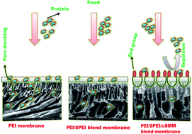
Sulfonated poly(ether imide) (SPEI) and charged surface modifying macromolecules (cSMM) were synthesized, characterized and blended into the casting solution of poly (ether imide) (PEI) with different compositions to develop surface modified ultra-filtration (UF) membranes by means of improved hydrophilicity. The membranes were prepared by a phase inversion technique and subjected to characterization experiments such as water permeation tests, equilibrium water content (WC), membrane hydraulic resistance (Rm) and protein rejection. Among others, a blend membrane containing 20 wt% SPEI and 5 wt% cSMM in PEI (called M12) exhibited the highest water permeation (440.6 L m−2 h−1 at 345 kPa), highest WC (86.3%) and lowest Rm (0.7 kPa/L m−2 h−1). The M12 membrane also exhibited the highest fluxes of 364.1 L m−2 h−1 and 230.3 L m−2 h−1 (at 345 kPa) with rejections of 31.3% and 62.1%, respectively, when the feed was aqueous trypsin and bovine serum albumin solution (1000 ppm). The difference in contact angle between the top and bottom surface confirmed surface migration of cSMM to the membrane top surface. SEM, AFM and tensile strength measurement revealed that the surface became more porous and rougher, and the mechanical strength was lowered by the blending of SPEI and addition of cSMM. That the M12 membrane achieved a lower internal fouling of 6% and 3.8% and higher flux recovery ratio (FRR) of 94% and 96.2% after UF of trypsin and bovine serum albumin solution explained its better antifouling properties as compared to a pristine PEI membrane.
1. Introduction

Membrane technology is becoming more and more important in water/wastewater treatment. With the help of ultra-filtration (UF), it is possible to separate particles, colloids and macromolecules, so that wastewater can be disinfected in this way. Thus, UF has been extensively used, as a separation process of high efficiency and low energy consumption, in drinking water treatment, environment protection, and in the pharmaceutical, textile and food industries.1,2 The UF process has become particularly important for separating proteins, peptide drugs from fermentation broths, protein recovery from blood plasma, reducing protein adsorption and cell adhesion for biomedical applications such as tissue engineering.3,4 However, successful application of UF membrane separation is seriously limited by membrane fouling.5 To overcome this drawback and to promote UF membrane applications, effective modification of the membrane surface is necessary.

Poly(ether imide) (PEI) is one of the most familiar membrane materials for the fabrication of micro-filtration (MF), UF, and nano-filtration (NF) membranes because of its excellent film formation, and mechanical properties as well as good thermal and chemical resistance. However, its hydrophobic nature has limited its applications especially in UF and NF. Therefore, the improvement in hydrophilicity of PEI membrane, particularly at the surface for fouling prevention, is necessary to widen the scope of PEI membrane applications.6 A variety of modification methods including physical adsorption and chemical bond formation were used by the researchers to prevent fouling at the membrane surface. Currently membrane surface modified materials are considered as effective means to influence the fouling properties of membranes, i.e., surface hydrophilicity, roughness, and charge.7 To achieve this goal, blending of hydrophilic additives into casting solutions has been studied recently due to its simple and low cost approach.8 In particular, blending of sulfonated poly(ether imide) (SPEI) into cellulose acetate was found to be an excellent surface modification method because of the blend polymer's film forming properties and chemical stability of the resulting membranes. Similarly, SPEI was blended to PEI membrane to make its surface negatively charged, which could successfully enhance the permeation rate and fouling resistance.9

Another approach of surface modification is to add surface modifying macromolecules (SMMs) to the host polymer matrix, targeting the modification of the membrane surface based on the theory of SMM surface migration. SMM when introduced as an additive in a casting solution will migrate to the membrane top surface and alter the chemistry of the surface without changing its bulk properties. Moreover, only a small amount of SMM additive is needed to cover the surface of membrane wholly.10 Blending of various additives such as poly(ethylene glycol) (PEG), poly(ethylene oxide) (PEO), poly(vinyl alcohol) (PVA), poly(vinyl pyrrolidone) (PVP), etc., into the casting solution may increases the surface hydrophilicity of membranes only temporarily and these additives may be leached out after a long period of operation due to their miscibility behavior in water.11 On the other hand, SMMs remained at the membrane surface for a much longer period due to the affinity between the central polyurethane segment and the host base polymer. The long chain of the polyurethane segment stays with the bulk polymer phase and end groups remained at the membrane surface.12,13 Hamza et al. reported that poly(ether sulfone) (PES)/SMMs blend membranes were less prone to fouling in oil–water separation.14

A new types of a charged SMM which having both end capped with sulfonate group was developed, probable to increases the hydrophilicity of the blend membrane. It is well established that the augmentation of hydrophilicity is crucial for a better fouling resistance performance in UF separation processes. Therefore, the aim of the present work is to investigate the effect of membrane surface modification by blending novel cSMM in the PEI and SPEI UF membranes. The prepared membranes were subjected to various characterization experiments such as pure water permeation (PWP) test, atomic force microscopy (AFM), contact angle (CA), scanning electron microscopy (SEM), and water content (WC) measurements. Finally, their performance in UF treatment of protein solutions was tested.

2. Experimental

2.1. Materials

PEI (Ultem® 1000) was supplied by GE Plastics, India as a gift sample. It was dried at 150 °C for 4 h before use. Chlorosulfonic acid (CSA), sodium hydroxide (NaOH), sodium chloride (NaCl) and phenolphthalein were obtained from Loba Chemie, India. 1,2-Dichloroethane (DCE), isopropanol (IPA), N-methyl-2-pyrrolidone (NMP), N,N-dimethyl acetamide (DMAc), and sodium lauryl sulphate (SLS) of analar grades were procured from Sigma Aldrich (USA). DEG, 4,4′-methylene bis(phenyl isocyanate) (MDI), hydroxyl benzene sulfonate (HBS) of analar grades, trypsin (20 kDa) and bovine serum albumin (BSA) (69 kDa) were procured from SRL Chemicals Ltd., India. Anhydrous sodium monobasic phosphate and sodium dibasic phosphate heptahydrate were also procured from Sigma Aldrich (USA) and used for the preparation of phosphate buffer solutions in the protein analysis. All chemicals were used as such without further purification. De-ionized and distilled water was employed for the preparation of the membranes and for the UF experiments.

2.2. Synthesis of SPEI

PEI was sulfonated using CSA as reported earlier.15 Twenty grams of PEI was dissolved in 100 mL of DCE at 60 °C and subsequently the PEI solution was kept at 30 °C. CSA (10 mL) mixed with 200 mL of DCE was slowly added to the PEI solution within 1 h with vigorous stirring. The SPEI precipitate was dissolved in DMAc at 50 °C, and coagulated by adding an excess amount of IPA. The precipitate was further filtered, washed with IPA, and dried at 40 °C in a vacuum oven for 24 h. Thus, SPEI in the hydrogen form was obtained. It was converted to the sodium form by soaking in NaOH (0.1 mol L−1) solution for 2 days.

2.3. Synthesis of charged surface modifying macromolecule

The cSMM preparation was done according to the method reported earlier.16 The cSMM, endcapped with HBS, was synthesized using a two-step solution polymerization method. The first step involved the reaction of MDI with DEG in a solvent of DMAc. This mixture produced a urethane prepolymer solution. Then the reaction was terminated by the addition of HBS resulting in a solution of sulfonated or charged SMM.
Brief procedure. A solution of 0.03 mol MDI (7.5 g) in 50 mL of degassed DMAc was loaded in a 1 L Pyrex round bottom flask. Then, a solution of 0.02 mol degassed DEG (2.122 g) in 100 mL of degassed DMAc was added drop-wise with stirring to react with MDI for 3 h. Then 0.02 mol of HBS (4.644 g) dissolved in 50 mL of degassed DMAc was added drop-wise and the solution was left under stirring for 24 h at 48–50 °C, resulting in a solution of cSMM. De-ionized water was added to cSMM solution in DMAC under vigorous stirring to precipitate the cSMM. Prepared cSMM was kept immersed in de-ionized water for 24 h under stirring to leach out residual solvent. Then, the cSMM was dried in hot air oven at 120 °C for 5 days. Finally it was stored and placed in desiccators. The scheme of the reaction and characterization of cSMM was reported earlier.17

2.4. Ion exchange capacity of SPEI and cSMM

About 1 g SPEI was soaked in a 2 M NaCl solutions for 1 day for the complete replacement of H+ with Na+. The remaining filtrate was titrated against 0.01 M of NaOH solution. The IEC was calculated by following equation:
 
image file: c4ra17097k-t1.tif(1)
where V is the volume of NaOH consumed by the titration (L) and Wd is the dry weight of the SPEI (g).

Degree of sulfonation (DS) was calculated18 by the following equation:

 
image file: c4ra17097k-t2.tif(2)
where 598 and 80 are, the molecular weight of PEI repeat unit and –SO3H respectively. IEC (mmol g−1 polymer) of cSMM polymer was determined by measuring the sulfur content via elemental analysis using CHNS analyzer model 2400 series II, Perkin-Elmer (Waltham, MA).

2.5. FT-IR spectroscopy

Fourier transform infra-red spectroscopy (FT-IR) was conducted to confirm the presence of functional groups in the polymer and in the membranes prepared in this study, using a FT-IR spectrometer (TENSOR 27, Bruker Optik GmbH, Germany). The spectra were recorded using KBr pellet in the frequency range of 500 to 4000 cm−1.

2.6. Membrane preparation

Pristine PEI membrane and SPEI and/or cSMM blended membranes were prepared by the phase inversion technique using NMP as solvent.19,20 The composition of the casting dopes is given in Table 1 together with the code of the membrane prepared from the dope and the chemical structure of PEI, SPEI and cSMM are shown in Fig. 1. The membrane-casting chamber was maintained at a temperature of 24 ± 1 °C and a relative humidity of 50 ± 2%. Before casting, a 2 L gelation bath, consisting of 2.5 wt% NMP solvent and 0.2 wt% surfactant, SLS in distilled water (non-solvent), was prepared and kept at 20 ± 1 °C. The blend solutions based on PEI, SPEI and additive were prepared in a solvent, NMP under constant mechanical stirring (300 rpm) in a round bottom flask for 4 h at 40 °C. The homogeneous solution that was allowed to stand at room temperature for at least one day in an air tight condition to get rid of air bubbles. The polymeric solution was cast on a glass plate by using a casting rod. Then, the casting solution film along with the glass plate was gently immersed into the gelation bath. Finally, the peeled off membrane was stored in a de-ionized water containing 0.1 wt% formalin solution to prevent microbial growth before it was used for UF experiments. The thickness of the prepared membrane was measured by digimatic micrometer (Mitutoyo, MDC-25SB, Japan) and it was maintained at 0.22 ± 0.02 mm.
Table 1 Composition of the casting solutions of the membranes
Membrane code Polymer (17.5 wt%) Additive, cSMM (wt%) Solvent, NMP (wt%)
PEI SPEI
M1 100 0 0 82.5
M2 90 10 0 82.5
M3 80 20 0 82.5
M4 100 0 1 81.5
M5 90 10 1 81.5
M6 80 20 1 81.5
M7 100 0 3 79.5
M8 90 10 3 79.5
M9 80 20 3 79.5
M10 100 0 5 77.5
M11 90 10 5 77.5
M12 80 20 5 77.5


image file: c4ra17097k-f1.tif
Fig. 1 Chemical structure of (A) PEI, (B) SPEI and (C) cSMM.

2.7. Pure water permeation and measurement of WC

The membranes were tested with a 400 mL batch type stirred cell (Amicon 8400-Model, Millipore, Billerica, MA) fitted with a Teflon-coated magnetic paddle. The effective membrane area available for UF was 38.5 cm2. Pure water permeation test was made at 345 kPa after the membranes were pressurized with distilled water at 414 kPa for 5 h. The pure water flux (PWF, Jw1) was calculated by21 the following equation:
 
image file: c4ra17097k-t3.tif(3)
where Jw1 is the pure water flux (L m−2 h−1), Q the permeate volume (L), A the membrane area (m2) and Δt is the sampling time (h).

The equilibrium water content (WC) of the membrane is defined as:

 
image file: c4ra17097k-t4.tif(4)
where Wd is weight of the dry membrane and Ww is weight of the membrane wetted with water.

To determine the membrane resistance for water permeation, the PWF (Jw1) was measured at pressures (ΔP) of 69, 138, 207, 276, and 345 kPa. The resistance of the membrane, Rm was evaluated22 from the slope of Jw1 versus ΔP using the following equation:

 
image file: c4ra17097k-t5.tif(5)

The tensile strength of the membrane was measured using uniaxial tensile test machine (Instron 3345, Bucks, UK). The sample was placed between the grips of the testing machine, and the speed of testing was set at the rate of 5 mm min−1. Six samples were taken from each type of membrane for the measurement and their averages are reported for their accuracy.

2.8. Membrane separation performance

Trypsin (20 kDa) and BSA (69 kDa) were chosen for the estimation of protein rejection by the prepared membranes. The feed solutions were prepared individually at a concentration of 0.1 wt% in phosphate buffer (pH ≈ 7.2) solution and permeation experiments were conducted at the pressure of 345 kPa and at the ambient temperature. The permeation flux (Jp) was calculated by an equation similar to eqn (3) but with Q that was the volume of permeates collected when the feed was the protein solution. The permeate protein concentration was measured using UV-visible double beam spectrophotometer (Systronics, 2201) at a wavelength of 280 nm. The percentage solute rejection (SR) was calculated23 by the following equation:
 
image file: c4ra17097k-t6.tif(6)
where Cp and Cf are the protein concentrations in the permeate and feed, respectively. After ultra-filtration experiments with the feed protein solution, the membrane was washed with de-ionized water for 30 min and the pure water flux of the cleaned membrane (Jw2) was determined again at 345 kPa. To evaluate the antifouling property of membranes, the flux recovery ratio (FRR) was calculated24 by equation:
 
image file: c4ra17097k-t7.tif(7)

Internal (Rif), surface (Rsf) and total (Rt) fouling were calculated by the following eqn (8)–(10) in order to analyze the antifouling properties25 more in detail:

 
image file: c4ra17097k-t8.tif(8)
 
image file: c4ra17097k-t9.tif(9)
 
image file: c4ra17097k-t10.tif(10)

2.9. Measurement of MWCO and average pore size

The molecular weight cut-off (MWCO) of a membrane was determined by the molecular weight of the solute the rejection percentage of which was greater than 80%. The average pore size of the membranes was estimated from the %SR of either trypsin (20 kDa) or BSA (69 kDa) by
 
image file: c4ra17097k-t11.tif(11)
where [R with combining macron] is the average pore size (radius) of the membrane (Å), and α is the Stokes radius (Å) of the solute. The Stoke radii of the solutes were obtained from the plot of solute molecular weight versus solute radius in aqueous solution.26

2.10. Membrane surface analysis

The contact angle (CA) (θ) between water and membrane surface was directly measured using a CA meter (VCA Optima Surface Analysis System, AST Products Inc., Billerica, MA). The CA was measured at five different locations on each membrane sample and the average values are reported. From the values of CA, the work of adhesion (ωA) i.e., the work required to pull the liquid away from the surface leaving the equilibrium27 absorbed film is given by the following equation:
 
ωA = γw(1 + cos[thin space (1/6-em)]θ) (12)
where γw is surface tension of water (7.2 × 10⁻² N m−1) and θ is the contact angle.

A scanning electron microscope (SEM, Bruker Nano, Ewing, NJ) was used to observe cross section of the membranes. The sample for the SEM was prepared by freezing the dry membrane in liquid nitrogen for 3 min and breaking it to produce a cross section. Then the sample was gold sputtered under vacuum conditions prior to SEM analysis.

Three dimensional AFM surface images of the control and modified PEI membranes were compared in terms of surface roughness by means of scanning probe microscopy (model 5500, Agilent Technologies Inc., Palo Alto, CA). The scan size was 25 μm × 25 μm. Surface roughness parameters were obtained from the AFM image at different locations of a membrane surface.

3. Results and discussion

3.1. FT-IR spectroscopy

The FT-IR spectra of the polymer (SPEI and cSMM) and membrane M12 that contains 20 wt% of SPEI and 5 wt% of cSMM in PEI are shown in Fig. 2. The presence of sulfonic groups in SPEI can be confirmed by the peak at 3458 cm−1. From the FT-IR spectrum of cSMM, the presence of sulfonic groups in cSMM can be confirmed by the absorption bands at 3411 cm−1 (O–H), 1235 cm−1 (asymmetric O[double bond, length as m-dash]S[double bond, length as m-dash]O), 1065 cm−1 (symmetric O[double bond, length as m-dash]S[double bond, length as m-dash]O), 1017 cm−1 (S[double bond, length as m-dash]O) and 700 cm−1 (S–O) and sulfonic group stretching vibration at 1376 cm−1. The characteristic peaks of M12 membrane confirm the presence of SPEI and cSMM in the membrane matrix.
image file: c4ra17097k-f2.tif
Fig. 2 FT-IR spectra of (a) SPEI polymer, (b) cSMM polymer and (c) 5 wt% cSMM composition with 80[thin space (1/6-em)]:[thin space (1/6-em)]20 ratio of PEI/SPEI (M12) membrane.

3.2. Ion exchange capacity

The ion exchange capacity and degree of sulfonation of SPEI were found to be 0.08 mmol g−1 and 51.1%, respectively and the IEC value of the polymer cSMM is 0.344 mmol g−1. The ion-exchange capacity (IEC) provides information on the charge density in the membranes, which is an imperative factor associated to the transport properties of the membranes. Charge density plays vital roles in separation performances of the membranes. The charged UF membranes can effectively reduce the adsorbed amount of proteins on the membrane surface and the adsorption depends on the strength of electrostatic interaction between the membrane and solute molecules at a specific pH value of the solution.28 Li and Chung have developed negatively charged hollow fiber UF membranes based on the blend of sulfonated poly (ether sulfone) (SPES) and unmodified PES. The selectivity of the membranes towards BSA and haemoglobin (Hb) was highly dependent on the charge density.29 Recently, Liu et al. have fabricated charged UF membranes using sulfonated polyphenylsulfone (SPPS) random copolymer and found that the hydrophilicity and antifouling capacity of the membranes were improved by addition of sulfonated copolymer.30

3.3. Membrane surface characterization

Fig. 3 showed that the SEM cross section image of the prepared membranes. As can be seen, all membranes have an asymmetric structure with a dense skin layer supported by a finger like macrovoids in the sub-layer. However, the morphology of the membrane has become different when SPEI or/and cSMM were added PEI matrix. The control PEI membrane (M1) consists of three layers, (1) the dense skin layer, (2) short vertical finger-like pores, and (3) horizontal macrovoids. When SPEI was added (M2), layer (2) becomes much thinner while layer (3) becomes thicker and the macrovoids become vertical. As the SPEI concentration increases (M3), the structure is practically the same as M2 but the macrovoids are larger. This result is ascribed to the hydrophilic nature of SPEI, which draws in more water into the membrane phase during the phase inversion process. The above trend is more intensified when SMM is added, as all macrovoids in (3) become larger and stand vertical.
image file: c4ra17097k-f3.tif
Fig. 3 SEM cross section photographs of the pure PEI (M1) and PEI/SPEI (M2 & M3), PEI/cSMM (M4, M7 & M10) and PEI/SPEI/cSMM (M5, M6, M8, M9, M11 & M12) blended membranes.

3D AFM surface images of the prepared membranes are shown in Fig. 4 and Table 2 summarizes the surface roughness values. The control membrane (M1) displayed a very smooth surface. The roughness of the membrane increases from M1 to M3 by blending SPEI but the change is relatively small. The blending of cSMM further increased the roughness progressively as the amount of cSMM increased (M1 < M4 < M7 < M10, M3 < M12). The trends observed in the roughness parameters are exactly the same as those found in WC; i.e. WC increases progressively with the amount of SPEI added (M1 < M2 < M3) as well as the amount of cSMM added (M1 < M4 < M7 < M10, M2 < M5 < M8 < M11, M3 < M6 < M9 < M12). Hence, the increase in roughness parameter is ascribed to the increase in porosity and also likely to the increase in pore size.31


image file: c4ra17097k-f4.tif
Fig. 4 3D AFM images of the pure PEI (M1) and PEI/SPEI (M3), PEI/cSMM (M4, M7 & M10) and PEI/SPEI/cSMM (M12) blended membranes.
Table 2 The contact angle, adhesion work, roughness, MWCO and pore size of the prepared membranes
Membrane code Contact angle (θ) (°) Adhesion work (ωA) (mN m−1) Roughness parameters (nm) MWCO (kDa) Pore size, [R with combining macron] (Å)
Top Bottom Top Bottom Ra Rq Rz
M1 93.7 93.0 67.4 68.2 11.4 16.6 89.7 20 28.0
M2 80.5 79.5 84.0 85.1    
M3 78.7 78.6 86.1 86.2 15.2 19.3 101 20 28.0
M4 77.3 78.6 88.0 86.2 20.6 30.4 110 20 28.2
M5 65.4 72.7 102.0 93.4    
M6 60.8 74.1 107.1 91.7    
M7 66.1 70.3 101.2 96.3 25.9 35.4 179 69 44.3
M8 57.6 68.2 110.6 98.7    
M9 50.0 71.5 118.3 94.8    
M10 61.0 63.0 107.0 104.7 44.5 49.3 282 69 44.1
M11 45.4 66.8 122.6 100.3    
M12 40.1 61.5 127.1 106.4 46.3 64.6 309 >69 >44


The surface hydrophilicity can be evaluated by measuring the CA.32 Table 2 summarizes CAs at the top and bottom side of the membranes. A trend exactly opposite to the roughness parameters and WC is found at the top surface; i.e. CA decreases progressively as the amount of SPEI is increased (M1 > M2 > M3) and the amount of cSMM is increased (M1 > M4 > M7 > M10, M2 > M5 > M8 > M11, M3 > M6 > M9 > M12). As a result, CA decreases considerably from 93.7° for M1 and 40.1° for M12. The trend is almost the same at the bottom layer with a notable exception of M2 < M5. Comparing the CAs of top and bottom surface, they are almost the same for M1, M2 and M3 membranes, indicating SPEI was uniformly distributed across the cross-section of SPEI blended membranes. On the other hand, when cSMM is added (from M4 to M12), CA of the top surface is significantly lower than the bottom layer, indicating the surface migration of cSMM. The surface energy (adhesion work, ωA) can also be used as a measure of hydrophilicity.33

In the present work, strength of liquid adheres to a solid membrane surface was estimated from CA data34 and the ωA results are listed in Table 2. The results indicate that the top ωA of pure PEI membrane (M1) increased from (67.4 mN m−1) to (127.1 mN m−1) in the PEI/SPEI containing 5 wt% cSMM membrane (M12). Both trends of the CA and ωA indicate the hydrophilicity of the PEI modified blended membrane which increases with the addition of both SPEI and cSMM composition but maximum ωA was observed in the M12 membrane. An increase in surface energy and a decrease in inter-facial tension resulted in an increase of wettability of the modified PEI membranes. Thus, M12 membrane is the most hydrophilic and thus had the highest surface energy as an evidenced by WC and CA test obtained in the present studies. Similar results have also been observed for PEI/N-phthaloyl chitosan blend UF system.35

3.4. Water permeation test

PWFs are summarized in Table 3. PWF shows a direct correlation to WC (and roughness parameters) and reverse correlation to CA. In other words, PWF is increased with an increase in the amount of SPEI added and is further increased by the addition of cSMM. This means that PWF increases as a result of increase of the porosity (or pore size) as well as the increase of hydrophilicity. As a result, PWF increased from 6.4 L m−2 h−1 for M1 and 440.6 L m−2 h−1 for M12 membrane, which is almost 70 times increased.
Table 3 The performances of modified poly(ether imide) membranes
Membrane code PWF (L m−2 h−1) Water content (%) Membrane hydraulic resistance (kPa/L m−2 h−1) Tensile strength (MPa) Trypsin solution BSA solution
Permeate flux (L m−2 h−1) Rejection (%) Permeate flux (L m−2 h−1) Rejection (%)
M1 6.4 61.0 52.6 6.4 5.2 80.0 2.1 94.6
M2 58.6 67.5 4.9
M3 71.4 70.8 4.4 5.2 8.2 80.2 3.8 92.5
M4 9.3 66.5 26.6 5.0 58.1 78.0 37.9 90.3
M5 97.5 72.8 3.0
M6 108.4 73.6 2.8 86.5 73.0 68.9 87.5
M7 25.5 69.4 12.4 4.7 21.6 51.5 10.5 90.3
M8 166.5 79.0 1.7
M9 205.3 81.4 1.5 182.4 61.6 126.5 73.5
M10 60.4 73.6 5.2 4.4 45.4 43.8 27.1 87.1
M11 326.6 83.6 0.9
M12 440.6 86.3 0.7 4.1 364.1 31.3 230.3 62.1


3.5. Hydraulic resistance and tensile strength

Rm is an indication of tolerance of membrane towards different hydraulic pressures.36 In Table 3, Rm of M12 is the lowest, which is natural since Rm is inversely proportional to PWF (eqn (5)). The data on tensile strength is summarized in Table 3.

The tensile strength decreases with the addition of SPEI (M1 > M3). It keeps decreasing with the addition of cSMM (M1 > M4 > M7 > M10 > M12). Thus, the membrane becomes mechanically less stable as porosity increases.

3.6. Ultrafiltration with protein solutions

Table 3 shows typical examples of experimental data for UF of aqueous trypsin and BSA solutions. Comparing the data for trypsin and BSA, the separation of BSA is much higher than that of trypsin. This result is common for UF membranes and can be ascribed to the sieving effect,37 by which BSA of higher molecular weight is rejected more than trypsin of lower molecular weight. On the other hand, the permeate flux is higher for trypsin than BSA, indicating severe pore blocking by larger BSA molecules. Comparing the data from different membranes, trypsin rejection decreases by adding SPEI (M1 > M3) and it further decreases progressively by adding the larger amount of cSMM (M1 ≈ M4 > M7 > M10, M3 > M6 > M9 >M12). The trend is more obvious for BSA rejection. As for the flux, even though they are significantly lower than PWF, the same trend is retained even in the presence of proteins in the feed; i.e. for both trypsin (M1 < M3, M1 < M4 < M7 < M10) and BSA (M3 < M6 < M9 < M12). It is interesting to note that there was a remarkable increase in flux from M1 to M3 by adding SPEI. The trend observed both in rejection and flux can be ascribed to the increase in hydrophilicity and the porosity (pore size) by adding SPEI and cSMM.

Based on the percentage solute rejection data given in Table 3, MWCO was obtained for each membrane. Molecular weight of trypsin (20 kDa) was used for the membranes (M1, M3 & M4). Trypsin rejection of M4 (78%) was considered nearly equal to 80%. Molecular weight of BSA (69 kDa) was used for the membranes (M7 & M10). As for M12, even BSA could not reach the solute rejection of 80%, therefore its MWCO was considered to be greater than 69 kDa. The MWCOs are summarized in Table 2. MWCO increases gradually from 20 to >69 kDa as the cSMM content increases from 0 to 5 wt% in the casting dope.

The pore sizes of the membranes are also summarized in Table 2. From the table, the pore size increases progressively from 28 to >44 Å as the cSMM content increases from 0 to 5 wt% in the casting dope. These data are in good agreement with results obtained in the earlier study.38 On the other hand, MWCO has a direct relationship with the pore size of the membrane.39 The results of both MWCO and pore size are in accordance with the SEM images shown in Fig. 3.

3.7. Antifouling studies

FRR values were introduced to assess the easiness of washing the fouled membrane. The higher the FRR value, better the recovery of the pure water flux.40 Fig. 5 illustrates the typical FRR values calculated from PWFs before and after UF of trypsin and BSA solution. Comparing to the control PEI membrane (M1), it is clearly seen that PEI/SPEI membrane (M3) has higher FRR values for both trypsin and BSA experiments. The trend is not quite obvious when cSMM is added. But when all the cSMM containing membranes (from M4 to M12) are averaged, FRRs for trypsin and BSA are, respectively, 90.3 and 87.8%, which values are significantly higher than the averages of M1 and M3 membranes, which are 85.8 and 74.8%. Hence, we can conclude that addition of both SPEI and cSMM could increase the capacity of hydraulic cleaning due to the increase in membrane surface hydrophilicity, which reduced protein/membrane surface interaction.
image file: c4ra17097k-f5.tif
Fig. 5 Flux recovery ratio of PEI/SPEI/cSMM blended UF membranes in trypsin and BSA solution.

The fouling behavior of all the prepared membranes was further studied in detail by calculating total fouling (Rt), the surface fouling (Rsf) and the internal fouling (Rif) as shown in Fig. 6. All modified PEI membrane had lower Rt and Rif than that of the pure PEI membrane (M1) for the trypsin and BSA solution. The lower Rt means that the flux loss by fouling is less and the membrane is less prone to protein adsorption. The lower Rif indicates that the irreversible fouling due to pore blocking is less. As for Rsf, for the fixed PEI/SPEI ratio of 100/0, the order is M1 ≈ M4 < M7 < M10, while for a fixed PEI/SPEI ratio of 80/20, the order is M3 ≈ M6 < M9 < M12 for the BSA solution. The fouling data obtained from protein solution of trypsin and BSA, the surface fouling seems to be enhanced by adding SMM. But this type of fouling is reversible41 and the flux can be recovered easily by washing. This is the reason why FRR became greater when cSMM was added.


image file: c4ra17097k-f6.tif
Fig. 6 The fouling properties of PEI/SPEI/cSMM blended UF membranes in trypsin and BSA solution.

4. Conclusions

Novel flat sheet UF membranes were prepared successfully from blend solutions of PEI/SPEI with different polymer compositions by phase inversion technique using NMP as solvent and cSMM as an additive. The effect of SPEI blend composition and the addition of cSMM on the surface morphology were investigated. It was found that all the membranes have an asymmetric structure. With an increase in SPEI in the casting solution more macrovoids were found vertically orientated and by the addition of cSMM the top surface became more porous. The 3D AFM images indicated that the membrane surface became rougher as cSMM content was increased. The WC and CA measurements confirmed that the overall porosity increased and the membrane surface became more hydrophilic by the blending of SPEI and the addition of cSMM. As well, PWF was correlated precisely to WC and CA. It is also interesting to note that cSMM migration to the surface was detected by the difference of CA between top and bottom surface. Tensile strength decreased by blending of SPEI and addition of cSMM. From the separation experiments of trypsin and BSA, it was found that the flux increased while protein rejection decreased by blending of SPEI and addition of cSMM. It was also concluded that the addition of cSMM increased the surface fouling and reduced the internal fouling. Since the surface fouling can be easily removed by washing with distilled water, addition of cSMM enhances the capacity of hydraulic membrane washing.

Acknowledgements

This work was supported by the Science and Engineering Research Board (SERB), Department of Science and Technology (DST), Government of India under project number SR/FT/CS-22-2011. This support is gratefully acknowledged. The authors also gratefully acknowledge financial support from the Natural Sciences and Engineering Research Council of Canada for the partial support of this work.

Notes and references

  1. Y. Wan, A. Prudente and S. Sathivel, Food Sci. Technol., 2012, 46, 574–579 CAS.
  2. P. Qin, X. Hong, M. Nazmul Karim, T. Shintani, J. Li and C. Chen, Langmuir, 2013, 29, 4167–4175 CrossRef CAS PubMed.
  3. A. Saxena, B. P. Tripathi, M. Kumar and V. K. Shahi, Adv. Colloid Interface Sci., 2009, 145, 1–22 CrossRef CAS PubMed.
  4. X. Chen, T. Chen, Z. Lin, X. Li, W. Wu and J. Li, Chem. Commun., 2015, 51, 487–490 RSC.
  5. M. Kumar and M. Ulbricht, RSC Adv., 2013, 3, 12190–12203 RSC.
  6. E. Celik, L. Liu and H. Choi, Water Res., 2011, 45, 5287–5294 CrossRef CAS PubMed.
  7. P. D. Peeva, T. Knoche, T. Pieper and M. Ulbricht, Sep. Purif. Technol., 2012, 92, 83–92 CrossRef CAS PubMed.
  8. H. T. Dang, C. Amelot, D. Rana, R. M. Narbaitz and T. Matsuura, J. Appl. Polym. Sci., 2010, 116, 2205–2215 CAS.
  9. L. Q. Shen, Z. K. Xu, Q. Yang, H. L. Sun, S. Y. Wang, Z. M. Liu and Y. Y. Xu, J. Appl. Polym. Sci., 2004, 92, 1709–1715 CrossRef CAS.
  10. Y. Fang, V. A. Pham, T. Matsuura, J. P. Santerre and R. M. Narbaitz, J. Appl. Polym. Sci., 1994, 54, 1937–1943 CrossRef CAS.
  11. L. Y. Lafreniere, F. D. F. Talbot, T. Matsuura and S. Sourirajan, Ind. Eng. Chem. Res., 1987, 26, 2385–2389 CrossRef CAS.
  12. D. Rana, R. M. Narbaitz, A.-M. Garand-Sheridan, A. Westgate, T. Matsuura, S. Tabe and S. Y. Jasim, J. Mater. Chem., 2014, 2, 10059–10072 RSC.
  13. Y. Kim, D. Rana, T. Matsuura and W.-J. Chung, Chem. Commun., 2012, 48, 693–695 RSC.
  14. A. Hamza, V. A. Pham, T. Matsuura and J. P. Santerre, J. Membr. Sci., 1997, 131, 217–227 CrossRef CAS.
  15. L. Q. Shen, Z. K. Xu, Z. M. Liu and Y. Y. Xu, J. Membr. Sci., 2003, 218, 279–293 CrossRef CAS.
  16. M. N. A. Mohd Norddin, A. F. Ismail, D. Rana, T. Matsuura, A. Mustafa and A. Tabe-Mohammadi, J. Membr. Sci., 2008, 323, 404–413 CrossRef PubMed.
  17. P. Kanagaraj, S. Neelakandan and A. Nagendran, J. Appl. Polym. Sci., 2014, 131, 40320 CrossRef.
  18. R. Y. M. Huang, P. Shao, M. Burns and X. Feng, J. Appl. Polym. Sci., 2001, 82, 2651–2660 CrossRef CAS.
  19. D. Rana, B. Scheier, R. M. Narbaitz, T. Matsuura, S. Tabe, S. Y. Jasim and K. C. Khulbe, J. Membr. Sci., 2012, 409–410, 346–354 CrossRef CAS PubMed.
  20. D. Y. Xing, S. Y. Chan and T. S. Chung, Chem. Eng. Sci., 2013, 87, 194–203 CrossRef CAS PubMed.
  21. M. F. Rabuni, N. M. K. Sulaiman, M. K. Aroua, C. Y. Chee and N. A. Hashim, Chem. Eng. Sci., 2015, 122, 426–435 CrossRef CAS PubMed.
  22. D. Bhattacharya, J. M. McCarthy and R. B. Grieves, AIChE J., 1974, 20, 1206–1212 CrossRef.
  23. M. Sivakumar, R. Malaisamy, C. J. Sajitha, D. Mohan, V. Mohan and R. Rangarajan, J. Membr. Sci., 2000, 169, 215–228 CrossRef CAS.
  24. A. L. Ahmad, A. A. Abdulkarim, B. S. Ooi and S. Ismail, Chem. Eng. J., 2013, 223, 246–267 CrossRef CAS PubMed.
  25. Y. Q. Wang, T. Wang, Y. L. Su, F. B. Peng, H. Wu and Z. Jiang, Langmuir, 2005, 21, 11856–11862 CrossRef CAS PubMed.
  26. M. N. Sarbolouki, Sep. Sci. Technol., 1982, 17, 381–386 CrossRef CAS.
  27. S. P. Nunes and K. V. Peinemann, J. Membr. Sci., 1992, 73, 25–35 CrossRef CAS.
  28. M. Kumar and M. Ulbricht, Polymer, 2014, 55, 354–365 CrossRef CAS PubMed.
  29. Y. Li and T. S. Chung, J. Membr. Sci., 2008, 309, 45–55 CrossRef CAS PubMed.
  30. Y. Liu, X. Yue, S. Zhang, J. Ren, L. Yang, Q. Wang and G. Wang, Sep. Purif. Technol., 2012, 98, 298–307 CrossRef CAS PubMed.
  31. Y. Kim, D. Rana, T. Matsuura, W. -J. Chung and K. C. Khulbe, Sep. Purif. Technol., 2010, 72, 123–132 CrossRef CAS PubMed.
  32. D. Rana and T. Matsurra, Chem. Rev., 2010, 110, 2448–2471 CrossRef CAS PubMed.
  33. L. Palacio and J. I. Calvo, J. Membr. Sci., 1999, 152, 189–201 CrossRef CAS.
  34. M. Khayet, M. M. Nasef and J. I. Mengual, J. Membr. Sci., 2005, 263, 77–95 CrossRef CAS PubMed.
  35. P. Kanagaraj, A. Nagendran, D. Rana, T. Matsuura, S. Neelakandan and T. Karthikkumar, Appl. Surf. Sci., 2015, 329, 165–173 CrossRef CAS PubMed.
  36. R. Mahendran, R. Malaisamy and D. Mohan, Polym. Adv. Technol., 2004, 15, 149–157 CrossRef CAS.
  37. W. Zhao, Y. Su, C. Li, Q. Shi, X. Ning and Z. Jiang, J. Membr. Sci., 2008, 318, 405–412 CrossRef CAS PubMed.
  38. P. Kanagaraj, A. Nagendran, D. Rana, T. Matsuura and S. Neelakandan, Int. J. Biol. Macromol., 2015, 72, 223–229 CrossRef CAS PubMed.
  39. R. Mahendran, R. Malaisamy and D. Mohan, Polym. Adv. Technol., 2004, 15, 149–157 CrossRef CAS.
  40. A. Nagendran, A. Vijayalakshmi, D. L. Arockiasamy, K. H. Shobana and D. Mohan, J. Hazard. Mater., 2008, 155, 477–485 CrossRef CAS PubMed.
  41. R. W. Baker, Membrane Technology and Applications, Wiley, Chichester, 2nd edn, 2004, ch. 6, pp. 242–246 Search PubMed.

This journal is © The Royal Society of Chemistry 2015
Click here to see how this site uses Cookies. View our privacy policy here.