Chiral, fluorescent microparticles constructed by optically active helical substituted polyacetylene: preparation and enantioselective recognition ability

Huajun Huangab, Wantai Yangab and Jianping Deng*ab
aState Key Laboratory of Chemical Resource Engineering, Beijing University of Chemical Technology, Beijing 100029, China. E-mail: dengjp@mail.buct.edu.cn; Fax: +86-10-6443-5128; Tel: +86-10-6443-5128
bCollege of Materials Science and Engineering, Beijing University of Chemical Technology, Beijing 100029, China

Received 16th December 2014 , Accepted 9th March 2015

First published on 9th March 2015


Abstract

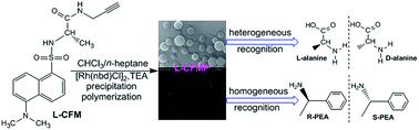
Microparticles simultaneously showing optical activity and fluorescence were prepared based on fluorescent, optically active helical polymers. Chiral and fluorescent substituted acetylene monomers (L- and D-CFM) were synthesized and then underwent precipitation polymerization in a solvent mixture of CHCl3/n-heptane in the presence of a Rh catalyst at room temperature. The CHCl3/n-heptane mixture at a suitable ratio provided spherical microparticles (L- and D-CFMPs) with uniform diameters of 910 nm in a high yield (ca. 90 wt%). The microparticles were comprised of polymer chains (number-average molecular weight, 8700 g mol−1) that were found to adopt chiral helical structures, according to circular dichroism and UV-vis absorption spectroscopy. The fluorescence property was measured using fluorescent microscopy and spectroscopy. Remarkably, the novel microparticles exhibited enantioselective recognition ability towards alanine and phenylethylamine enantiomers. However, L- and D-CFMPs behaved differently in the enantioselective recognition processes. Possible mechanisms were proposed for the observed enantioselective recognition.


1. Introduction

Fluorescent technology has been extensively used in a diverse number of significant fields such as diagnostics,1 biological imaging,2 recognition/sensing,3 detection,4 analytical techniques,5 and environmental sciences.6 The generally utilized fluorophores include organic dyes,7,8 quantum dots (QDs),9,10 metal particles,11 and conjugated polymers.12 For the first three categories, their dispersion properties are not highly satisfactory, so they are frequently encapsulated by or immobilized on other materials (polymers, silica, etc.) for practical uses.13 Nonetheless, encapsulation processes give rise to another problem, that is, fluorophores easily leak out of the host systems due to the simple physical incorporation. In addition, encapsulation may weaken the fluorophores' fluorescent intensity because of the presence of host. To solve the problems, encapsulation of fluorophores can be accomplished by covalently chemical bonds, but this process may change the molecular structure of, e.g. small-molecular organic dyes and in turn attenuate their fluorescence property. Alternatively, fluorescent conjugated polymers have been gathering ever-growing attention in last years.14 For monomers bearing fluorescent groups, the resulting polymers are advantageous in terms of the uniform distribution of fluorescent moieties.15 However, there is also a problem in fluorescent conjugated polymers, namely, a majority of them, typically exemplified by polyacetylenes,16–22 are hydrophobic, which remarkably limits their practical applications particularly in aqueous media. To overcome this problem, conjugated polymer-based particles23–25 (nano- and micro-scaled) being able to be dispersed in water are one of the good choices. Furthermore, fluorescent particles are also attractive for their controllable loading of fluorophores, uniform size, spherical morphology, and ready surface functionalization.26,27 Additionally, fluorescent polymer particles with more complex structures and multiple functions for instance magnetic fluorescent particles are especially attractive due to the sensitivity toward external magnetic field.28,29

Chirality is ubiquitous in nature. Even though we do not yet know the exact driving force for this fascinating phenomenon, chiral sciences, chiral technologies, and chiral materials have led to tremendous impact on biology, medicine, pharmaceutical, agrochemical, materials, and chemistry. If we combine chirality and fluorescent technology in one entity, novel, fascinating, and significant materials shall be developed.30–34 They will undoubtedly lead to more far-reaching implications. Investigations under this theme have been well documented in literature.35 The chiral fluorescent materials so far investigated range from small organic molecules (including cyclic molecules),36–39 (bio)macromolecules,40–42 supermolecules,43 metal particle complexes11 to metal organic frameworks (MOFs) consisting of chiral ligands.44 The chiral fluorescent architectures demonstrated interesting enantioselective recognition functions by the enantioselective formation of hydrogen bonds,45 complex,46 or inclusion complex.47 Regrettably, a unique type of chiral polymers, that is, chiral helical polymers, have been little studied in this respect. Helical polymers are expected to behave differently from usual chiral polymers due to the well-known chiral amplification effect.48,49 Nevertheless, no chiral fluorescent particles have been established in literature based on such interesting helical polymers, even though synthetic helical polymers have constructed an active research area.50–60 The essential reason is due to the lacking of effective techniques for preparing the anticipated particles.

Nowadays it is still a big challenge for preparing polymeric particles derived from acetylenics,61–63 primarily because of the specific polymerization mechanism and the requirement of metal catalyst. In earlier studies, we successfully prepared optically active nano- and micro-particles based on helical substituted polyacetylenes.64–66 The chiral polymer particles demonstrated remarkable potentials in asymmetric catalysis, enantioselective crystallization, enantioselective release, chiral adsorption, etc. We also prepared fluorescent polymeric particles derived from optically inactive helical polyacetylene.67 The fluorescent particles helped us to follow the nucleation and growth of substituted acetylene-derived particles in the course of precipitation polymerization, vividly justified our hypothesis on the formation mechanism and growth of the particles. Based on the original investigations above, we in the present study designed and prepared a novel type of polymeric particles, which were constructed by fluorescent, optically active helical substituted polyacetylene and excitingly demonstrated chirality and fluorescent properties simultaneously. Remarkably, such polymer particles enantioselectively recognized towards alanine and phenylethylamine enantiomers. Accordingly, novel sensors with enantioselective recognition ability can be anticipated from them in the future studies.

2. Experimental

2.1 Materials

Dansyl chloride and [Rh(nbd)Cl]2 were purchased from Alfa Aesar. Propargylamine was obtained from Acros Organics. L- and D-alanine, Boc-L- and Boc-D-alanine, and (R)-(+)- and (S)-(−)-1-phenylethylamine (PEA) were purchased from Aladdin Reagent Co. (Shanghai, China). All the reagents above were of analytical grade and used without further purification. CHCl3, tetrahydrofuran (THF), and dimethyl formamide (DMF) were distilled under reduced pressure before use. Deionized water was used in all the recognition experiments.

2.2 Measurements

FT-IR spectra were recorded using a Nicolet NEXUS 870 infrared spectrometer. 1H and 13C NMR spectra were recorded on a Bruker AV 400 spectrometer. The morphology of the microparticles was observed with a HITACHI S-4700 scanning electron micro-scope (SEM). Circular dichroism (CD) and UV-vis absorption spectra were measured using a Jasco-810 spectropolarimeter. The molecular weights and molecular weight polydispersities (Mw/Mn) were determined by GPC/SEC (Agilent Technologies 1200 Series) with DMF as eluent. Fluorescence spectra were measured on a HITACHI F-7000 Fluorescence Spectrophotometer (HITACHI, Japan). For all the measurements of fluorescence spectra, the excitation wavelength was kept at 400 nm with a slit width of 5 nm. Fluorescence images were observed with a fluorescence microscope (Olympus IX81). Elemental analysis was performed on an Elementar vario EL cube element analyzer. Thermogravimetric analysis (TGA) was carried out using a Q50 TGA at a scanning rate of 10 °C min−1 under N2. Differential scanning calorimetry (DSC) was performed at a scanning rate of 10 °C min−1 under N2 on a DSC1 (Mettler-Toledo).

2.3 Synthesis of monomers

Preparation of the chiral fluorescent monomers (L- and D-CFM) is presented in Scheme 1. The monomers were synthesized by a three-step procedure according to our earlier method.64–67 All the reagents (analytic grade) used in this section were bought from Aldrich and directly used without further purification. The major processes are briefly stated below, with L-CFM as example.
image file: c4ra16466k-s1.tif
Scheme 1 Schematic for preparing chiral fluorescent monomer (L-CFM). D-CFM was synthesized in the same manner by using Boc-D-alanine.

In the first step, Boc-(L)-alanine (1.89 g, 10 mmol), isobutyl chloroformate (1.3 ml, 10 mmol), and 4-methylmorpholine (1.1 ml, 10 mmol) were added in THF (100 ml) sequentially. Then the mixture solution was stirred at room temperature for about 30 min. Propargylamine (0.65 ml, 10 mmol) was dropwise added in the solution. The reaction lasted for 4 h at room temperature. For collecting the product, the white precipitate was filtered off. The filtrate was washed with 2 M HCl aqueous solution thrice and then washed with saturated aqueous NaHCO3 to neutralize the solution. Afterwards, the solution was dried over anhydrous MgSO4, filtered, and concentrated to give the first step product bearing BOC group. In the second step, the product from the first step was deprotected for 3 h by using acetic acid and 2 M HCl aqueous solution as a buffer, and then a certain amount of K2CO3 was added in the solution to neutralize the residual acid. Subsequently, the surplus K2CO3 was filtered off. In the third step, the filtrate was concentrated by rotary evaporator and then dissolved in DMF and CH2Cl2 mixture (DMF, 3 ml; CH2Cl2, 30 ml). After adding triethylamine (2 ml) and stirring for 30 min, dansyl chloride (1 g, 3.8 mmol) was added in the solution. The reaction was kept at 0 °C for 24 h. The white precipitate was filtered off, and the coarse monomer was purified further by recrystallization thrice from CHCl3/hexane. The monomer was structurally identified with FT-IR, 1H and 13C NMR, and elemental analysis techniques. The FT-IR spectra are presented in Fig. 1, while the NMR spectra and elemental analysis data are presented in ESI (Fig. S1). The discussion on FT-IR spectra will be provided later, together with the spectrum of the corresponding polymeric microparticles. All the characterizations clearly verified the successful preparation of the designed monomers (L- and D-CFM).


image file: c4ra16466k-f1.tif
Fig. 1 Typical FT-IR spectra of monomer (L-CFM) and the polymeric microparticles (KBr tablet).

2.4 Fabrication of chiral fluorescent microparticles (CFMP)

According to our earlier successful preparation of helical substituted polyacetylene-based microparticles by precipitation polymerization,68,69 L- and D-CFM in the present study underwent precipitation polymerization for obtaining the anticipated microparticles. A typical process for the precipitation polymerization is briefly presented below, as schematically illustrated in Scheme 2.
image file: c4ra16466k-s2.tif
Scheme 2 Schematic illustration for preparing the chiral fluorescent microparticles (including both D- and L-CFMPs).

All the processes in precipitation polymerization were carried out under N2. For performing the precipitation polymerization, a certain amount of monomer (L-CFM, [M] = 0.025 mol L−1; as representative) and catalyst [Rh(nbd)Cl]2 ([Rh] = 2.5 × 10⁻⁴ mol L−1) were separately charged into two tubes. 2 ml CHCl3 was added into the monomer tube, and 0.5–2 ml CHCl3 and 0.1 ml triethylamine (TEA) were charged into the catalyst tube. After complete dissolution of the chemicals, the Rh catalyst solution was immediately transferred into the tube containing the monomer. A certain amount of n-heptane was slowly added in the reaction vessel. The polymerization occurred at 30 °C and lasted for 3 h, during which polymeric microparticles were formed. After filtration, washing with n-hexane three times and drying in a vacuum, microparticles as greenyellow precipitate was collected for further measurements and application experiments. D-CFM was polymerized in the same way. The resulting microparticles are defined as L-CFMPs (from L-CFM) and D-CFMPs (from D-CFM).

2.5 Chiral recognition experiments

Chiral recognition experiments were conducted in two routes: (1) by using microparticles directly dispersed in aqueous media towards (D)- and (L)-alanine enantiomers (heterogeneous recognition); (2) by dissolving the microparticles in CHCl3 towards (R)-(+)- and (S)-(−)-1-phenylethylamine (homogeneous recognition). More details are provided next.
By directly using microparticles. A certain amount of chiral fluorescent microparticles (L-CFMP or D-CFMP) was separately put into eight bottles (3.6 mg each bottle), in which deionized water (10 ml) was added. Afterwards, L-alanine and D-alanine in varied ratios (0/5, 1/4, 2/3, 2.5/2.5, 3/2, 4/1, 5/0, and 0/0 in mol mol−1) were dissolved in the dispersion mixtures above. After 24 h, the dispersion mixtures were subjected to CD, UV-vis absorption, and fluorescence emission spectroscopy measurements.
By using microparticles solution. A certain amount of chiral fluorescent microparticles (L-CFMP or D-CFMP) was put into two bottles (1.8 mg per bottle), and then dissolved by CHCl3 (20 ml). Afterwards, R-PEA and S-PEA were separately added into the solutions above in a gradually increasing manner (0, 0.25, 0.5, 0.75, 1 ml). After stirring for 10 min, the mixtures were subjected to CD, UV-vis absorption, and fluorescence emission spectra measurements.

3. Results and discussion

3.1 Preparation and characterization of microparticles

Compared to other widely used approaches for preparing polymeric microparticles, precipitation polymerization is advantageous in providing pure product, since no emulsifiers and stabilizers are required, which are indispensable for emulsion polymerizations and suspension polymerizations, respectively. We have established precipitation polymerization technique for preparing microparticles constructed by optically active helical substituted polyacetylene.69 Following the same strategy, we also prepared fluorescent microparticles derived from substituted polyacetylene.67 Furthermore, we observed the growing process of the microparticles by both SEM and fluorescent microscopy in the studies mentioned above. The formation mechanism of spherical microparticles was proposed.67 Briefly, the formation of spherical microparticles consists of three major steps: chain propagation, nucleation, and growth of particles.

The present study intends to prepare helical substituted polyacetylene-based microparticles which are expected to simultaneously possess optical activity and fluorescence property. The designed microparticles were successfully obtained, and more remarkably, they showed both optical activity and fluorescence, as to be reported below.

For preparing the microparticles, new monomers (L- and D-CFM) were firstly synthesized, as illustratively presented in Scheme 1. The monomers were obtained in a yield of 30 wt%. Both NMR (1H and 13C NMR) spectra and elemental analysis demonstrated the formation of CFM, as presented in Fig. S1 in ESI. The detailed analyses of the spectra, together with the data from elemental analysis, are also presented therein in ESI. The FT-IR spectrum of the monomer (L-CFM as example) is illustrated in Fig. 1, together with the FT-IR spectrum of the microparticles thereof. In the spectrum of the monomer, the characteristics for C[triple bond, length as m-dash]C bond (2120 cm−1), amide (I and II, 1655, 1568 cm−1), S[double bond, length as m-dash]O (1323 cm−1), and C–N (1143 cm−1) are clearly observed. The characterizations in combination clearly identified the structure of the monomer, according to our earlier studies on substituted acetylenes.58–61

Subsequently, the monomers (L- and D-CFM) underwent precipitation polymerization for preparing microparticles. We tried several solvent mixture systems (DMF/n-heptane, ethanol/acetone, methanol/acetone, etc.) as solvent to conduct the precipitation polymerizations. The solvent mixture was found to play key roles in forming the anticipated microparticles. Fortunately, we found that among the examined solvent mixtures, CHCl3/n-heptane mixture can be used as the solvent. Furthermore, the solvent composition exerted large influence on the formation of microparticles. The mixture of appropriate ratio provided the expected microparticles, as can be seen in the SEM images presented in Fig. 2.


image file: c4ra16466k-f2.tif
Fig. 2 SEM images of microparticles prepared in different solvent mixtures of CHCl3/n-heptane: (A) 2.5/5.5; (B) 3.0/5.0; (C) 3.5/4.5; (D) 4.0/4.0 (v/v). For detailed conditions, see Table 1.

To find the appropriate solvent mixture, we maintained the concentration of both monomer (taking L-CFM as model) and Rh catalyst, and adjusted the ratio of CHCl3/n-heptane (CHCl3 and n-heptane: good solvent and poor solvent for the resulting polymer, respectively). When the solvent ratio of CHCl3/n-heptane was lower than 2.5/5.5 (ml ml−1, the same below), spherical microparticles (L-CFMPs, the polymeric microparticles from monomer L-CFM) could not be formed (data not included here). When the CHCl3/n-heptane ratio was 2.5/5.5, some spherical L-CFMPs appeared but seriously coagulated together in the SEM image (Fig. 2A). When the solvent ratio of CHCl3/n-heptane was adjusted to 3/5, we found this case led to L-CFMPs with the best morphology (Fig. 2B). Further increase of the solvent ratio of CHCl3/n-heptane again led to nonspherical microparticles (Fig. 2C and D).

It thus seems that even for the CHCl3/n-heptane solvent mixtures, only the one of CHCl3/n-heptane being 3.0/5.0 (ml ml−1) offered rather satisfying microparticles. Relevant data are listed in Table 1. The microparticles (average diameter, 910 nm) showed regular spherical morphology. The CV (coefficient of variance) of them was 8.33. Herein, it should be noted that the best morphology and the lowest CV did not appear in the same group of microparticles. For other solvent mixtures, both a higher content of the good solvent (CHCl3) and a higher content of the poor solvent (n-heptane) resulted in particles with pronounced agglomeration. Accordingly, the solvent mixture was determined at CHCl3/n-heptane being 3.0/5.0 (ml ml−1) for subsequently preparing microparticles. In addition, under the same conditions, D-CFM also underwent precipitation polymerization, and formed satisfactory microparticles. The SEM image for the resulting microparticles (D-CFMPs) is presented in Fig. S2 (ESI).

Table 1 Effects of solvent mixture on microparticlesa
CHCl3/n-heptane (ml ml−1) Mnb Mw/Mnb Yieldc (wt%) Particles diameterd (nm) CVd (%)
a Polymerizations were performed under conditions: monomer concentration [M], 0.05 M; the ratio of [Rh(nbd)Cl]2/[M], 1/100 (in mol); the ratio of thiethylamine [TEA]/[M], 300 (mol%); 30 °C for 3 h.b Determined by GPC/SEC (PS standards, DMF as eluent) after washing with deionized water.c Determined gravimetrically (the solid product, the same below).d Particle diameter and CV (coefficient of variance), determined according to SEM images (presented below).e Monomer, L-CFM.f Monomer, D-CFM.g Only spherical particles were calculated.
2.5/5.5e 7900 2.25 84 550 4.35g
3.0/5.0e 8700 1.73 88 910 8.33
3.5/4.5e 9100 1.65 90 950 6.10
4.0/4.0e 7300 2.05 81 780 7.12
3.0/5.0f 9050 1.69 83 970 6.73


Since the monomers contain structures showing fluorescent property, we expect that the microparticles thereof to show fluorescence accordingly. To elucidate it, we observed the microparticles (L-CFMPs) with a fluorescence microscope by directly dispersing them in water. The fluorescent images are presented in Fig. 3. Just as expected, the microparticles demonstrated apparent fluorescence (Fig. 3B). Additionally, the image indicates that no large aggregation occurred to the microparticles, indicating the good dispersion ability in water. This is desirable for the subsequent recognition tests to be discussed later.


image file: c4ra16466k-f3.tif
Fig. 3 Fluorescent images of pure water (A) and chiral fluorescent microparticles (L-CFMPs, prepared with CHCl3/n-heptane = 3.0/5.0, in ml) dispersed in water (B).

The obtained polymeric microparticles can be dissolved in CHCl3 and DMF, but they cannot be totally dissolved in THF, and insoluble in water. So we further characterized the L-CFMPs by GPC/SEC with DMF as the eluent to analyze the molecular weight and molecular weight distribution (PDI, Mw/Mn) of the polymer chains in the microparticles. The relevant data are presented in Table 1. For the L-CFMPs derived from the solvent mixture of CHCl3/n-heptane = 3.0/5.0, number-average molecular weight (Mn) and PDI of the corresponding macromolecules are 8700 and 1.73, respectively. The microparticles were obtained in a quite high yield (ca. 90 wt%). For D-CFMPs, similar results were also obtained (Table 1).

The obtained polymeric L-CFMPs were further subjected to FT-IR spectroscopy measurement, and the spectrum is presented in Fig. 1. When compared to the spectrum of the monomer, the peak at 2120 cm−1 (for C[triple bond, length as m-dash]C bond) disappeared, strongly demonstrating the formation of polymer chains. The polymer backbones are oil-soluble, and the dansyl groups in the pendant chains are also soluble in CDCl3. Therefore, the polymeric L-CFMPs could be further subjected to 1H and 13C NMR measurement with CDCl3 as solvent, as shown in Fig. S3 in ESI. Since the signal assigned to the vinyl H's along the polymer main chains are rather sharp (6.6 ppm), the polymer chains in the microparticles are considered having high cis content, referring to the earlier intensive studies.70 Therefore, the above essential characterizations, including NMR, FT-IR, and GPC measurements, demonstrated clearly the chemical structures of the polymeric species.

The thermo property of the L-CFMPs was assessed by TGA and DSC, as presented in Fig. S4 and S5 in ESI. It shows that the polymer chains began to decompose at approximately 160 °C. They exhibited a Tg (glass transition temperature) around 90 °C. This is similar to the earlier microparticles derived from substituted polyacetylene.67

We designed and prepared the monomers (L- and D-CFM), as shown in Scheme 1, and then successfully polymerized them by precipitation polymerization to fabricate polymeric microparticles. The chiral structure of the monomer (L- and D-CFM) in principle could render the microparticles with optical activity, referring to our earlier studies.64–66,69 More importantly, the great number of studies from other groups52,53,58,59 and from us64–69,71 definitely demonstrate that substituted polyacetylenes with suitable pendant groups can form helical structures. Accordingly, we further expect that the microparticles to show large optical activity. Therefore, they were characterized by CD and UV-vis absorption spectroscopy, as illustrated in Fig. 4.


image file: c4ra16466k-f4.tif
Fig. 4 CD and UV-vis absorption spectra of chiral fluorescent monomer (L- and D-CFM), polymer solution in CHCl3 by dissolving the corresponding microparticles (L- and D-CFP), and microparticles directly pressed after swelling with THF (L- and D-CFMPs). All the spectra were recorded at room temperature. Concentration of the polymer solutions, 2.5 × 10⁻⁴ mol L−1 (by monomer units).

CD and UV-vis absorption spectroscopy, especially the former, have been proved to be efficient to explore the helical structures of polymers.58,59,72 The recorded CD and UV-vis spectra are illustrated in Fig. 4. Both L- and D-CFMPs were characterized herein, and the corresponding monomers were also examined for a comparison. In Fig. 4A, both the two monomers did not show noticeable CD effect in the UV-vis absorption wavelength range. Nonetheless, both the two types of microparticles, i.e. L- and D-CFMPs showed pronounced CD signals around 340 nm. Corresponding with it, the microparticles also showed large UV-vis absorptions around the same wavelength. According to earlier studies on helical substituted polyacetylenes, we conclude that the polymer chains in the microparticles took helical structures; furthermore, the helical structures were of predominant one chirality. This observation was in well agreement with our earlier studies.64–69

Since the microparticles were not cross-linked, they were further dissolved in a good solvent, e.g. CHCl3, and the solution was also characterized by CD and UV-vis spectroscopy. The obtained spectra are also presented in Fig. 4. Intense CD signal and UV-vis absorption were also observed around 340 nm, further verifying that the polymer chains both in the solid state and in the solution adopted helical conformations of predominant one helicity.72,73

We know that the helical structures formed in substituted polyacetylenes may show a dynamic feature, i.e. the helical structures may undergo reversible transition between helical structures and disordered structures, resulting from the variation in temperature or solvents with different polarity.67 To explore the present microparticles, they were dissolved in CHCl3 and DMF, because the two solvents have different polarity. The relevant spectra are presented in Fig. 5A and B. Under the identical conditions, using CHCl3 as solvent provided relatively higher CD and UV-vis absorption, due to the lower polarity of CHCl3, which could not destroy the intramolecular hydrogen bonds formed in the polymer chains. It is widely recognized that such intramolecular hydrogen bonds are helpful to keep the helical conformations stable. In a sharp contrast, DMF destroyed at least part of the hydrogen bonds, which is not favourable for the polymer chains to remain the helical structures.


image file: c4ra16466k-f5.tif
Fig. 5 CD and UV-vis spectra of microparticles (L-CFMPs) dissolved in CHCl3 and DMF (A and B) measured at 25 °C; and microparticles dissolved in DMF, measured at increasing (C and D) and decreasing temperature (E and F). Concentration of the polymer solutions, 2.5 × 10⁻⁴ mol L−1 (by monomer units).

DMF was further utilized as solvent to dissolve the microparticles, and the polymer solution was then measured by CD and UV-vis absorption spectroscopy at varied temperature. Herein, DMF was utilized to ensure a large temperature range due to its high boiling point. The results are presented in Fig. 5C and D. It shows that both the CD and UV-vis absorption intensity hardly changed with increasing/decreasing temperature, reflecting that the helical structures were highly stable against heat under the examined conditions. This is different from the helical polyacetylene studied earlier.72 The reason for this observation should be due to the larger stereo-hindrance caused by the bulky pendant chains; furthermore, the double hydrogen bonds formed intramolecularly in the present polymer chains (as described in Scheme 3) also contributed to it.


image file: c4ra16466k-s3.tif
Scheme 3 Illustrative representation for the enantioselective recognition observed in L-CFMPs towards (A) alanine and (B) phenylethylamine enantiomers.

Next, we measured fluorescence spectra on the microparticles (L-CFMPs), as illustrated in Fig. 6. Both the monomer and microparticles showed two excitation wavelengths, around 300 and 390 nm, respectively. For the emission spectra, both the monomer and the corresponding microparticles showed an intense fluorescence peak with the maximum wavelength at 510 nm. This result is accordant with the observation in the fluorescent image (Fig. 3B).


image file: c4ra16466k-f6.tif
Fig. 6 Fluorescence excitation (A) and emission (B) spectra of monomer (L-CFM) and microparticles (L-CFMPs). Both the monomer and microparticles were directly used in the solid state.

3.2 Chiral recognition

With the chiral fluorescent microparticles (L- and D-CFMPs) in hand, we next examined their chiral recognition ability. In theory, such polymeric microparticles might enantioselectively recognize chiral compounds. The recognition experiments were carried out through two distinct routes, as described in detail in Experimental section.

In the first case, the microparticles were directly dispersed in water to recognize water-soluble chiral compounds, by taking alanine as model of chiral guests. We firstly measured CD and UV-vis absorption spectra during the chiral recognition process. However, we found both L-CFMPs and D-CFMPs failed to show regular change towards either D- or L-alanine (Fig. S6 and S7). The possible reason for this observation will be provided later on. For the fluorescence spectra, D-CFMPs did not show the expected recognition ability (Fig. 7A). However, we excitingly found that L-CFMPs demonstrated pronounced recognition towards alanine enantiomers, as presented in Fig. 7B. Taking the sample without adding alanine as a control sample, the fluorescence intensity enhanced with increasing the amount of D-alanine, but weakened with increasing L-alanine amount. In the presence of the same amount of alanine enantiomers, the fluorescence kept nearly unchanged. To make the observations more clear, the fluorescence intensity was specifically plotted as a function of D-alanine/L-alanine (or L-alanine/D-alanine) ratio, as shown in Fig. 7C and D. From the two diagrams, the change in fluorescence intensity can be clearly observed. Fig. 7B–D tell us that L-CFMPs possessed the anticipated enantioselective recognition ability. Since the polymeric microparticles were just dispersed in aqueous media, the recognition assessments above just provided qualitative analyses.


image file: c4ra16466k-f7.tif
Fig. 7 Fluorescence emission spectra of D-CFMPs (A) and L-CFMPs (B) dispersed in water containing L-alanine/D-alanine at different ratio: 0/5, 1/4, 2/3, 2.5/2.5, 3/2, 4/1, 5/0, and 0/0 (mol mol−1). The total concentration of alanine, 0.01 mol L−1; CFMPs, 3.6 mg/10 ml water.

To keep the microparticles intact, the recognition experiments above were accomplished by directly using the particles, namely, in heterogeneous systems as discussed above. We subsequently performed the recognition experiments in homogeneous systems (the second case). For this purpose, the microparticles were firstly dissolved in a good solvent, i.e. CHCl3. Then (R)-(+)- and (S)-(−)-1-phenylethylamine (PEA) were taken as the model chiral compounds to explore the enantioselective recognition of the polymers. Just like the heterogeneous systems, both L- and D-CFP did not show any obvious change at around 340 nm in both CD spectra and UV-vis absorption towards PEA enantiomers, as presented in Fig. S8 and S9 in ESI. The results mean that the helical conformation of the polymers did not change upon adding chiral PEA. In the case of D-CFMPs, they did not show noticeable enantioselective fluorescent recognition ability towards (R)-(+)- or (S)-(−)-1-PEA (Fig. S10 in ESI). All these observations are similar to the observations in the heterogeneous recognition tests in aqueous media (case one), as discussed above.

However, to our delighted, L-CFMPs showed enantioselective recognition ability towards PEA enantiomers, as presented in Fig. 8. When the amount of S-PEA increased, fluorescence of the sample increased gradually (Fig. 8A and C). For R-PEA, just in a sharp contrast to S-PEA, an increase in it led to a progressive decrease in fluorescence (Fig. 8B and D). The observation in the two cases, namely heterogeneous recognition (Fig. 7C and D) and homogeneous recognition (Fig. 8C and D), are identical. In more detail, L-CFMPs exhibited considerable enantioselective recognition towards alanine enantiomers in the dispersed system (Fig. 7, heterogeneous recognition), while the corresponding polymer enantioselectively recognized towards PEA enantiomers (Fig. 8, homogeneous). For D-CFMPs and the corresponding polymer, no enantioselective recognition was observed.


image file: c4ra16466k-f8.tif
Fig. 8 Fluorescence emission spectra of chiral fluorescent microparticles dissolved in CHCl3 upon adding of different amount of R-PEA and S-PEA: 0, 0.25, 0.5, 0.75, and 1 ml. The content of L-CFMPs, 2.5 × 10⁻⁴ mol L−1.

Summarily, for the L- and D-CFMPs prepared in the present study, unexpected phenomena were observed: (1) in both L- and D-CFMPs, only the former and the corresponding polymer showed enantioselective recognition ability, while the D-CFMPs and the corresponding polymer failed; (2) in the observed enantioselective recognition, only the fluorescence spectra exhibited response in the enantioselective recognition, but for CD and UV-vis absorption spectra, such a response was not observed. Based on the observations above and referring to our earlier studies dealing with helical polyacetylenes,72 we tentatively put forward one possible explanation for the enantioselective recognition observed in the L-CFMPs, as illustratively presented in Scheme 3.

Along the polymer main chains, there are simultaneously amide groups and sulfamide groups in the pendant chains (Scheme 3A). We also know that the intramolecular hydrogen bonds along the polymer main chains are necessary to stabilize the helical conformations,58,59,72 as shown in Scheme 3. Both the neighbouring amide groups and sulfamide groups can form hydrogen bonds individually, so there are two kinds of hydrogen bonds formed along the polymer main chains. Moreover, the hydrogen bonds formed by the amide groups are sited more closely to the polymer backbones, while the hydrogen bonds formed by the sulfamide groups are relatively farther. We assume that although both the two types of hydrogen bonds contribute to stabilizing the helical polymer structures, the interior ones, i.e. the hydrogen bonds formed by the amide groups made larger contribution. When polar chiral compounds are added, e.g. alanine and PEA enantiomers in the present study, the enantiomer of a certain chirality may form hydrogen bonds with the sulfamide groups, by which to destroy the original hydrogen bonds formed thereby. This will further lead to two outcomes: (1) since the destroyed hydrogen bonds are relatively farther from the helical polymer backbones, they cannot not affect largely the helical structures, which can be supported by the little changes in CD and UV-vis absorption. (2) Since the sulfamide groups are next to the chiral carbon, when the chiral guest (alanine and PEA enantiomers) are added, only the enantiomers of a certain chirality can form hydrogen bonds with the sulfamide groups due to the stereo-effect of the chiral carbon. This leads to the enantioselective recognition observed in L-CFMPs.

When a comparison is made between the two chiral targets, namely alanine and PEA, difference can be found in the L-CFMPs with respect to fluorescence (Fig. 7 and 8). We think that the difference relies on the molecular structures of alanine and PEA, as illustratively shown in Scheme 3A and B. For L-alanine, it can form two hydrogen bonds with two sulfamide groups, which results in concentrated fluorophores. Thus self-quenching74,75 may occur, and leads to the decrease in fluorescence (Fig. 7D). Nonetheless, each D-alanine just forms one hydrogen bond with a sulfamide group due to the stereo-hindrance. The residual –NH2 groups are helpful to improve the fluorescence because of the electron-pushing effect.76,77

For D-CFMPs and the corresponding polymer, the interspaces between the pendent chains may be too small for enantiomers of alanine to affect the original hydrogen bonds formed along the polymer chains. As a consequence, little change occurred to the fluorescence spectra (Fig. 7A). For both PEA enantiomers, they behave similarly in forming only one hydrogen bond with the free S[double bond, length as m-dash]O bond (not forming hydrogen bond) in the sulfamide group, leading to the increase of fluorescence in both cases (Fig. S10). Accordingly, D-CFMPs could not enantioselectively recognize PEA. However, more studies are still required to make the hypotheses above exactly clear. We will continue the studies along the interesting direction.

4. Conclusions

We prepared two novel chiral substituted acetylene monomers (L- and D-CFM) based on propargylamine, Boc-(L or D)-alanine, and dansyl chloride. The monomers underwent precipitation polymerization in a solvent mixture of chloroform/n-heptane with suitable ratio, providing the anticipated polymeric microparticles in high yield. The chemical structures of the polymeric species were clearly identified by FT-IR, 1H and 13C NMR measurements. The resulting microparticles (L- and D-CFMPs) were rather regular in size and morphology. They demonstrated both optical activity and fluorescence property. Also interestingly, the microparticles (L-CFMPs) could be dispersed in water and demonstrated enantio-differentiating ability in recognizing alanine enantiomers (a heterogeneous system). When dissolved in organic solvent (CHCl3), the polymer chains from L-CFMPs also enantioselectively recognized phenylethylamine enantiomers. The enantioselective recognition was demonstrated by the change in fluorescence, but not in CD and UV-vis absorption intensity. Unexpectedly, D-CFMPs did not show the enantioselective recognition ability. A possible mechanism for the observed enantioselective recognition performance in L-CFMPs was proposed. The chiral fluorescent polymer and the microparticles thereof provide opportunities for developing novel sensors for detecting chiral enantiomers in both aqueous and organic systems.

Acknowledgements

This work was supported by the National Natural Science Foundation of China (21474007, 21274008, 21174010, 20974007), the Funds for Creative Research Groups of China (51221002), and the “Specialized Research Fund for the Doctoral Program of Higher Education” (SRFDP 20120010130002).

References

  1. R. M. Gaikwad, M. E. Dokukin, K. S. Iyer, C. D. Woodworth, D. O. Volkov and I. Sokolov, Analyst, 2011, 136, 1502–1506 RSC.
  2. J. Moraes, K. Ohno, T. Maschmeyer and S. Perrier, Chem. Mater., 2013, 25, 3522–3527 CrossRef CAS.
  3. C. Paquet, S. Ryan, S. Zou, A. Kell, J. Tanha, J. Hulse, L.-L. Tay and B. Simard, Chem. Commun., 2012, 48, 561–563 RSC.
  4. S. H. Choi, J. Yang, C. Suh and J. Cho, J. Membr. Sci., 2011, 367, 306–313 CrossRef CAS PubMed.
  5. P. Jana, S. K. Maity, S. Bera, P. K. Ghorai and D. Haldar, CrystEngComm, 2013, 15, 2512–2518 RSC.
  6. S. J. Granger, R. Bol, J. M. B. Hawkins, S. M. White, P. S. Naden, G. H. Old, J. K. Marsh, G. S. Bilotta, R. E. Brazier, C. J. A. Macleod and P. M. Haygarth, Sci. Total Environ., 2011, 409, 1095–1103 CrossRef CAS PubMed.
  7. W. Wang, P. D. Nallathamby, C. M. Foster, J. L. Morrell-Falvey, N. P. Mortensen, M. J. Doktycz, B. Gu and S. T. Retterer, Nanoscale, 2013, 5, 10369–10375 RSC.
  8. R. Marinez-Manez and F. Sancenon, Chem. Rev., 2003, 103, 4419–4476 CrossRef PubMed.
  9. Y.-Q. Wang, Y.-Y. Zhang, F. Zhang and W. Y. Li, J. Mater. Chem., 2011, 21, 6556–6562 RSC.
  10. F. Jin, M. L. Zheng, M. L. Zhang, Z. S. Zhao and X. M. Duan, RSC Adv., 2014, 4, 33206–33214 RSC.
  11. T. Liu, Y. Su, H. Song and Y. Lv, Analyst, 2013, 138, 6558–6564 RSC.
  12. D. T. McQuade, A. E. Pullen and T. M. Swager, Chem. Rev., 2000, 100, 2537–2574 CrossRef CAS PubMed.
  13. D. Zhang and T. E. Hogen-Esch, Macromolecules, 2010, 43, 8170–8176 CrossRef.
  14. W. Samuel, I. Thomas and D. Guy, Chem. Rev., 2007, 107, 1339–1386 CrossRef PubMed.
  15. F. Thielbeer, S. V. Chankeshwara and M. Bradley, Biomacromolecules, 2011, 12, 4386–4391 CrossRef CAS PubMed.
  16. Y. Hu, Z. Zhao, X. Bai, X. Yuan, X. Zhang and T. Masuda, RSC Adv., 2014, 4, 55179–55186 RSC.
  17. X. Lou, Q. Zeng, Y. Zhang, Z. Wan, J. Qin and Z. Li, J. Mater. Chem., 2012, 22, 5581–5586 RSC.
  18. R. Sivkova, J. Vohlídal, M. Bláha, J. Svoboda, J. Sedláček and J. Zednik, Macromol. Chem. Phys., 2012, 213, 411–424 CrossRef CAS.
  19. B. A. S. Jose, S. Matsushita, Y. Moroishi and K. Akagi, Macromolecules, 2011, 44, 6288–6302 CrossRef.
  20. W. Z. Yuan, H. Zhao, X. Y. Shen, F. Mahtab, J. W. Y. Lam, J. Z. Sun and B. Z. Tang, Macromolecules, 2009, 42, 9400–9411 CrossRef CAS.
  21. G. Kwak, W.-E. Lee, H. Jeong, T. Sakaguchi and M. Fujiki, Macromolecules, 2009, 42, 20–24 CrossRef CAS.
  22. K. Tamura, T. Fujii, M. Shiotsuki, F. Sanda and T. Masuda, Polymer, 2008, 49, 4494–4501 CrossRef CAS PubMed.
  23. S. Wang, W. Zhao, J. Song, S. Cheng and L. J. Fan, Macromol. Rapid Commun., 2013, 34, 102–108 CrossRef CAS PubMed.
  24. J. Huber and S. Mecking, Macromolecules, 2010, 43, 8718–8723 CrossRef CAS.
  25. J. Pecher and S. Mecking, Chem. Rev., 2010, 110, 6260–6279 CrossRef CAS PubMed.
  26. W. Liu, X. Huang, H. Wei, X. Tang and L. Zhu, Chem. Commun., 2011, 47, 11447–11449 RSC.
  27. S. Ghosh, S. M. Bachilo, R. A. Simonette, K. M. Beckingham and R. B. Weisman, Science, 2010, 330, 1656–1659 CrossRef CAS PubMed.
  28. H. Zhang, J. Chen, Y. Zhang, P. Pan and Q. Zhang, Mater. Lett., 2014, 119, 143–145 CrossRef CAS PubMed.
  29. Y. Liu, G. Cheng, Z. Wang, J. Zhang, D. Sun, G. Hong and J. Ni, Mater. Lett., 2013, 97, 187–190 CrossRef CAS PubMed.
  30. J. Jiao, G. Wei, F. Li, X. Mao, Y. Cheng and C. Zhu, RSC Adv., 2014, 4, 5887–5892 RSC.
  31. J. Jiao, X. Liu, X. Mao, J. Li, Y. Cheng and C. Zhu, New J. Chem., 2013, 37, 317–322 RSC.
  32. Z. Huang, S. Yu, K. Wen, X. Yu and L. Pu, Chem. Sci., 2014, 5, 3457–3462 RSC.
  33. X. Zhang, J. Yin and J. Yoon, Chem. Rev., 2014, 114, 4918–4959 CrossRef CAS PubMed.
  34. L. Pu, Acc. Chem. Res., 2012, 45, 150–163 CrossRef CAS PubMed.
  35. L. Pu, Chem. Rev., 2004, 104, 1687–1716 CrossRef CAS PubMed.
  36. Y. Wu, H. Guo, X. Zhang, T. D. James and J. Zhao, Chem.–Eur. J., 2011, 17, 7632–7644 CrossRef CAS PubMed.
  37. S. Yu, W. Plunkett, M. Kim and L. Pu, J. Am. Chem. Soc., 2012, 134, 20282–20285 CrossRef CAS PubMed.
  38. G. Qing, Y. He, F. Wang, H. Qin, C. Hu and X. Yang, Eur. J. Org. Chem., 2007, 1768–1778 CrossRef CAS.
  39. F. Li, L. Li, W. Yang, L.-S. Zheng, Z.-J. Zheng, K. Jiang, Y. Lu and L.-W. Xu, Tetrahedron Lett., 2013, 54, 1584–1588 CrossRef CAS PubMed.
  40. D. Chen, W. Lu, G. Du, L. Jiang, J. M. Ling and Z. Q. Shen, J. Polym. Sci., Part A: Polym. Chem., 2012, 50, 4191–4197 CrossRef CAS.
  41. G. Fukuhara and Y. Inoue, Chem. Commun., 2012, 48, 1641–1643 RSC.
  42. J. Li, Y. Wu, F. Song, G. Wei, Y. Cheng and C. Zhu, J. Mater. Chem., 2012, 22, 478–482 RSC.
  43. W. Wei, K. Qu, J. Ren and X. Qu, Chem. Sci., 2011, 2, 2050–2056 RSC.
  44. M. M. Wanderley, C. Wang, C.-D. Wu and W. Lin, J. Am. Chem. Soc., 2012, 134, 9050–9053 CrossRef CAS PubMed.
  45. K.-X. Xu, H.-J. Kong, P. Li, L. Yang, J.-L. Zhang and C.-J. Wang, New J. Chem., 2014, 38, 1004–1010 RSC.
  46. J. Zhao, M. G. Davidson, M. F. Mahon, G. Kociok-Köhn and T. D. James, J. Am. Chem. Soc., 2004, 126, 16179–16186 CrossRef CAS PubMed.
  47. S. P. Upadhyay, R. R. S. Pisurlenkar, E. C. Coutinho and A. V. Karnik, J. Org. Chem., 2007, 72, 5709–5714 CrossRef CAS PubMed.
  48. M. M. Green, J. W. Part, T. Sato, A. Teramoto, S. Lifson, R. L. B. Selinger and J. V. Seling, Angew. Chem., Int. Ed., 1999, 38, 3138–3154 CrossRef.
  49. A. R. A. Palmans and E. W. Meijer, Angew. Chem., Int. Ed., 2007, 46, 8948–8968 CrossRef CAS PubMed.
  50. J. X. Cui, J. Zhang and X. H. Wan, Chem. Commun., 2012, 48, 4341–4343 RSC.
  51. F. Freire, J. M. Seco, E. Quiñoá and R. Riguera, J. Am. Chem. Soc., 2012, 134, 19374–19383 CrossRef CAS PubMed.
  52. T. Aoki, T. Kaneko, N. Maruyama, A. Sumi, M. Takahashi, T. Sato and M. Teraguchi, J. Am. Chem. Soc., 2003, 125, 6346–6347 CrossRef CAS PubMed.
  53. C. Zhang, H. Wang, Q. Geng, T. Yang, L. Liu, R. Sakai, T. Satoh, T. Kakuchi and Y. Okamoto, Macromolecules, 2013, 46, 8406–8415 CrossRef CAS.
  54. T. Shiraki, A. Dawn, Y. Tsuchiya and S. Shinkai, J. Am. Chem. Soc., 2010, 132, 13928–13935 CrossRef CAS PubMed.
  55. J. J. L. M. Cornelissen, A. E. Rowan and R. J. M. Nolte, Chem. Rev., 2001, 101, 4039–4070 CrossRef CAS PubMed.
  56. D. J. Hill, M. J. Mio, R. B. Prince, T. S. Haghes and J. S. Moore, Chem. Rev., 2001, 101, 4013–4038 CrossRef PubMed.
  57. R.-M. Ho, Y.-W. Chiang, S.-C. Lin and C.-K. Chen, Prog. Polym. Sci., 2011, 36, 376–453 CrossRef CAS PubMed.
  58. J. C. Liu, J. W. Y. Lam and B. Z. Tang, Chem. Rev., 2009, 109, 5799–5867 CrossRef CAS PubMed.
  59. E. Yashima, K. Maeda, H. Iika, Y. Furusho and K. Nagai, Chem. Rev., 2009, 109, 6102–6211 CrossRef CAS PubMed.
  60. J. G. Rudick and V. Percec, Acc. Chem. Res., 2008, 41, 1641–1652 CrossRef CAS PubMed.
  61. H. Chen, L. Li, D. Liu, H. Huang, J. Deng and W. Yang, RSC Adv., 2014, 4, 63611–63619 RSC.
  62. M. Rassetti, I. Fratoddi, L. Lilla, C. Pasquini, M. V. Russo and O. Ursini, J. Polym. Sci., Part A: Polym. Chem., 2012, 50, 5097–5106 CrossRef.
  63. F. Freire, J. M. Seco, E. Quiñoá and R. Riguera, Angew. Chem., Int. Ed., 2011, 50, 11692–11696 CrossRef CAS.
  64. W. Li, X. Liu, G. Qian and J. P. Deng, Chem. Mater., 2014, 26, 1948–1956 CrossRef CAS.
  65. X. F. Luo, J. P. Deng and W. T. Yang, Angew. Chem., Int. Ed., 2011, 50, 4909–4912 CrossRef CAS PubMed.
  66. B. Chen, J. P. Deng and W. T. Yang, Adv. Funct. Mater., 2011, 21, 2345–2350 CrossRef CAS.
  67. H. Huang, C. Chen, D. Zhang, J. P. Deng and Y. Wu, Macromol. Rapid Commun., 2014, 35, 908–915 CrossRef CAS PubMed.
  68. D. Zhang, H. Zhang, C. Song, W. Yang and J. P. Deng, Synth. Met., 2012, 162, 1858–1863 CrossRef CAS PubMed.
  69. D. Y. Zhang, C. Song, J. P. Deng and W. T. Yang, Macromolecules, 2012, 45, 7329–7338 CrossRef CAS.
  70. J. P. Deng, J. Tabei, M. Shiotsuki, F. Sanda and T. Masuda, Macromolecules, 2004, 37, 1891–1896 CrossRef CAS.
  71. M. Shiotsuki, S. Sanda and T. Masuda, Polym. Chem., 2011, 2, 1044–1058 RSC.
  72. C. Song, X. Liu, D. Liu, C. Ren, W. Yang and J. P. Deng, Macromol. Rapid Commun., 2013, 34, 1426–1445 CrossRef CAS PubMed.
  73. J. P. Deng, B. Chen, X. Luo and W. Yang, Macromolecules, 2009, 42, 933–938 CrossRef CAS.
  74. C. Munkholm, D. R. Parkinson and D. R. Walt, J. Am. Chem. Soc., 1990, 112, 2608–2612 CrossRef CAS.
  75. R. L. Phillips, I. B. Kim, L. M. Tolbert and U. H. F. Bunz, J. Am. Chem. Soc., 2008, 130, 6952–6954 CrossRef CAS PubMed.
  76. R. O. Loutfy and E. R. Menzel, J. Am. Chem. Soc., 1980, 102, 4967–4970 CrossRef CAS.
  77. H. Langhals, A. Hofer, S. Bernhard, J. S. Siegel and P. Mayer, J. Org. Chem., 2011, 76, 990–992 CrossRef CAS PubMed.

Footnote

Electronic supplementary information (ESI) available: The NMR spectra of monomer and polymeric microparticles; the elemental analysis data of monomer; the TGA and DSC curve of polymeric microparticles; CD and UV-vis absorption spectra of L- and D-CFMPs. SEM image and fluorescence emission spectrum of D-CFMPs. See DOI: 10.1039/c4ra16466k

This journal is © The Royal Society of Chemistry 2015
Click here to see how this site uses Cookies. View our privacy policy here.