
Simultaneous colorimetric detection of four drugs in their pharmaceutical formulations using unmodified gold nanoparticles as a probe†
Karuna A. Rawata,
Hirakendu Basub,
Rakesh Kumar Singhalb and
Suresh Kumar Kailasa*a
aDepartment of Applied Chemistry, S. V. National Institute of Technology, Surat-395007, Gujarat, India. E-mail: sureshkumarchem@gmail.com; skk@ashd.svnit.ac.in; Fax: +91-261-2227334; Tel: +91-261-2201730
bAnalytical Chemistry Division, Bhabha Atomic Research Center, Trombay, Mumbai 400085, India
First published on 6th February 2015
Abstract
We have developed a simple UV-visible spectrometric method for parallel detection of four drugs (venlafaxine, imipramine, amlodipine and alfuzosin) by using unmodified gold nanoparticles (Au NPs) as a colorimetric probe. The citrate was self-assembled onto the Au NPs to form a probe that undergoes a color change from red to blue by the addition of the four drugs. It is presumed that the color change is a result of the aggregation of the Au NPs induced by the four drugs, resulting a red-shift in their absorption spectra from 521 to 653, 695, 688 and 636 nm for venlafaxine, imipramine, amlodipine and alfuzosin, respectively. The method was validated in the concentration range of 0.001–100 μM, where it demonstrated good linearity (R2 = 0.997, 0.997, 0.995 and 0.998) with limits of detection in the range of 0.9–9.3 nM. The aggregation of the Au NPs induced by the four drugs was confirmed by dynamic light scattering (DLS) and transmission electron microscopy (TEM). The applicability of the method was demonstrated by determining the drugs contents in pharmaceutical and biological samples (urine and plasma).
1. Introduction
Venlafaxine (1-(2-(dimethylamino)-1-(4-methoxy-phenyl)ethyl)cyclohexanol hydrochloride), a second generation antidepressant drug, is used as a serotonin–noradrenaline reuptake inhibitor in the treatment of major depressive disorder patients.1 It is metabolized into two minor metabolites, N-desmethylvenlafaxine, N,O-didesmethylvenlafaxine, and the major active metabolite is O-desmethylvenlafaxine which presents an activity profile similar to that of venlafaxine.2 Imipramine (10,11-dihydro-N,N-dimethyl-5H-dibenz(b,f)azepine-5-propanamine) is a tricyclic antidepressant of the dibenzazepine group, and is used in the treatment of major depression and in panic disorder.3 Amlodipine (2-((2-aminoethoxy)methyl)-4-(2-chlorophenyl)-3-ethoxycarbonyl-5-methoxycarbonyl-6-methyl-1,4-dihydropyridine) is a dihydro-pyridine calcium channel blocker and is used as an antihypertensive in the treatment of angina pectoris.4 Alfuzosin hydrochloride (N-(3-((4-amino-6,7-dimethoxy-2-quinozolinyl)methyl amino)propyl)tetrahydro-2-furanocarboxamide hydrochloride), a quinazoline derivative, is used in the benign prostatic hyperplasia to relieve symptoms of urinary obstruction.4 It is also used as an antagonist of α1 post-synaptic adrenergic receptors, showing some myorelaxant effects α-adreno receptor blocker with actions similar to prozosin.6 Due to their widespread use in the treatment of various disorders, a simple, selective and sensitive analytical technique is essentially needed for assay of the above drugs in pharmaceutical formulations, biological matrixes and also for toxicological and pharmacokinetic studies.1–6Identification and quantification of four drugs (venlafaxine; imipramine; amlodipine; alfuzosin) require a simple, specific and sensitive method for routine analysis of individual drug in biological samples. So far, various analytical techniques including high performance liquid chromatography (HPLC),7–10 liquid chromatography with mass spectrometry (LC-MS),14 capillary electrophoresis,15 fluorescence16 and UV-visible spectrometry17–19 have been used for the analysis of four drugs independently in pharmaceutical and biological samples. However, some of these methods are expensive, somewhat time consuming, they require derivatization steps and expensive instruments, which makes it unavailable in many laboratories for the analysis of four drugs in biological samples. Importantly, these methods either can detect one or two drugs but are not capable to detect four drugs simultaneously. Literature review indicates that to date no studies have reported on the simultaneous detection of four drugs by spectrophotometry in pharmaceutical samples. This encouraged us to develop a simple and sensitive spectrophotometric method for the simultaneous detection of four drugs without derivatization at minimal sample volumes and preparations.
Over the past decades, integration of nanomaterials in sample preparations and in analytical instrumentation has become an important research topic, and successfully applied to monitor a wide variety of chemical species in environmental and biological samples.20–22 Among these attractive nanomaterials, gold nanoparticles (Au NPs) have been widely adopted as potential candidates for colorimetric sensing of various species (metal ions, anions, drugs, pesticides and biomolecules) with reduced sample preparations.23,24 The concentration of target analytes is easily monitored by the color change of Au NPs that is highly sensitive to the size, shape, capping agents, and medium refractive index, as well as the aggregation state of Au NPs, which can be observed by the naked eye and quantified by UV-visible spectrometry.25 In this connection, Au NPs were functionalized with various organic molecules including oleylamine,26 carboxymethyl cellulose,27 4-aminoantipyrine,28 quercetin,29 and dithiocarbamate derivatives30–32 and used as probes for the colorimetric detection of various target analytes. The above Au NPs-based colorimetric assays are based on the control dispersion and aggregation of Au NPs, which usually relies on affinities between Au NPs and analytes. As a result, surface plasmon coupling is observed in the UV-visible spectra of Au NPs, resulting a red-shift in their spectra and a color change from red to blue. The key point of the above Au NPs-based methods is the surface modification of Au NPs with various organic molecules, which can alter their sensing abilities to specific target analytes. Even though, these probes successfully detected target analytes with high selectivity and sensitivity, unfortunately these methods involve multi-step synthetic procedures and required multidentate ligands to probe target analytes.
Instead of functionalized Au NPs as a probe, it is anticipated that unmodified Au NPs act as probes for colorimetric sensing of various target analytes with good selectivity and sensitivity.25 For example, Zheng's group developed a simple colorimetric method for the detection of dopamine using unmodified Au NPs as a probe.33 Li and co-workers described the use of unmodified Au NPs as a sensor for the colorimetric detection of trace bleomycin in biological samples.34 Wei et al. reported a similar colorimetric strategy for the determination of melamine using unmodified Au NPs as a probe.35 Apart from these, unmodified Au NPs have been used as probes for the detection of various metal ions including Hg2+ (ref. 36) and Cu2+ (ref. 37) ions in complex matrices. Kim's group demonstrated the use of unmodified Au NPs as a probe for the colorimetric assay of matrix metalloproteinase activity via metal-affinity coordination.38 Although the results of unmodified Au NP-based colorimetric assays can be observed by the naked eye, the above-mentioned methods either can detect one or two target analytes; therefore, the feasibility of unmodified Au NPs as a probe for simultaneous detection of multiple target analytes (four) has not been explored.
In this paper, we describe a simple colorimetric sensing strategy for simultaneous detection of four drugs (venlafaxine, imipramine, amlodipine and alfuzosin) in their pharmaceutical formulations using unmodified Au NPs as a probe. The sensing mechanism is based on the surface plasmon resonance (SPR)-induced color variance of unmodified Au NPs, which is dependent on the interparticle distance of Au NPs. The aggregation of unmodified Au NPs was induced by four drugs via electrostatic interactions and hydrogen bonding, resulting a red-shift in absorption spectra as well as color change (ESI of Scheme S1†). This colorimetric strategy displayed good sensitivity and excellent selectivity for the simultaneous colorimetric detection of four drugs in their pharmaceutical formulations with reduced sample preparations at minimal volume of sample.
2. Experiments
2.1. Chemicals and reagents
All reagents used were of analytical reagent grade. Chloroauric acid (HAuCl4·xH2O), HCl, tris(hydroxymethyl)aminomethane (tris buffer), and phosphate buffered saline (PBS) were purchased from Sigma Aldrich, (St. Louis, MO). Venlafaxine, imipramine hydrochloride, amlodipine besylate, alfuzosin hydrochloride, flucanazole, metformin, lisinopril, citrizine and carbidopa were obtained from Torrent Pharmaceutical Limited, Ahmedabad, India as gift samples. Topiramate and Levocetrizine were obtained from Sun Pharma, Vadodara, India as gift samples. Ammonium acetate was procured from Labort Ltd., India. Milli-Q-pure water was used throughout the experiments. Venlor-XR (venlafaxine-37.5 mg, Cipla Ltd., India), Antidep-75 (imipramine-75 mg, Torrent Pharma Ltd., India), Aginal-5 (amlodipine-5 mg, Alembic Ltd, India) and Alfusin-10 (alfuzosin-10 mg, Cipla Ltd., India) were purchased from local medical stores in Surat, Gujarat, India.2.2. Instrumentation
UV-visible spectra were measured on a Maya Pro 2000 spectrophotometer (Ocean Optics, USA) at room temperature. Transmission electron microscopy (TEM) images were taken on Tecnai 20 (Philips, Holland) at an acceleration voltage of 100 kV. DLS measurements were performed by using Zetasizer Nano ZS90 (Malvern, UK). Fourier transform infrared (FT-IR) spectra were recorded on a Perkin Elmer (FT-IR spectrum BX, Germany).2.3. Synthesis of unmodified Au NPs
Unmodified Au NPs were synthesized by known citrate-reduction procedure.39 Briefly, 50 mL of aqueous solution of hydrogen tetrachloroaurate(III) tetrahydrate (1 mM) was taken into a 100 mL round-bottom flask and boiled under stirring for 10 min. To this, 5.0 mL of 38.8 mM trisodium citrate solution was added rapidly and then the solution was boiled for another 20 min with the color of the solution changing from yellow to cherry red. The obtained cherry red Au NPs were allowed to cool down to room temperature and stored at 4 °C. The diameter of Au NPs was about 5.3 nm. The synthesized Au NPs shows the absorption maximum at 521 nm with an extinction coefficient (2.7 × 10⁸ M−1 cm−1), and the concentration of Au NPs solution was about 8.75 nM.402.4. Detection of four drugs (venlafaxine, imipramine, amlodipine and alfuzosin) using unmodified Au NPs as a probe
Colorimetric detection of four drugs was carried out by using unmodified Au NPs as a probe. Briefly, different concentrations of four drugs (venlafaxine, imipramine, amlodipine and alfuzosin, 100 μL, 0.1 mM) were added separately into 0.5 mL of unmodified Au NPs to find the limit of detection from the calibration graph. After vortexing the above samples, the color changes of the solutions were studied by UV-visible spectrometry and photographs were recorded using a digital camera. The DLS and TEM were used to measure the hydrodynamic diameters and morphological changes of unmodified Au NPs in the presence of four drugs, which confirms the aggregation of unmodified Au NPs induced by four drugs. The structures and pKa values of four drugs are shown in ESI of Table S1.†2.5. Analysis of four drugs in their pharmaceutical formulations
The present method was applied for the quantification of drugs and common excipients in the commercially available various dosage forms. The procured tablets were crushed, dissolved in water and then stirred for 5 min at 500 rpm. The obtained solutions were decanted and filtered using Whatman filter paper to remove suspended solid particles. The concentrations of drugs were further ascertained by using calibration graphs from standard solutions of four drugs. The samples are analyzed for the analytes of interest by adding a specified amount of pure drugs to the sample, thus increasing its concentration. The solutions were prepared by spiking preanalyzed tablets with pure drugs at three different levels (5, 7.5 and 10 μM) and then analyzed by the aforesaid procedure.3. Results and discussion
3.1. Specificity of unmodified Au NPs
To investigate the selectivity of the proposed method, aqueous solutions were spiked with 12 different kinds of drugs (venlafaxine, imipramine, amlodipine, alfuzosin, indapamide, flucanazole, metformin, lisinopril, topiramate, cetrizine, levocetrizine and carbidopa, 100 μL, 0.1 mM) separately and then added 0.5 mL of unmodified Au NPs into the above solutions. The main reason to select the above drugs in this study is due to they have either similar structures or same mode of action. As shown in Fig. 1, the absorption spectra and color of unmodified Au NPs were changed only by the addition of four drugs (venlafaxine, imipramine, amlodipine, alfuzosin), confirming a decrease in the interparticle distance of unmodified Au NPs, which yields both a substantial red-shift in the plasmon band energy to longer wavelength and a red-to-blue color change. As a result, the characteristic SPR peak of unmodified Au NPs at 521 nm was red-shifted to 653, 695, 688, and 636 nm for venlafaxine, imipramine, amlodipine, and alfuzosin, respectively. Furthermore, the absorption ratios (at A653/A521, A695/A521, A688/A521, and A636/A521) of unmodified Au NPs were much larger than that of other drug molecules, indicating that the four drugs are very selective to induce the aggregations of unmodified Au NPs (ESI of Fig. S1a†). These results indicated that unmodified Au NPs acted as a probe for simultaneous colorimetric detection of four drugs (venlafaxine, imipramine, amlodipine, and alfuzosin) in aqueous solutions. | ||
| Fig. 1 UV-visible spectra of unmodified Au NPs upon the addition of different drugs separately (indapamide, flucanazole, metformin, lisinopril, topiramate, cetrizine, levocetrizine, carbidopa, venlafaxine, imipramine, amlodipine and alfuzosin, 0.1 mM, 100 μL). (b) UV-visible absorption spectra of unmodified Au NPs in the presence of 0.1 mM of 100 μL venlafaxine at ammonium acetate buffer pH from 2.0 to 12.0. Insets: photographic images of corresponding solutions. | ||
3.2. Effect of pH
Solution pH affects not only the stability of unmodified Au NPs and the binding of citrate onto the surfaces of Au NPs, but also the formation of nanostructured networks on the surfaces of Au NPs. The effect of pH on the absorbance ratios (at A653/A521, A695/A521, A688/A521, and A636/A521 for venlafaxine, imipramine, amlodipine, and alfuzosin) were investigated in different buffer media pHs such as ammonium acetate, PBS and Tris–HCl from 2.0 to 12.0 (ESI of Fig. S1b–S3†). Since, this parameter has a critical effect on the aggregation of unmodified Au NPs was induced by four drugs.28,29 It can be observed that the absorption ratios were found to be highest by using ammonium acetate buffer, which indicates that the high degree of four drugs-induced aggregation of unmodified Au NPs in ammonium acetate buffer. We also studied the effect of ammonium acetate pH on the absorption ratios of A653/A521 (venlafaxine), A695/A521 (imipramine), A688/A521 (amlodipine), and A636/A521 (alfuzosin) in ammonium acetate buffer pH from 2.0 to 12.0 (Fig. 1b and ESI of Fig. S1b and S2†). These results showed that the absorbance ratios were found to be highest at ammonium acetate pH 2.0. This is due to the surface neutralization of Au NPs surfaces, yielding a decrease in the distances of Au NPs, which results a red-shift and a color change.41 However, the absorption ratio (at A653/A521, A695/A521, A688/A521, and A636/A521) reaches a maximum at ammonium acetate buffer pH 4.0, indicating that the strong interaction between the surfaces of Au NPs and four drugs. Thus, we chose ammonium acetate buffer pH 4.0 as the best pH for simultaneous colorimetric detection of four drugs.3.3. Sensing mechanism
As shown in ESI Scheme S1,† citrate was used as a reducing agent and stabilizer for the preparation of Au NPs without further modifications. The surface charge of Au NPs is negative; therefore, the coulombic force-induced repulsion makes Au NPs disperse in aqueous solution. The unmodified Au NPs were prepared in a single step without modification, which avoids the usage of expensive dual-labeling. At ammonium acetate pH 4.0, the surfaces of unmodified Au NPs get negative charges, since first pKa value of citrate is <4.0. Meanwhile, amino groups of four drugs exhibit positive charges, because their pKa values are 9.4 (venlafaxine and imipramine), 8.7 (amlodipine), and 8.13 (alfuzosin) (ESI of Table S1†), which facilitate the strong electrostatic interaction between negative charges of Au NPs and positive charges of drugs molecules. Furthermore, the target analytes (venlafaxine, imipramine, amlodipine and alfuzosin) are having active functional groups (–NH2, and –OH etc.) and aromatic rings, which yields to form hydrogen bonding and to form π–π interactions in between target analytes-assembled Au NPs (ESI Scheme S1†). Thus, the four drugs have shown high affinity to induce the aggregation of Au NPs, which results a change in absorption spectra and color. Therefore, we believe that electrostatic, π–π interactions and hydrogen bonding to mediate the aggregation of unmodified Au NPs induced by four drugs. This colorimetric strategy displayed high sensitivity and excellent selectivity for the simultaneous detection of four drugs because of high efficiency and great specificity of nanostructured supramolecular chemistry.To confirm the sensing mechanism between unmodified Au NPs and four drugs, we studied the FT-IR spectra of pure drugs and the aggregation of Au NPs induced by four drugs (ESI of Fig. S4–S7†). It can be noticed that a sharp peak at 3351 cm−1 corresponds to the –OH group stretching of venlafaxine (ESI of Fig. S4†). Similarly, the peaks at 1613 and 2936 cm−1 are assigned to the stretching vibrations of phenyl group and aliphatic C–H, respectively, while a characteristic peak at 1036 cm−1 corresponds to stretching and vibrations of –C–O–C group in venlafaxine. It can be noticed that the intensity of –OH group stretching was completely reduced at 3351 cm−1, indicating that the involvement of OH group in the aggregation of Au NPs induced by venlafaxine. As shown in ESI of Fig. S5,† the characteristic peaks of CH2 symmetric and asymmetric stretching vibrations were completely disappeared at 2930 and 2851 cm−1, which is indicative of the aggregation of Au NPs induced by imipramine via electrostatic and π–π interactions. Furthermore, the peaks at ∼3371, ∼3155 and 3065 cm−1 correspond to N–H stretching (symmetric and asymmetric) of primary amine group of amlodipine and alfuzosin (ESI of Fig. S6 and S7†). However, the characteristic peaks were drastically reduced in the spectra of aggregation of Au NPs induced by amlodipine and alfuzosin (ESI of Fig. S6 and S7†), indicating that –NH2 groups of amlodipine and alfuzosin play key role to induce the aggregation of Au NPs. Importantly, it can be noticed that the intensity of aromatic –C![[double bond, length as m-dash]](https://www.rsc.org/images/entities/char_e001.gif) C– and C–H stretchings of four drugs were extensively reduced in the spectra of aggregation of Au NPs induced by four drugs, which indicate that the deformation of benzene ring via π–π interactions in between target analytes and assembled Au NPs. Based on the above results, we believe that the aggregations of Au NPs were induced by four drugs through electrostatic, π–π interactions and hydrogen bonding (Table 1).
C– and C–H stretchings of four drugs were extensively reduced in the spectra of aggregation of Au NPs induced by four drugs, which indicate that the deformation of benzene ring via π–π interactions in between target analytes and assembled Au NPs. Based on the above results, we believe that the aggregations of Au NPs were induced by four drugs through electrostatic, π–π interactions and hydrogen bonding (Table 1).
| Parameter | Unmodified Au NPs | Drugs | |||
|---|---|---|---|---|---|
| Venlafaxine | Imipramine | Amlodipine | Alfuzosin | ||
| Color | Red | Blue | Blue | Blue | Blue |
| SPR (nm) | 521 | 653 | 695 | 688 | 636 |
| Size (nm) | 5.3 | 132.2 | 92.9 | 301.6 | 194.0 |
| Functional groups in sensing | — | –OH group | π–π interactions | –NH2 | –NH2 |
| Linear range (μM) | — | 0.01–100 | 0.001–100 | 0.01–100 | 0.01–100 |
| Slope | — | 0.547 | 0.408 | 0.372 | 0.343 |
| R2 | — | 0.997 | 0.997 | 0.995 | 0.998 |
| LOD (nM) | — | 6.3 | 0.9 | 8.4 | 9.3 |
In order to investigate the changes in average size and morphology of Au NPs, we studied the DLS and TEM of unmodified Au NPs and the aggregation of Au NPs induced by four drugs. As shown in Fig. 2a, the average hydrodynamic diameter of unmodified Au NPs is found to be 5.3 nm and the Au NPs are well dispersed with uniform size distribution. However, the average hydrodynamic diameter of unmodified Au NPs was greatly increased to 132.2, 92.9, 301.6 and 194.0 nm by the addition of four drugs (Fig. 2b–e). As a result, the morphology of unmodified Au NPs was greatly affected by the addition of four drugs, which is indicative of a decrease in interparticle distance of Au NPs (Fig. 3). The DLS data of Au NPs were in good harmony with TEM images regarding size distribution of Au NPs in the presence of four drugs. These results strongly suggested that the four drugs are strongly induced the aggregation of Au NPs, which allows us to develop a simple colorimetric sensing platform for the simultaneous detection of four drugs by UV-visible spectrometry, which can be observed with naked-eye.
 | ||
| Fig. 2 DLS data of (a) unmodified Au NPs and the aggregation of unmodified Au NPs induced by (a) venlafaxine, (b) imipramine (c) amlodipine and (d) alfuzosin. | ||
 | ||
| Fig. 3 TEM images of (a) unmodified Au NPs and the aggregation of unmodified Au NPs was induced by (a) venlafaxine, (b) imipramine (c) amlodipine and (d) alfuzosin. | ||
3.4. Colorimetric assay of four drugs
To evaluate the minimum detectable concentrations of four drugs (venlafaxine, imipramine, amlodipine, and alfuzosin) in aqueous solution by color change, different concentrations of four drugs were added separately into unmodified Au NPs solutions at ammonium acetate buffer pH 4.0. We recorded the UV-visible absorption spectra of Au NPs and their corresponding colorimetric responses to monitor the progress of aggregation upon the addition of different concentrations of four drugs (Fig. 4 and 5). With the increase of target analytes concentrations, the color of unmodified Au NPs solutions was gradually turned blue, suggesting an increase of the aggregation of Au NPs. Accordingly, the characteristic SPR band at 521 nm was decreased gradually with the generation of new absorption peaks at 653, 695, 688 and 636 nm for venlafaxine, imipramine, amlodipine, and alfuzosin, respectively (Fig. 4 and 5). As shown in Fig. 4 and 5, the absorption ratios at A653/A521, A695/A521, A688/A521, and A636/A521 were increased proportionally with the concentration of four drugs in the range of 0.01–100 μM for venlafaxine, amlodipine, and alfuzosin and 0.001–100 μM for imipramine, which indicates that the increase of the degree of unmodified Au NPs aggregation induced by four drugs via electrostatic, π–π interactions and hydrogen bonding. As a result, the spectroscopic observations were in consistent with the change of color from cherry red through purple and then to blue. The linear equation is fitted as A653/A521 = 0.547x + 1.120![[thin space (1/6-em)]](https://www.rsc.org/images/entities/char_2009.gif) logc (R2 = 0.997), A695/A521 = 0.408x + 0.267logc (R2 = 0.997), A688/A521 = 0.372x + 0.907logc (R2 = 0.995), and A636/A521 = 0.343x + 0.792logc (R2 = 0.998) for venlafaxine, imipramine, amlodipine, and alfuzosin, respectively (ESI of Fig. S8†). The limits of detection (LOD) were found to be 6.3, 0.9, 8.4 and 9.3 nM for venlafaxine, imipramine, amlodipine, and alfuzosin, respectively. Using this probe, the lowest detection concentrations of venlafaxine, imipramine, amlodipine, and alfuzosin were 0.1, 0.05, 0.01 and 0.1 μM by the naked eye (Fig. 4 and 5). Table 1 shows the analytical data of the present method for simultaneous colorimetric detection of four drugs using unmodified Au NPs as a probe. Compared with the performance of chromatographic7–14 and spectrophotometric16–19 methods for detection of four drugs, the present method demonstrated a lowered detection limit and a simplified procedure (without the any sample pretreatment at minimal volume of samples) (Table 2). Although LC-MS is an efficient tool for separation and determination of four drugs independently, the unmodified Au NPs-based colorimetric method is particularly attractive for simultaneous detection of four drugs because the color changes can be easily discerned with the naked-eye and then quantified by UV-visible spectrometry, which avoids tedious sample preparations and complicated instrumentation.
logc (R2 = 0.997), A695/A521 = 0.408x + 0.267logc (R2 = 0.997), A688/A521 = 0.372x + 0.907logc (R2 = 0.995), and A636/A521 = 0.343x + 0.792logc (R2 = 0.998) for venlafaxine, imipramine, amlodipine, and alfuzosin, respectively (ESI of Fig. S8†). The limits of detection (LOD) were found to be 6.3, 0.9, 8.4 and 9.3 nM for venlafaxine, imipramine, amlodipine, and alfuzosin, respectively. Using this probe, the lowest detection concentrations of venlafaxine, imipramine, amlodipine, and alfuzosin were 0.1, 0.05, 0.01 and 0.1 μM by the naked eye (Fig. 4 and 5). Table 1 shows the analytical data of the present method for simultaneous colorimetric detection of four drugs using unmodified Au NPs as a probe. Compared with the performance of chromatographic7–14 and spectrophotometric16–19 methods for detection of four drugs, the present method demonstrated a lowered detection limit and a simplified procedure (without the any sample pretreatment at minimal volume of samples) (Table 2). Although LC-MS is an efficient tool for separation and determination of four drugs independently, the unmodified Au NPs-based colorimetric method is particularly attractive for simultaneous detection of four drugs because the color changes can be easily discerned with the naked-eye and then quantified by UV-visible spectrometry, which avoids tedious sample preparations and complicated instrumentation.
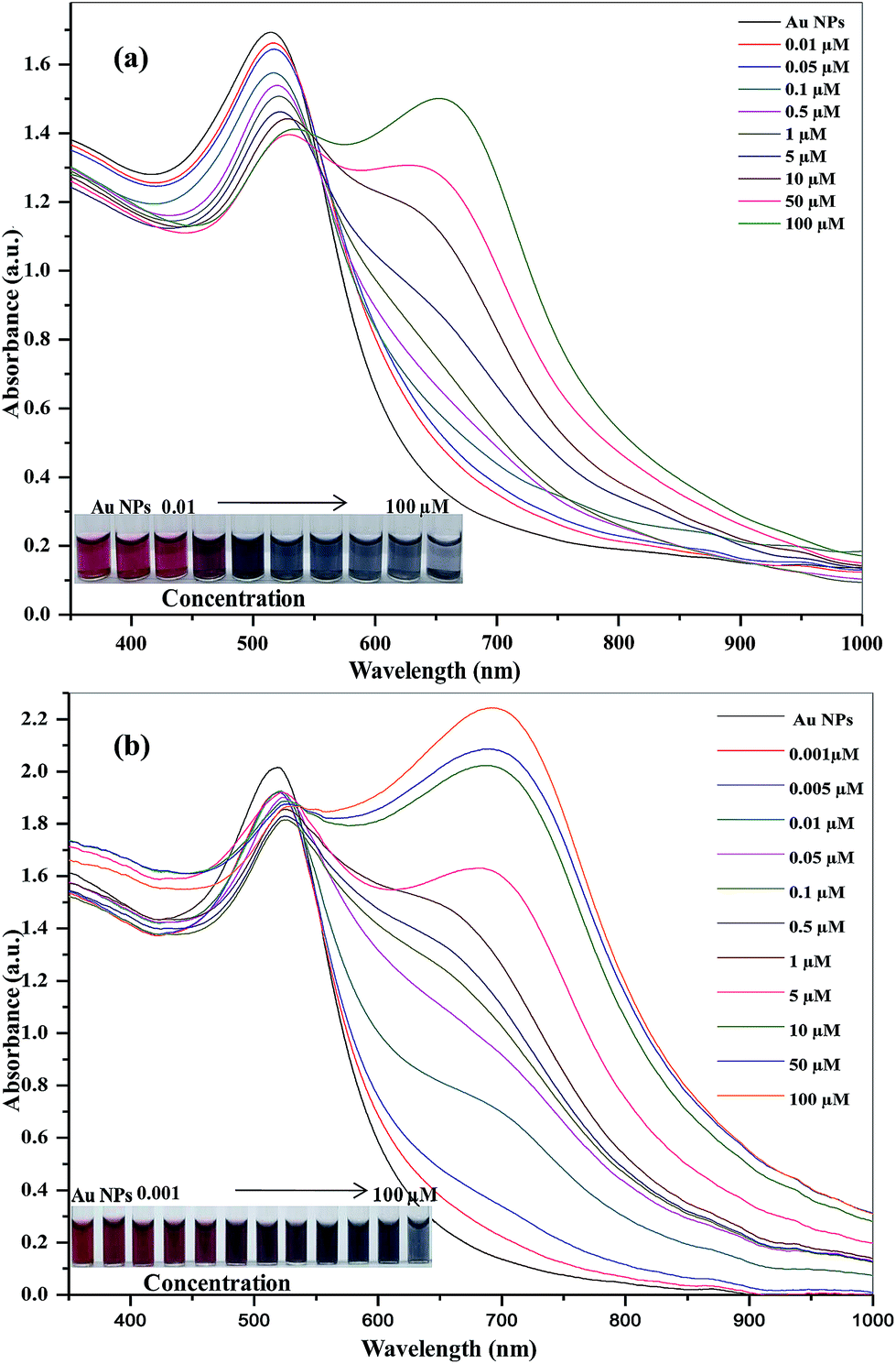 | ||
| Fig. 4 UV-visible absorbance spectra of unmodified Au NPs upon the addition of (a) venlafaxine in the range of 0.01–100 μM and (b) imipramine in the range of 0.001–100 μM. Insets: photographic images of corresponding solutions. On the basis of the absorption ratios at A653/A521 and at A695/A521 plots, the limits of detection were found to be 6.3 and 0.9 nM for venlafaxine and imipramine (n = 3). The calibration graph was shown in ESI of Fig. S8a and b.† | ||
 | ||
| Fig. 5 UV-visible absorbance spectra of unmodified Au NPs upon the addition of (a) amlodipine and (b) alfuzosin in the range of 0.01–100 μM. Insets: photographic images of corresponding solutions. On the basis of the absorption ratios at A688/A521 and at A636/A521 plots, the limits of detection were found to be 8.4 and 9.3 nM for amlodipine and alfuzosin (n = 3). The calibration graph was shown in ESI of Fig. S8c and d.† | ||
| Name of the drug | Linear range (μM) | Limit of detection (μM) | Technique | Reference |
|---|---|---|---|---|
| Venlafaxine | 37.2–2969.5 | 7.4 | HPLC | 7 |
| Imipramine | 0.0007–0.01 | — | HPLC | 8 |
| Amlodipine | 0.6–38.6 | 0.6 | HPLC | 9 |
| Alfuzosin | 0.3–87.0 | — | HPLC | 10 |
| Venlafaxine | 0.01–0.11 | 0.006 | LC-MS | 11 |
| Imipramine | — | — | LC-MS | 12 |
| Amlodipine | 0.0001–0.02 | — | LC-MS | 13 |
| Alfuzosin | 0.9–69.6 | 0.9 | LC-MS | 14 |
| Amlodipine | 0.2–30.2 | 0.2 | Fluorescence | 16 |
| Venlafaxine | 12.9–116.5 | 6.1 | Fluorescence | 17 |
| Imipramine | 0.002–0.03 | — | UV-visible | 18 |
| Alfuzosin | 0.006–0.1 | — | UV-visible | 19 |
| Venlafaxine | 0.01–100.0 | 0.0063 | Unmodified Au NPs-based UV-visible | Present method |
| Imipramine | 0.001–100.0 | 0.0090 | ||
| Amlodipine | 0.01–100.0 | 0.0084 | ||
| Alfuzosin | 0.01–100.0 | 0.0093 |
3.5. Selectivity of unmodified Au NPs for four drugs
The specificity of the proposed analytical platform was investigated by comparing the degrees of color variance of solution mixtures containing four drugs separately and other drugs (ESI of Fig. S9†). In these tests, the concentrations of four drugs and other drugs (50.0 and 1000 μM, 1:20) were added to unmodified Au NPs. It can be observed that the unmodified Au NPs solutions remained cherry red with the addition of other interfering drugs (1000 μM) except for four drugs (venlafaxine, imipramine, amlodipine, and alfuzosin). However, upon the addition of four drugs (50 μM) separately to solutions of Au NPs containing the mixture of other drugs, the color of unmodified Au NPs solution became blue, resulting a red-shift in the absorption spectra of unmodified Au NPs. These results show that unmodified Au NPs-based colorimetric sensing mechanism possesses good specificity toward four drugs. Even at high concentrations, the color was red for all other drugs solutions, and the mean absorption ratios (at A653/A521, A695/A521, A688/A521, and A636/A521) were <1 (0.17–0.42) for other drugs. In contrast, the color of unmodified Au NPs with four drugs (50 μM) in the presence of other drugs (indapamide, flucanazole, metformin, lisinopril, topiramate, cetrizine, levocetrizine and carbidopa) was blue, and the absorption ratios (at A653/A521, A695/A521, A688/A521, and A636/A521) of Au NPs were >1 (∼1.12, 1.14, 1.34 and 1.19 for venlafaxine, imipramine, amlodipine, and alfuzosin).
To study the influence of increasing concentration of other drugs with sensing ability of Au NPs towards analytes, we studied the UV-visible absorption spectra of unmodified Au NPs by the addition of analyte (100 μM) along with different concentrations of other drugs (other drugs:analyte; 10:1 to 100:1). As shown in ESI of Fig. S9b† analyte gives good spectral changes at lower ratios, but at higher concentration of other drugs, the intensity of peak at 653 nm is decreased. This suggests that the tolerable limit of probe for sensing of target analytes in presence of other drugs. ESI of Fig. S10 and S11a† show the UV-visible absorption spectra of unmodified Au NPs by the addition of analytes (imipramine, amlodipine and alfuzosin) in the presence of other drugs, which indicates that the probe effectively detected the target analytes in the presence of other drugs (1.5–3.0 mM). In order to define the selectivity of the probe in the coexistence of target analytes, we studied the UV-visible absorption spectra of Au NPs by the addition of each target drug (100 μM) in the presence of other three target drugs at different concentrations (0–10 μM) (ESI of Fig. S12–S16†). I can be observed that the absorption ratios at A653/A521, A695/A521, A688/A521, and A636/A521 are still generated for venlafaxine (Vnl), imipramine (Imp), amlodipine (Amd), and alfuzosin (Alf) in the coexistence of other three target drugs independently as well as mixture of three drugs (0–10 μM), which indicates that the coexistence of mixture of three drugs did not influence the absorption spectra of aggregation of Au NPs induced by each target drug. Therefore, these results indicated that the unmodified Au NPs were successfully acted as a probe for the simultaneous colorimetric detection of target drugs in the coexistence of remaining three target drugs.
3.6. Determination of four drugs in their pharmaceutical and biological samples
In order to test the applicability and matrix interferences of the present method for the analysis of four drugs in their pharmaceutical formulations, we applied the present method for the quantification of four drugs in four tablets such as Venlor-XR (venlafaxine), Antidep-75 (imipramine-75 mg), Aginal-5 (amlodipine-5 mg) and Alfusin-10 (alfuzosin-10 mg). ESI of Fig. S17 and S18† show the UV-visible absorption spectra of unmodified Au NPs in the presence of target drugs dilutions in the range of 0.01–100 μM, indicating that the present method successfully detected the four drugs in their pharmaceutical formulations. The accuracy of the proposed method was further ascertained by performing recovery studies via standard addition method. The samples are analyzed for the analytes of interest by adding specified amount of each drug to the sample, thus increasing its concentration. The recovery of the known amount of added analyte was computed. The percentage recoveries of four drugs from pharmaceutical dosages were ranged from 108.1 to 125.0%, with relative standard deviation < 2.1% (ESI of Table S2†).To investigate whether the colorimetric sensor can be used in real samples analysis, we applied this probe for detection of four drugs in biofluids (urine nad plasma). Urine samples were acquired from healthy volunteer of SVNIT, Surat, India while plasma samples were collected from Iyyer Pathology Laboratory, Surat, Gujarat, India. The collected samples were diluted 50 times and spiked with different concentrations of each drug (25, 50 and 75 μM), and then analyzed by the aforesaid procedure. Results from the determination of four drugs from urine and plasma are shown in ESI of Table S3† (n = 3 for each concentration). The mean recoveries for four drugs (25, 50 and 75 μM) were 97.7–102.4%, with 0.5–3.5% precision and −2.2–2.4% accuracy for four drugs, indicating that the present method exhibits good precision and accuracy. The good agreement between these results and known values indicates that this method can be useful for the simultaneous determination of venlafaxine, imipramine, amlodipine, and alfuzosin in pharmaceutical formulations and biological samples.
4. Conclusions
We have developed a novel and convenient colorimetric technique for simultaneous detection of four drugs (venlafaxine, imipramine, amlodipine, and alfuzosin) in their pharmaceutical formulations using unmodified Au NPs as a probe. The probe was prepared in single-step reaction without further modifications. The stable unmodified Au NPs is a mean diameter of 5.3 nm with a quite uniform size distribution and showed a colorimetric response upon exposure to four drugs, which is associated with the π–π interactions and hydrogen bonding between Au NPs and four drugs. The aggregation of unmodified Au NPs induced by four drugs was confirmed by DLS and TEM, which results a change in absorption spectra and in color. This method was free form the interference and exhibited low detection limits and high selectivity to the four drugs. This colorimetric sensor was successfully applied to detect four drugs in their pharmaceutical formulations, which indicates that the probe would hold promising potential for simultaneous detection of four drugs in pharmaceutical and biological samples.Acknowledgements
This research was supported by the DST, India under DST-Inspire Fellowship Ph. D. Programme. We also thank Department of Science and Technology for providing Maya Pro 2000 spectrophotometer under the Fast-Track Young Scientist Scheme (2011–2014).References
- J. M. Andrews, P. T. Ninan and C. B. Nemeroff, Depression, 1996, 4, 48–56 CrossRef CAS.
- D. R. Hicks, D. Wolaniuk, A. Russell, N. Cavanaugh and M. Kraml, Ther. Drug Monit., 1994, 16, 100–107 CrossRef CAS PubMed.
- U. Lepola, M. Arató, Y. Zhu and C. Austin, J. Clin. Psychiatry, 2003, 64, 654–662 CrossRef CAS.
- M. Haria and A. J. Wagstaff, Drugs, 1995, 50, 560–586 CrossRef CAS PubMed.
- British Pharmacopoeia, The Stationary office under license from the controller of Her Majesty's Stationary Office for the Department of Health on behalf of the Health Ministers, 2003, vol. I, p. 73.
- I. Cavero, F. Lefevre-Borg and P. Manoury, Fed. Am. Soc. Exp. Biol., Fed. Proc., 1984, 43, 3 Search PubMed.
- X. Y. Qin, J. Meng, X. Y. Li, J. Zhou, X. L. Sun and A. D. Wen, J. Chromatogr. B: Anal. Technol. Biomed. Life Sci., 2008, 872, 38–42 CrossRef CAS PubMed.
- H. N. Deepakumari, K. B. Vinay and H. D. Revanasiddappa, ISRN Anal. Chem., 2013, 1–10 CrossRef PubMed.
- G. Bahrami and S. Mirzaeei, J. Pharm. Biomed. Anal., 2004, 36, 163–168 CrossRef CAS PubMed.
- A. K. Shakya, T. A. Arafar, A. Abuawaad, H. Al-Hroub and M. Melhim, Jordan J. Pharm. Sci., 2010, 3, 25–27 CAS.
- S. K. Dubey, R. N. Saha, H. Jangala and S. Pasha, J. Pharm. Anal., 2013, 3, 466–471 CrossRef PubMed.
- L. Sun and J. A. Stenken, J. Chromatogr. A, 2007, 1161, 261–268 CrossRef CAS PubMed.
- R. R. Kallem, R. Mullangi, K. K. Hotha, L. K. Ravindranath, Y. N. Spoorthy and J. V. L. N. Seshagirirao, Bioanalysis, 2013, 5, 827–837 CrossRef CAS PubMed.
- N. A. Gomes, A. Pudage, S. S. Joshi, V. V. Vaidya, S. A. Parekh and A. V. Tamhankar, Chromatographia, 2009, 69, 9–18 CAS.
- H. Wu, S. K. Kailasa, J. Yan, C. Chin and H. Ku, J. Ind. Eng. Chem., 2014, 20, 2071–2076 CrossRef CAS PubMed.
- Y. Kadioglu and M. Ozturk, Braz. J. Pharm. Sci., 2012, 48, 719–725 CrossRef CAS PubMed.
- L. Du, X. Wei, X. Lei, L. Wang, Q. Gong and X. Wang, Anal. Methods, 2014, 6, 1108–1113 RSC.
- J. M. G. Fraga, A. I. J. Abizanda, F. J. Moreno and J. J. A. León, J. Pharm. Biomed. Anal., 1991, 9, 109–115 CrossRef.
- S. A. Al-Tamimi, F. A. Aly and A. M. Almutairi, J. Anal. Chem., 2013, 68, 313–320 CrossRef CAS.
- S. K. Kailasa, V. N. Mehta and H. Wu, RSC Adv., 2014, 4, 16188–16205 RSC.
- S. K. Kailasa and H. Wu, Microchim. Acta, 2014, 181, 853–864 CrossRef CAS.
- S. K. Kailasa and H. Wu, Trend. Anal. Chem., 2015, 65, 54–72 CrossRef CAS PubMed.
- D. Vilela, M. C. González and A. Escarpa, Anal. Chim. Acta, 2012, 751, 24–43 CrossRef CAS PubMed.
- K. Saha, S. S. Agasti, C. Kim, X. Li and V. M. Rotello, Chem. Rev., 2012, 112, 2739–2779 CrossRef CAS PubMed.
- S. He, D. B. Liu, Z. Wang, K. Y. Cai and X. Y. Jiang, Sci. China: Phys., Mech. Astron., 2011, 54, 1757–1765 CrossRef.
- M. Aslam, L. Fu, M. Su, K. Vijayamohanan and V. P. Dravid, J. Mater. Chem., 2004, 14, 1795–1797 RSC.
- X. Wei, L. Qi, J. Tan, R. Liu and F. Wang, Anal. Chim. Acta, 2010, 671, 80–84 CrossRef CAS PubMed.
- K. A. Rawat, K. R. Surati and S. K. Kailasa, Anal. Methods, 2014, 6, 5972–5980 RSC.
- K. A. Rawat and S. K. Kailasa, Microchim. Acta, 2014, 181, 1917–1929 CrossRef CAS.
- S. K. Kailasa and H. Wu, Analyst, 2012, 137, 1629–1638 RSC.
- V. N. Mehta, S. K. Kailasa and H. Wu, New J. Chem., 2014, 38, 1503–1511 RSC.
- V. N. Mehta, J. N. Solanki and S. K. Kailasa, Microchim. Acta, 2014, 181, 1905–1915 CrossRef CAS.
- Y. Zheng, Y. Wang and X. Yang, Sens. Actuators, B, 2011, 156, 95–99 CrossRef CAS PubMed.
- F. Li, Y. Feng, C. Zhao and B. Tang, Biosens. Bioelectron., 2011, 26, 4628–4631 CrossRef CAS PubMed.
- F. Wei, L. Qi, R. Lam, S. Cheng, S. Lu, D. Ho and N. Li, Appl. Phys. Lett., 2010, 96, 133702 CrossRef PubMed.
- G. Chen, W. Chen, Y. Yen, C. Wang, H. Chang and C. Chen, Anal. Chem., 2014, 86, 6843–6849 CrossRef CAS PubMed.
- Q. Shen, W. Li, S. Tang, Y. Hu, Z. Nie, Y. Huang and S. Yao, Biosens. Bioelectron., 2103, 41, 663–668 CrossRef PubMed.
- G. B. Kim, K. H. Kim, Y. H. Park, S. Ko and Y. Kim, Biosens. Bioelectron., 2013, 41, 833–839 CrossRef CAS PubMed.
- J. Turkevich, P. C. Stevenson and J. Hillier, Discuss. Faraday Soc., 1951, 11, 55–75 RSC.
- M. M. Maye, L. Han, N. N. Kariuki, N. K. Ly, W. B. Chan, J. Luo and C. J. Zhong, Anal. Chim. Acta, 2003, 496, 17–27 CrossRef CAS.
- S. Basu, S. K. Ghosh, S. Kundu, S. Panigrahi, S. Praharaj, S. Pande, S. Jana and T. Pal, J. Colloid Interface Sci., 2007, 313, 724–734 CrossRef CAS PubMed.
Footnote |
| † Electronic supplementary information (ESI) available. See DOI: 10.1039/c4ra16109b |
| This journal is © The Royal Society of Chemistry 2015 |