Synthesis, molecular modeling, and biological evaluation of quinazoline derivatives containing the 1,3,4-oxadiazole scaffold as novel inhibitors of VEGFR2

Fang Qiao b, Yong Yinb, Yu-Ning Shenb, She-Feng Wangb, Shao Shab, Xun Wub, Ai-Min Luc, Chen Xu*b, Wei-Ming Zhang*a and Hai-Liang Zhu*b
aNanjing Institute for the Comprehensive Utilization of Wild Plant, Nanjing 210042, People’s Republic of China. E-mail: botanyzh@163.com; Fax: +86 25 83592672; Tel: +86 25 83592572
bState Key Laboratory of Pharmaceutical Biotechnology, Nanjing University, Nanjing 210093, People’s Republic of China. E-mail: zhuhl@nju.edu.cn
cCollege of Science, Nanjing Agriculture University, Nanjing 210095, People’s Republic of China

Received 4th October 2014 , Accepted 4th February 2015

First published on 5th February 2015


Abstract

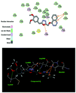
A series of 4-alkoxyquinazoline derivatives containing the 1,3,4-oxadiazole scaffold have been designed and synthesized, and their inhibitory activities were also tested against A549, MCF-7 and Hela. Of these compounds, 2-(3,4-dimethoxybenzyl)-5-((quinazolin-4-yloxy)methyl)-1,3,4-oxadiazole (compound 4j) showed the most potent inhibitory activity (IC50 = 0.23 μM for MCF-7, IC50 = 0.38 μM for A549 and IC50 = 0.32 μM for Hela) and the effect was better than the positive control drug Tivozanib (IC50 = 0.38 μM for MCF-7, IC50 = 0.62 μM for A549 and IC50 = 0.34 μM for Hela). A docking simulation was performed to position compound 4j into the VEGFR active site to determine the probable binding model. These results suggested that compound 4j with potent inhibitory activity in tumor growth inhibition may be a potential anticancer agent.


1. Introduction

Malignant tumors threaten human health and are currently a leading cause of death throughout the world.1,2 So the development of anti-cancer drugs is considered an important task by the WHO and the Health Departments of many countries.3 Although several classes of anti-cancer agents are presently available, severe side-effects and drug resistance constantly emerge.4 Therefore, the elaboration of new types of anticancer drugs is a critical task that can prevent this serious medical problem. Recently, different targets in key steps of the growth and proliferation of cancer cells have been studied which could lead to new weapons against tumors.5

Tumour cell growth or hyperplasia is a fairly complex process, and involves a series of steps.6 Angiogenesis, which is the formation of new blood vessels, is not only a pivotal step during the formation of metastases but also the focus of researchers all over the world.7 Vascular endothelial growth factor (VEGF) plays a significant role in tumor angiogenesis, vascular permeability, endothelial cell activation, proliferation, and migration.8 The VEGF family consists of VEGF-A, VEGF-B, VEGF-C, VEGF-D, and placenta growth factor (PlGF).9 The biological functions of VEGF are achieved through interaction with the VEGF receptor family. In mammals, the VEGFR family are transmembrane tyrosine kinases and consist of three members, VEGFR1, VEGFR2 and VEGFR3.10 The VEGFRs consist of three parts: an extracellular ligand-binding domain, an intracellular tyrosine kinase domain and a single transmembrane segment.11,12 Binding of VEGF to VEGFR results in receptor dimerization, which leads to phosphorylation and dimerization on downstream signal transducer proteins.10,11 This VEGFR tyrosine kinase-mediated cell growth signaling pathway is an important signaling pathway in regulating the proliferation of many types of tumors, such as breast cancer,13 non small cell lung cancer,14 head and neck cancer,15 glioblastomas.16 VEGFR2 plays a major role in the regulation of VEGF-driven responses in endothelial cells and it is proven to be a prerequisite signal transducer in tumorigenesis.17 Therefore, VEGFR2 is a hot target in current research. In recent years, a number of compounds have been reported as being potent inhibitors of VEGFR2 in vitro or possessing antiangiogenic activity (Fig. 1), such as Vandetanib,18 Sorafenib (BAY-43-9006),19 Tivozanib,20 ZD4190.21


image file: c4ra11780h-f1.tif
Fig. 1 Various VEGFR tyrosine kinase inhibitors.

As shown in Fig. 1, the scaffolds of the known inhibitors of VEGFR2 can be divided into two main groups: those that contain a core of 4-anilino-quinazoline and others that contain a core of 4-ol-quinoline. Given the above information our group have designed and synthesized a series of new compounds by employing a similar template with a scaffold 4-ol-quinzoline core,22 with the presumption that the quinazoline core is a common scaffold of VEGFR2 inhibitors which inhibits VEGFR2 by binding to the kinase domain via the gatekeeper Cys919 and Phe918 of VEGFR2.23,24 The results showed that compound S1 (4-(2-(2-methyl-5-nitro-1H-imidazol-1-yl)ethoxy) quinazoline) has obvious inhibitory activities against VEGFR2 (Fig. 1), the IC50 value of S1 was 0.43 μM for MCF-7.

Besides, 1,3,4-oxadiazole is a heterocyclic structure with extensive biological activity.25,26 In particular, a few 2,5-disubstituted 1,3,4-oxadiazoles have been found with some extent of anticancer activity.27,28 Furthermore, the special structure of 1,3,4-oxadiazole heterocycles as good bioisosteres of amides and esters, can improve pharmacological activity via hydrogen bonding interactions with the receptors.29,30

Considering the above mentioned facts, on the basis of our proceeding work, we designed and synthesized a series of new 4-alkoxyquinazoline derivatives containing the 1,3,4-oxadiazole moiety and studied their antitumor activities against A549, MCF-7 and Hela cell lines, respectively. The interactions of these quinazoline-based analogs with the ATP binding site of VEGFR2 (PDB code: 4ASE) were studied via molecular docking.

2. Results and discussion

2.1. Chemistry

Compound 3 was synthesized by the route outlined in Scheme 1 according to Mirzaei et al.’s method.31 Compound 3 was prepared by the reaction of 4-hydroxyquinazoline in excess ethyl bromoacetate and then hydrazinolysis of the ethyl ester group by hydrazine hydrate with a yield of 78%.
image file: c4ra11780h-s1.tif
Scheme 1 Synthesis of compounds 4a–4v. Reagents and conditions: (i) ethyl bromoacetate, Cs2CO3, DMF, reflux, 24 h; (ii) hydrazine hydrate, methanol, 0 °C, 4.5 h; (iii) substituted carboxylic acid, phosphoryl chloride, reflux, 10–16 h.

2-(Quinazolin-4-yloxy)acetohydrazide was then reacted with substituted benzoic acids and phenylacetic acids to prepare the corresponding quinozoline derivatives 4a–4v. The chemical structures of these quinozoline derivatives are displayed in Table 1. All these compounds gave satisfactory elementary analyses (±0.4%). 1H NMR and ESI MS spectra data was consistent with the assigned structures.

Table 1 Structure of compounds 4a–4v

image file: c4ra11780h-u1.tif

Compound R
4a image file: c4ra11780h-u2.tif
4b image file: c4ra11780h-u3.tif
4c image file: c4ra11780h-u4.tif
4d image file: c4ra11780h-u5.tif
4e image file: c4ra11780h-u6.tif
4f image file: c4ra11780h-u7.tif
4g image file: c4ra11780h-u8.tif
4h image file: c4ra11780h-u9.tif
4i image file: c4ra11780h-u10.tif
4j image file: c4ra11780h-u11.tif
4k image file: c4ra11780h-u12.tif
4l image file: c4ra11780h-u13.tif
4m image file: c4ra11780h-u14.tif
4n image file: c4ra11780h-u15.tif
4o image file: c4ra11780h-u16.tif
4p image file: c4ra11780h-u17.tif
4q image file: c4ra11780h-u18.tif
4r image file: c4ra11780h-u19.tif
4s image file: c4ra11780h-u20.tif
4t image file: c4ra11780h-u21.tif
4u image file: c4ra11780h-u22.tif
4v image file: c4ra11780h-u23.tif


2.2. Biological activity

2.2.1. Antiproliferative effects against cancer cells. The target compounds were evaluated by in vitro anti-proliferation assays against three human cancer cell lines A549, MCF-7 (with VEGFR protein over expression) and Hela to test the antitumour activities of the synthesized compounds. The results are shown in Table 2. Most compounds showed a remarkably potent antitumor activity, indicating that 4-alkoxyquinazoline derivatives containing the 1,3,4-oxadiazole scaffold could possess a significant anticancer potency. Among these compounds, it was observed that 4j showed the most potent biological activity (IC50 = 0.23 μM for MCF-7), comparable to the positive control Tivozanib (IC50 = 0.38 μM for MCF-7).
Table 2 Inhibition of cancer cell proliferation by compounds 4a–4v
Compound IC50 ± SD (μM)
MCF-7a A549b Helac
a Inhibition of the growth of MCF-7 cell lines.b Inhibition of the growth of A549 cell lines.c Inhibition of the growth of Hela cell lines.
4a 2.05 ± 0.07 1.51 ± 0.04 2.16 ± 0.07
4b 0.87 ± 0.06 0.96 ± 0.03 0.87 ± 0.03
4c 2.55 ± 0.16 2.06 ± 0.09 2.73 ± 0.16
4d 0.67 ± 0.07 0.94 ± 0.04 0.84 ± 0.02
4e 4.59 ± 0.24 5.49 ± 0.36 4.65 ± 0.19
4f 0.77 ± 0.04 0.87 ± 0.05 0.79 ± 0.03
4g 1.89 ± 0.02 0.86 ± 0.03 1.51 ± 0.08
4h 2.54 ± 0.08 1.87 ± 0.02 1.75 ± 0.06
4i 16.23 ± 0.67 14.96 ± 0.97 14.21 ± 0.71
4j 0.23 ± 0.04 0.32 ± 0.02 0.38 ± 0.03
4k 4.63 ± 0.26 5.25 ± 0.23 4.92 ± 0.14
4l 7.36 ± 0.12 8.96 ± 0.16 8.65 ± 0.18
4m 7.19 ± 1.64 8.07 ± 0.89 8.67 ± 0.85
4n 9.58 ± 0.57 8.27 ± 0.85 9.98 ± 1.36
4o >50 44.32 ± 0.07 48.31 ± 1.46
4p 18.73 ± 1.69 17.52 ± 1.47 17.47 ± 1.36
4q 22.94 ± 1.12 19.39 ± 1.86 17.7 ± 1.83
4r 7.3 ± 1.04 6.32 ± 0.75 7.43 ± 0.77
4s 13.12 ± 0.63 13.17 ± 0.36 15.44 ± 0.27
4t 8.94 ± 0.22 8.38 ± 0.63 8.35 ± 0.69
4u 34.2 ± 1.57 32.87 ± 1.19 35.07 ± 1.23
4v 16.1 ± 1.25 19.35 ± 0.83 16.35 ± 1.49
Tivozanib 0.38 ± 0.060 0.62 ± 0.030 0.34 ± 0.020


From the data listed in Table 2, we can draw the conclusion that the activity of the tested compounds may be correlated to the variation and modification of the structure. Among the 22 new synthetic 4-alkoxyquinazoline derivatives, the phenylacetic acid derivatives 4a–4k, whose IC50 values range from 0.23 to 16.23 μM, displayed higher antitumor potencies than benzoic acid derivatives 4l–4v, with IC50 values ranging from 6.32 to 54.35 μM, which demonstrates that the introduction of a phenylacetic acid resulted in the decline of anticancer activity. For instance, with the p-nitro group, compound 4d with a benzyl group exhibited superior activity compared with compound 4v which has a phenyl group. This conclusion that a longer carbon chain increased the anticancer activity of quinazoline derivatives can provide a significant contribution to design the most effective length of the carbon chain of the quinazoline derivative with the best antitumor activity. Moreover, among the eleven quinazoline derivatives (4a–4k), compound 4j showed the most potent activities with an IC50 value of 0.23 μM against MCF-7 (breast cancer), which is superior to the positive control Tivozanib with a corresponding IC50 value of 0.38 μM. In addition, the data show that compound 4j displayed significant activities with IC50 values of 0.32, 0.38 μM against A549 and Hela, respectively, indicating that it possesses potent anticancer activity. Also varying the substituent on the phenylacetic acid, for instance, halogen, nitro and methoxy, also leads to different anticancer activities. Among them, we can observe that compounds with electron-donating substituents (such as OCH3) improved the VEGFR2 inhibitory activity compared to those with halogen substituents (such as F, Cl, Br). In addition, a comparison of the p-position on phenylacetic acid demonstrated that a p-position halogen group (4e, 4c, 4g) improved VEGFR2 inhibitory activity and the potency order is F < Cl < Br and we found the same law for the p-position on benzoic acid (4q, 4s, 4t). Other p-position substituents such as 4p with a methyl group substituent have slightly improved activity. Last but not least, compounds with the same group on the o-, m- and p-position lead to different activity. We found that derivatives with a methoxy group on the m-position and p-position of phenylacetic acid displayed higher antitumor activity with IC50 values of 0.87, 0.77 μM, respectively, against MCF-7 than the compound with the methoxy group on the o-position of phenylacetic acid with an IC50 value of 1.64 μM against MCF-7. Given the above results, we designed and evaluated the compound with 3,4-dimethoxy group substitution (4j), and the activity was significantly enhanced up to 0.23 μM against MCF-7.

2.2.2. Kinase selectivity. In order to validate that the anti-proliferative effect was produced by interaction of VEGFR protein with the synthesized compounds, all compounds of the series were subjected to in vitro VEGFR2, EGFR (epidermal growth factor receptor), bFGF (basic fibroblast growth factor) and PDGFR (platelet-derived growth factor receptor) kinase inhibitory assays. As shown in Table 3, all synthesized compounds showed much higher inhibitory activity for VEGFR2 than for EGFR, bFGF and PDGFR, like the reference Tivozanib. Additionally, good agreement was found between the IC50 values of these compounds and their relevant IC50 values in the anti-proliferative assay. Hence, a further study comparing the anti-proliferative activity against the MCF-7 cell line with the VEGFR2 inhibitory activity of the top 10 compounds (4j, 4d, 4f, 4b, 4g, 4a, 4h, 4c, 4l, 4e) was performed and the results revealed that there was a moderate correlation between VEGFR2 inhibition and the inhibition of cancer cellular proliferation, as evidenced in Fig. 2, with a correlation coefficient of 7.4071 and an R2 value of 0.9345. In conclusion, the synthesized compounds can inhibit the function of VEGFR2 and the anti-proliferative effect was produced partly by interaction of VEGFR2 protein and the molecular inhibitors.
Table 3 Inhibition of selected kinases IC50a (nM)
Compound VEGFR2 EGFR bFGF PDGFR
a IC50 values were averaged values determined by at least two independent experiments. Variation was generally 5%.
4a 12.31 310.58 164.49 73.52
4b 5.83 302.79 152.33 66.13
4c 19.05 335.96 182.17 82.06
4d 4.73 293.57 136.32 55.33
4e 33.25 357.76 201.25 100.16
4f 5.28 295.51 146.76 63.69
4g 11.43 305.20 154.23 70.75
4h 15.07 332.58 179.06 80.49
4i 55.16 510.72 490.12 273.39
4j 2.32 216.43 98.55 39.64
4k 36.31 363.04 232.39 102.64
4l 29.50 320.89 194.86 90.76
4m 40.53 394.81 216.44 116.14
4n 43.65 462.27 386.21 152.08
4o 98.75 587.42 685.01 325.12
4p 60.38 507.76 517.94 334.11
4q 67.08 496.61 561.69 287.52
4r 41.13 414.37 213.52 140.73
4s 43.08 476.68 421.19 162.58
4t 50.14 453.56 338.62 185.28
4u 78.81 548.06 624.84 296.41
4v 59.44 493.23 516.52 241.78
Tivozanib 3.40 116.64 108.15 45.93


image file: c4ra11780h-f2.tif
Fig. 2 Correlation between the anti-proliferative activity against MCF-7 and the VEGFR inhibitory activity.
2.2.3. Molecular docking. In order to gain more understanding of the structure–activity relationships observed at VEGFR2, molecular docking studies of the most potent inhibitor 4j and the positive control compound Tivozanib into the active site of the VEGFR2 complex structure (PDB code: 4ASE) were performed.32

All docking runs were applied using the LigandFit Dock protocol of Discovery Studio 3.5. The CDOCKER_INTERACTION_ENERGY of the synthesized compounds is shown in Table 4. The CDOCKER_INTERACTION_ENERGY of the synthesized compounds demonstrated that these compounds have good affinity for the active site of 4ASE, which further proved the positive correlation between the CDOCKER_INTERACTION_ENERGY and their anticancer activity. Among the docking calculation results for the synthesized compounds, compound 4j showed the lowest interaction energy −50.5831 kcal mol−1 (Table 4). The binding model of compound 4j and VEGFR2 is depicted in Fig. 3 and 4. Fig. 3 and 4 show the binding mode of compound 4j interacting with VEGFR2 protein and the docking results revealed that four amino acids Lys868, Cys919, His1026 and Asp1046 located in the binding pocket of the protein played vital roles in the conformation with compound 4j, which were stabilized by three π–π bonds and two hydrogen bonds, which are shown in the 2D and 3D diagrams. Two π–π interactions were formed between His1026 and the quinazoline ring of 4.87682 Å and 6.00074 Å while the other π–π interaction of 5.05787 Å involved Lys868. One hydrogen bond of 2.5 Å was formed between Cys919 and the phenylacetic acid ring. Another hydrogen bond was formed between Asp1046 and 4j. Fig. 5 and 6 show the interaction of the positive control compound Tivozanib and 4ASE. From the two pictures, we can draw a conclusion that two amino acid residues Cys919 and Asp1046 located in the binding pocket of 4ASE played a key role in the conformation. As illustrated in Fig. 4 and 6, there was a hydrogen bond between the hydrogen of the quinazoline ring of Tivozanib and the oxygen of the carboxyl group of Cys919, while a hydrogen bond is seen between the oxygen of the methoxy group of compound 4j and the hydrogen of the amino group of Cys919. The figures also suggest that compound 4j and Tivozanib appear to interact with the hydrogen of the amino group of Asp1046 through hydrogen bonds. The docking results of Tivozanib and 4ASE revealed that compound 4j had a high binding affinity for 4ASE. In addition, there were three π–π stacking interactions between the quinazoline ring and the benzene ring of Lys868, His1026. The π–π interaction energies are of the same order of magnitude as hydrogen bonds and make the complex more stable. Therefore, compound 4j showed an interaction energy of −50.5831 kcal mol−1 lower than the −47.6633 kcal mol−1 of Tivozanib. The enzyme assay data and the molecular docking results demonstrated that compound 4j is a potential inhibitor of VEGFR2.

Table 4 The docking calculation for the synthesized compounds (4a–4v)
Compound CDOKER INTERACTION ENERGY/kcal mol−1 Compound CDOKER INTERACTION ENERGY/kcal mol−1
4a −47.2314 4l −45.6047
4b −48.9269 4m −43.7809
4c −46.4385 4n −43.4841
4d −47.7327 4o −39.4104
4e −44.6077 4p −42.3049
4f −47.0279 4q −42.2217
4g −47.4352 4r −43.904
4h −47.1029 4s −42.9932
4i −42.8375 4t −43.55
4j −50.5831 4u −41.4659
4k −45.1589 4v −42.4093
Tivozanib −47.6633    


image file: c4ra11780h-f3.tif
Fig. 3 2D molecular docking model of compound 4j with 4ASE.

image file: c4ra11780h-f4.tif
Fig. 4 3D molecular docking model of compound 4j with 4ASE.

image file: c4ra11780h-f5.tif
Fig. 5 2D molecular docking model of Tivozanib with 4ASE.

image file: c4ra11780h-f6.tif
Fig. 6 3D molecular docking model of Tivozanib with 4ASE.

3. Conclusion

To sum up, a series of 4-alkoxyquinazoline derivatives containing the 1,3,4-oxadiazole scaffold have been designed and synthesized, and their inhibitory activities were tested against A549, MCF-7 and Hela cell lines. Compound 4j exhibited the most potent inhibitory activity (IC50 = 0.23 μM for MCF-7, IC50 = 0.38 μM for A549 and IC50 = 0.32 μM for Hela), which was compared with the positive control Tivozanib. Preliminary SARs and molecular modeling studies provided further insight into interactions between the enzyme and its ligand. Analysis of compound 4j’s binding conformation at the active site showed that compound 4j was stabilized by the interactions with Lys868, Cys919, His1026 and Asp1026. The results provide valuable information for the design of VEGFR2 inhibitors as antitumor agents.

4. Experiments

4.1. Materials and measurements

All chemicals (reagent grade) used were commercially available. All of the synthesized compounds were chemically characterized by thin layer chromatography (TLC), proton nuclear magnetic resonance (1H NMR) and elemental microanalyses (CHN). Analytic thin-layer chromatography (TLC) was performed on glass-backed silica gel sheets (silica gel 200 Å GF254). All compounds were detected using UV light (254 nm or 365 nm). Melting points were determined using an XT4 MP apparatus (Taike Corp, Beijing, China) and are as read. 1H NMR spectra were measured using a Bruker AV-400 spectrometer at 25 °C and referenced to Me4Si. Chemical shifts are reported in ppm (δ) using the residual solvent line as an internal standard. ESI-MS spectra were recorded using a Mariner System 5304 mass spectrometer. Elemental analyses were performed using a CHN-O-Rapid instrument and were within ± 0.4% of the theoretical values.

4.2. Chemistry

4.2.1. General procedure for the synthesis of compound 2. A mixture of 4-hydroxyquinazoline (0.01 mol), ethyl bromoacetate (0.015 mol) and Cs2CO3 (0.02 mol) was dissolved in 30 mL DMF at 80 °C for 24 h. The mixture was poured into water. The products were extracted with ethyl acetate. The extract was dried over anhydrous Na2SO4, filtered and evaporated to give compound 2.
4.2.2. General procedure for the synthesis of compound 3. To a stirred solution of hydrazine hydrate (6.5 mL) in an ice bath, a solution of compound 2 (0.03 mol) in methanol (40 mL) was added slowly (10 min). The stirring was continued for 4.5 h at 0 °C under argon atmosphere and then put it into −10 °C cold trap for 3 hours. The white precipitate which formed at −10 °C was filtered and recrystallized from chloroform to give compound 3.
4.2.3. General procedure for synthesis of compounds 4a–4v. An equimolar mixture of compound 3 (0.001 mol) and substituted carboxylic acid (0.001 mol) in phosphoryl chloride (15 mL) was refluxed for 10–16 h. Then reaction mixture was cooled, poured into ice-cold water and neutralized with 20% NaHCO3 solution. The resultant solid was filtered, washed with water and recrystallized from ethanol to give 1,3,4-oxadiazole derivatives 4a–4v.
4.2.3.1. 2-(3-Bromobenzyl)-5-((quinazolin-4-yloxy)methyl)-1,3,4-oxadiazole (4a). Yield: 59%; m.p. 116–118 °C. 1H NMR (400 MHz, DMSO): 4.27 (s, 2H), 5.45 (s, 2H), 7.27 (d, J = 8.32 Hz, 2H), 7.57 (m, 3H), 7.73 (d, J = 8.08 Hz, 1H), 7.88 (m, 1H), 8.14 (m, 1H), 8.49 (s, 1H). 13C NMR (101 MHz, DMSO) δ: 166.60, 163.24, 160.37, 148.34, 148.06, 140.53, 135.36, 130.28, 128.45, 128.30, 127.99, 127.85, 126.75(2), 124.23, 121.76, 41.15, 30.55. MS (ESI): 398.53 (C18H14BrN4O2, [M + H]+). Anal. calcd for C18H13BrN4O2: C, 54.43; H, 3.30; N, 14.10%. Found: C, 54.45; H, 3.32; N, 14.08%.
4.2.3.2. 2-(3-Methoxybenzyl)-5-((quinazolin-4-yloxy)methyl)-1,3,4-oxadiazole (4b). Yield: 53%; m.p. 197–199 °C. 1H NMR (400 MHz, DMSO): 3.71 (s, 3H), 4.23 (s, 2H), 5.46 (s, 2H), 6.84 (d, J = 7.88 Hz, 3H), 7.24 (t, J = 7.52 Hz, 1H), 7.59 (m, 1H), 7.73 (d, J = 7.92 Hz, 1H), 7.88 (m, 1H), 8.14 (m, 1H), 8.50 (s, 1H). 13C NMR (101 MHz, DMSO) δ: 169.11, 166.18, 160.58, 159.61, 148.95, 148.49, 137.54, 134.97, 129.69, 127.71, 127.60, 126.44, 121.68(2), 115.15, 112.37, 55.39, 41.19, 30.79. MS (ESI): 349.39 (C19H17N4O3, [M + H]+). Anal. calcd for C19H16N4O3: C, 65.51; H, 4.63; N, 16.08%. Found: C, 65.53; H, 4.61; N, 16.11%.
4.2.3.3. 2-(4-Chlorobenzyl)-5-((quinazolin-4-yloxy)methyl)-1,3,4-oxadiazole (4c). Yield: 48%; m.p. 248–251 °C. 1H NMR (400 MHz, DMSO): 4.28 (s, 2H), 5.46 (s, 2H), 7.33 (m, 2H), 7.39 (d, J = 8.48 Hz, 2H), 7.58 (m, 1H), 7.72 (d, J = 8.12 Hz, 1H), 7.87 (m, 1H), 8.14 (m, 1H), 8.50 (s, 1H). 13C NMR (101 MHz, DMSO) δ: 168.38, 163.51, 160.36, 150.58, 148.04, 134.33(2), 130.85(2), 128.01, 127.85, 127.25(2), 126.57, 123.11, 121.76, 41.12, 30.57. MS (ESI): 353.23 (C18H14ClN4O2, [M + H]+). Anal. calcd for C18H13ClN4O2: C, 61.28; H, 3.71; N, 15.88%. Found: C, 61.25; H, 3.72; N, 15.86%.
4.2.3.4. 2-(4-Nitrobenzyl)-5-((quinazolin-4-yloxy)methyl)-1,3,4-oxadiazole (4d). Yield: 40%; m.p. 129–130 °C. 1H NMR (400 MHz, DMSO): 4.47 (s, 2H), 5.47 (s, 2H), 7.58 (m, 3H), 7.72 (d, J = 8.50 Hz, 1H), 7.87 (m, 1H), 8.14 (m, 1H), 8.20 (d, J = 8.76 Hz, 2H), 8.50 (s, 1H). 13C NMR (101 MHz, DMSO) δ: 165.58, 163.28, 160.37, 148.26, 148.07, 147.23, 142.56, 135.35, 130.88(2), 127.99, 127.85, 126.57(2), 124.21, 121.76, 41.12, 30.70. MS (ESI): 364.83 (C18H14N5O4, [M + H]+). Anal. calcd for C18H13N5O4: C, 59.50; H, 3.61; N, 19.28%. Found: C, 59.51; H, 3.63; N, 19.31%.
4.2.3.5. 2-(4-Fluorobenzyl)-5-((quinazolin-4-yloxy)methyl)-1,3,4-oxadiazole (4e). Yield: 53%; m.p. 109–110 °C. 1H NMR (400 MHz, DMSO): 4.27 (s, 2H), 5.46 (s, 2H), 7.16 (t, J = 8.84 Hz, 2H), 7.35 (m, 2H), 7.58 (t, J = 7.16 Hz, 1H), 7.73 (d, J = 8.04 Hz, 1H), 7.88 (m, 1H), 8.14 (m, 1H), 8.50 (s, 1H). 13C NMR (101 MHz, DMSO) δ: 166.45, 163.22, 160.38, 150.39, 148.91, 148.40, 135.35, 130.57(2), 127.89, 126.93, 126.51, 124.21, 121.74, 113.41(2), 41.13, 30.69. MS (ESI): 337.92 (C18H14FN4O2, [M + H]+). Anal. calcd for C18H13FN4O2: C, 64.28; H, 3.90; N, 16.66%. Found: C, 64.25; H, 3.91; N, 16.68%.
4.2.3.6. 2-(4-Methoxybenzyl)-5-((quinazolin-4-yloxy)methyl)-1,3,4-oxadiazole (4f). Yield: 62%; m.p. 110–111 °C. 1H NMR (400 MHz, DMSO): 3.72 (s, 3H), 4.18 (s, 2H), 5.45 (s, 2H), 6.88 (d, J = 8.64 Hz, 2H), 7.21 (d, J = 8.60 Hz, 2H), 7.58 (t, J = 7.88 Hz, 1H), 7.73 (d, J = 8.04 Hz, 1H), 7.88 (m, 1H), 8.15 (m, 1H), 8.49 (s, 1H). 13C NMR (101 MHz, DMSO) δ: 166.75, 163.03, 160.37, 158.89, 148.27, 148.09, 135.32, 130.44(2), 127.98, 127.84, 126.59, 126.51, 121.77, 114.55(2), 55.53, 41.15, 30.18. MS (ESI): 349.83 (C19H17N4O3, [M + H]+). Anal. calcd for C19H16N4O3: C, 65.51; H, 4.63; N, 16.08%. Found: C, 65.52; H, 4.65; N, 16.11%.
4.2.3.7. 2-(4-Bromobenzyl)-5-((quinazolin-4-yloxy)methyl)-1,3,4-oxadiazole (4g). Yield: 54%; m.p. 115–117 °C. 1H NMR (400 MHz, DMSO): 4.26 (s, 2H), 5.45 (s, 2H), 7.27 (d, J = 8.44 Hz, 2H), 7.57 (m, 3H), 7.73 (d, J = 7.96 Hz, 1H), 7.88 (m, 1H), 8.14 (m, 1H), 8.49 (s, 1H). 13C NMR (101 MHz, DMSO) δ: 166.09, 163.16, 160.37, 148.27, 135.34, 134.19, 132.03(2), 131.68(2), 127.99, 127.86, 126.59, 121.77, 120.97, 41.39, 30.36. MS (ESI): 398.98 (C18H14BrN4O2, [M + H]+). Anal. calcd for C18H13BrN4O2: C, 54.43; H, 3.30; N, 14.10%. Found: C, 54.40; H, 3.32; N, 14.11%.
4.2.3.8. 2-(2-Methoxybenzyl)-5-((quinazolin-4-yloxy)methyl)-1,3,4-oxadiazole (4h). Yield: 48%; m.p. 101–102 °C. 1H NMR (400 MHz, DMSO): 3.70 (s, 3H), 4.15 (s, 2H), 5.44 (s, 2H), 6.91 (t, J = 7.40 Hz, 1H), 6.99 (d, J = 8.16 Hz, 1H), 7.26 (m, 2H), 7.59 (t, J = 7.16 Hz, 1H), 7.73 (d, J = 7.96 Hz, 1H), 7.88 (m, 1H), 8.14 (m, 1H), 8.50 (s, 1H). 13C NMR (101 MHz, DMSO) δ: 166.35, 162.76, 160.35, 157.45, 148.19(2), 135.34, 130.84, 129.45, 128.00, 127.85, 126.57, 122.60(2), 120.92, 111.54, 55.87, 41.15, 30.06. MS (ESI): 349.79 (C19H17N4O3, [M + H]+). Anal. calcd for C19H16N4O3: C, 65.51; H, 4.63; N, 16.08%. Found: C, 65.50; H, 4.62; N, 16.05%.
4.2.3.9. 2-Benzyl-5-((quinazolin-4-yloxy)methyl)-1,3,4-oxadiazole (4i). Yield: 56%; m.p. 108–109 °C. 1H NMR (400 MHz, DMSO): 4.26 (s, 2H), 5.46 (s, 2H), 7.30 (m, 5H), 7.58 (m, 1H), 7.73 (d, J = 7.92 Hz, 1H), 7.88 (m, 1H), 8.15 (m, 1H), 8.50 (s, 1H). 13C NMR (101 MHz, DMSO) δ: 166.63, 163.08, 160.36, 155.33, 148.21, 135.35, 134.25, 129.98(2), 128.75(2), 127.85, 127.78, 125.76, 124.26, 121.76, 41.13, 30.60. MS (ESI): 319.63 (C18H15N4O2, [M + H]+). Anal. calcd for C18H14N4O2: C, 67.91; H, 4.43; N, 17.60%. Found: C, 67.93; H, 4.42; N, 17.62%.
4.2.3.10. 2-(3,4-Dimethoxybenzyl)-5-((quinazolin-4-yloxy)methyl)-1,3,4-oxadiazole (4j). Yield: 57%; m.p. 96–98 °C. 1H NMR (400 MHz, DMSO): 3.70 (d, J = 10.48 Hz, 6H), 4.17 (s, 2H), 5.45 (s, 2H), 6.80 (m, 1H), 6.88 (d, J = 8.28 Hz, 2H), 7.59 (m, 1H), 7.73 (d, J = 7.96 Hz, 1H), 7.88 (m, 1H), 8.14 (m, 1H), 8.50 (s, 1H). 13C NMR (101 MHz, DMSO) δ: 166.65, 163.04, 160.39, 149.22, 148.46, 148.07, 135.30, 127.96, 126.56(2), 121.77, 121.33, 112.96(2), 112.34, 55.94, 55.81, 41.16, 30.60. MS (ESI): 379.86 (C20H19N4O4, [M + H]+). Anal. calcd for C20H18N4O4: C, 63.48; H, 4.79; N, 14.81%. Found: C, 63.45; H, 4.78; N, 14.79%.
4.2.3.11. 2-(2-Chlorobenzyl)-5-((quinazolin-4-yloxy)methyl)-1,3,4-oxadiazole (4k). Yield: 46%; m.p. 92–93 °C. 1H NMR (400 MHz, DMSO): 4.29 (s, 2H), 5.38 (s, 2H), 7.26 (m, 2H), 7.38 (m, 2H), 7.50 (m, 1H), 7.64 (d, J = 7.92 Hz, 1H), 7.80 (m, 1H), 8.06 (m, 1H), 8.42 (s, 1H). 13C NMR (101 MHz, DMSO) δ: 166.37, 163.19, 160.35, 156.79, 148.42, 138.13, 135.36, 134.56, 130.85(2), 128.65, 128.01, 127.83, 124.23, 121.69(2), 41.25, 26.37. MS (ESI): 353.45 (C18H14ClN4O2, [M + H]+). Anal. calcd for C18H13ClN4O2: C, 61.28; H, 3.71; N, 15.88%. Found: C, 61.26; H, 3.73; N, 15.90%.
4.2.3.12. 2-(2-Methoxyphenyl)-5-((quinazolin-4-yloxy)methyl)-1,3,4-oxadiazole (4l). Yield: 67%; m.p. 162–164 °C. 1H NMR (400 MHz, DMSO): 3.80 (s, 3H), 5.58 (s, 2H), 7.11 (m, 1H), 7.23 (d, J = 8.40 Hz, 1H), 7.59 (m, 2H), 7.77 (m, 2H), 7.89 (m, 1H), 8.17 (m, 1H), 8.57 (s, 1H). 13C NMR (101 MHz, DMSO) δ: 164.59, 163.27, 160.35, 156.36, 148.49, 148.15, 135.36, 129.95(2), 127.83, 127.05, 126.52(2), 124.33, 115.65, 112.94, 55.63, 41.15. MS (ESI): 335.93 (C18H15N4O3, [M + H]+). Anal. calcd for C18H14N4O3: C, 64.66; H, 4.22; N, 16.76%. Found: C, 64.64; H, 4.23; N, 16.78%.
4.2.3.13. 2-(3-Chlorophenyl)-5-((quinazolin-4-yloxy)methyl)-1,3,4-oxadiazole (4m). Yield: 55%; m.p. 101–103 °C. 1H NMR (400 MHz, DMSO): 5.59 (s, 2H), 7.61 (m, 2H), 7.72 (m, 2H), 7.93 (m, 3H), 8.16 (m, 1H), 8.58 (s, 1H). 13C NMR (101 MHz, DMSO) δ: 164.81, 163.28, 160.47, 148.30, 148.14, 135.35, 132.49, 132.06, 128.00(2), 127.88(2), 126.62, 125.78, 125.42, 121.83, 41.13. MS (ESI): 339.25 (C17H12ClN4O2, [M + H]+). Anal. calcd for C17H11ClN4O2: C, 60.28; H, 3.27; N, 16.54%. Found: C, 60.25; H, 3.25; N, 16.55%.
4.2.3.14. 2-(3-Bromophenyl)-5-((quinazolin-4-yloxy)methyl)-1,3,4-oxadiazole (4n). Yield: 53%; m.p. 108–109 °C. 1H NMR (400 MHz, DMSO): 5.52 (s, 2H), 7.51 (m, 2H), 7.68 (d, J = 8.04 Hz, 1H), 7.80 (m, 2H), 7.92 (d, J = 7.84 Hz, 1H), 8.05 (m, 1H), 8.11 (m, 1H), 8.51 (s, 1H). 13C NMR (101 MHz, DMSO) δ: 164.36, 163.38, 160.46, 148.96, 148.30, 135.35, 132.23, 130.59, 129.33, 128.00(2), 127.87(2), 126.63, 125.62, 121.83, 41.12. MS (ESI): 384.94 (C17H12BrN4O2, [M + H]+). Anal. calcd for C17H11BrN4O2: C, 53.28; H, 2.89; N, 14.62%. Found: C, 53.30; H, 2.91; N, 14.61%.
4.2.3.15. 2-(3-Fluorophenyl)-5-((quinazolin-4-yloxy)methyl)-1,3,4-oxadiazole (4o). Yield: 35%; m.p. 117–118 °C. 1H NMR (400 MHz, DMSO): 5.52 (s, 2H), 7.43 (m, 1H), 7.56 (m, 2H), 7.69 (m, 2H),7.75 (d, J = 7.80 Hz, 1H), 7.81 (m, 1H), 8.09 (m, 1H), 8.51 (s, 1H). 13C NMR (101 MHz, DMSO) δ: 163.95, 163.36, 160.47, 148.31, 148.14, 135.35, 132.45, 128.00(2), 127.88(2), 126.62, 125.54, 123.35, 121.83, 119.37, 41.17. MS (ESI): 323.84 (C17H12FN4O2, [M + H]+). Anal. calcd for C17H11FN4O2: C, 63.35; H, 3.44; N, 17.38%. Found: C, 63.38; H, 3.45; N, 17.40%.
4.2.3.16. 2-((Quinazolin-4-yloxy)methyl)-5-p-tolyl-1,3,4-oxadiazole (4p). Yield: 47%; m.p. 131–134 °C. 1H NMR (400 MHz, DMSO): 2.38 (s, 3H), 5.57 (s, 2H), 7.40 (d, J = 8.04 Hz, 2H), 7.59 (m, 1H), 7.75 (d, J = 7.96 Hz, 1H), 7.87 (m, 3H), 8.16 (m, 1H), 8.57 (s, 1H). 13C NMR (101 MHz, DMSO) δ: 164.39, 163.36, 160.47, 150.13, 148.15, 135.35, 132.39, 127.88, 127.45(2), 126.62, 126.23(2), 121.42, 120.29, 117.74, 41.13, 23.15. MS (ESI): 319.75 (C18H15N4O2, [M + H]+). Anal. calcd for C18H14N4O2: C, 67.91; H, 4.43; N, 17.60%. Found: C, 67.92; H, 4.45; N, 17.63%.
4.2.3.17. 2-(4-Fluorophenyl)-5-((quinazolin-4-yloxy)methyl)-1,3,4-oxadiazole (4q). Yield: 65%; m.p. 114–116 °C. 1H NMR (400 MHz, DMSO): 5.53 (s, 2H), 7.40 (t, J = 8.88 Hz, 2H), 7.55 (m, 1H), 7.70 (d, J = 8.12 Hz, 1H), 7.84 (m, 1H), 7.99 (m, 2H), 8.12 (m, 1H), 8.53 (s, 1H). 13C NMR (101 MHz, DMSO) δ: 164.17, 163.40, 160.46, 148.31, 148.14, 135.35, 129.84, 129.75, 128.00(2), 127.88(2), 126.62, 121.83, 117.31, 117.14, 41.15. MS (ESI): 323.97 (C17H12FN4O2, [M + H]+). Anal. calcd for C17H11FN4O2: C, 63.35; H, 3.44; N, 17.38%. Found: C, 63.38; H, 3.45; N, 17.37%.
4.2.3.18. 2-(3-Methoxyphenyl)-5-((quinazolin-4-yloxy)methyl)-1,3,4-oxadiazole (4r). Yield: 48%; m.p. 116–117 °C. 1H NMR (400 MHz, DMSO): 3.80 (s, 3H), 5.54 (s, 2H), 7.18 (m, 1H), 7.51 (m, 4H), 7.71 (d, J = 8.00 Hz, 1H), 7.86 (m, 1H), 8.13 (m, 1H), 8.54 (s, 1H). 13C NMR (101 MHz, DMSO) δ: 164.55, 163.37, 160.47, 148.29, 135.35, 129.69, 128.00(2), 127.65(2), 126.46, 121.87, 121.69, 119.86, 115.17, 1121.36, 55.39, 41.17. MS (ESI): 335.63 (C18H15N4O3, [M + H]+). Anal. calcd for C18H14N4O3: C, 64.66; H, 4.22; N, 16.76%. Found: C, 64.68; H, 4.25; N, 16.73%.
4.2.3.19. 2-(4-Chlorophenyl)-5-((quinazolin-4-yloxy)methyl)-1,3,4-oxadiazole (4s). Yield: 43%; m.p. 101–103 °C. 1H NMR (400 MHz, DMSO): 5.54 (s, 1H), 7.56 (m, 1H), 7.63 (d, J = 8.64 Hz, 2H), 7.71 (d, J = 7.84 Hz, 1H), 7.85 (m, 1H), 7.94 (d, J = 8.64, 2H), 8.13 (m, 1H), 8.53 (s, 1H). 13C NMR (101 MHz, DMSO) δ: 163.85, 163.37, 160.45, 148.33, 148.14, 135.35, 130.67, 129.75(2), 128.35(2), 127.87, 126.62, 124.32, 121.83, 117.24, 41.17. MS (ESI): 339.25 (C17H12ClN4O2, [M + H]+). Anal. calcd for C17H11ClN4O2: C, 60.08; H, 3.27; N, 16.54%. Found: C, 60.06; H, 3.26; N, 16.57%.
4.2.3.20. 2-(4-Bromophenyl)-5-((quinazolin-4-yloxy)methyl)-1,3,4-oxadiazole (4t). Yield: 52%; m.p. 134–135 °C. 1H NMR (400 MHz, DMSO): 5.54 (s, 2H), 7.56 (t, J = 7.08 Hz, 1H), 7.70 (m, 1H), 7.78 (m, 2H), 7.87 (m, 3H), 8.13 (m, 1H), 8.53 (s, 1H). 13C NMR (101 MHz, DMSO) δ: 164.28, 163.21, 160.46, 148.30, 148.13, 135.35, 132.18, 131.76, 128.00(2), 127.88(2), 126.62, 126.28, 122.71, 121.82, 41.15. MS (ESI): 384.39 (C17H12BrN4O2, [M + H]+). Anal. calcd for C17H11BrN4O2: C, 53.28; H, 2.89; N, 14.62%. Found: C, 53.30; H, 2.90; N, 14.60%.
4.2.3.21. 2-Phenyl-5-((quinazolin-4-yloxy)methyl)-1,3,4-oxadiazole (4u). Yield: 49%; m.p. 112–113 °C. 1H NMR (400 MHz, DMSO): 5.54 (s, 2H), 7.57 (m, 4H), 7.70 (d, J = 7.92 Hz, 1H), 7.85 (m, 1H), 7.93 (m, 2H), 8.12 (m, 1H), 8.53 (s, 1H). 13C NMR (101 MHz, DMSO) δ: 164.90, 163.03, 160.47, 148.32, 148.15, 135.35, 132.65, 129.98(2), 128.00, 127.89(2), 127.03, 126.62, 123.50, 121.83, 41.19. MS (ESI): 305.87 (C17H13N4O2, [M + H]+). Anal. calcd for C17H12N4O2: C, 67.10; H, 3.97; N, 18.41%. Found: C, 67.11; H, 3.99; N, 18.38%.
4.2.3.22. 2-(4-Nitrophenyl)-5-((quinazolin-4-yloxy)methyl)-1,3,4-oxadiazole (4v). Yield: 37%; m.p. 167–168 °C. 1H NMR (400 MHz, DMSO): 5.63 (s, 2H), 7.59 (m, 1H), 7.74 (d, J = 7.80 Hz, 1H), 7.88 (m, 1H), 8.16 (m, 1H), 8.22 (m, 2H), 8.40 (m, 2H), 8.58 (s, 1H). 13C NMR (101 MHz, DMSO) δ: 164.85, 163.33, 160.47, 148.32, 148.15, 135.35, 132.25, 128.00(2), 127.88(2), 126.62, 123.50, 121.83, 117.18, 41.17. MS (ESI): 350.90 (C17H12N5O4, [M + H]+). Anal. calcd for C17H11N5O4: C, 58.45; H, 3.17; N, 20.05%. Found: C, 58.44; H, 3.19; N, 20.02%.

4.3. Antiproliferation assay

The antiproliferative activity of the prepared compounds 4a–4v against A549, MCF-7 and Hela was evaluated as described in the literature with some modifications.33 Target tumor cells were grown to log phase in DMEM medium supplemented with 10% fetal bovine serum. After reaching a dilution of 3 × 10⁴ cells per mL with the medium, 100 μL of the obtained cell suspension was added to each well of 96-well culture plates. Subsequently, incubation was performed at 37 °C in 5% CO2 atmosphere for 24 h before the cytotoxicity assessment. Tested samples at pre-set concentrations were added to wells with Tivozanib being employed as a positive reference. After 24 h exposure period, 10 μL of PBS containing 5 mg mL−1 of MTT was added to each well. After 4 h, the medium was replaced by 150 μL DMSO to dissolve the purple formazan crystals produced. The plates were read on a Victor-V multilabel counter (Perkin-Elmer) using the default europium detection protocol. Percent inhibition or IC50 values of compounds were calculated by comparison with DMSO treated control wells. The results are shown in Table 2.

4.4. Kinase assay

The full-length coding sequence of human KDR/GST fusion protein was constructed from the human KDR (NP_002244) (Asp807-Val1356) and the N-terminal polyhistidine tagged GST tag. The fusion protein consists of 787 amino acids and has a molecular weight of 89.3 kDa. The VEGFR 2 kinase assay was performed by the method described in the literature.22

96-well plates were coated at room temperature for 1–2 h with 100 μL per well of 25 μg mL−1 poly-(Glu4–Tyr) peptide (Sigma) in Tris-buffered saline (TBS) (25 mM Tris, pH 7.2, 150 mM NaCl). Unbound peptide was washed three times with PBS. The cytoplasmic domains of VEGFR2, PDGFR, EGFR, bFGF enzymes were diluted (depending on the specific activity of the batch, from 10- to 20- fold) in 0.1% BSA/4 mM HEPES. A master mix of enzyme plus kinase buffer was prepared: (per well) 10 μL of diluted enzyme, 10 μL of kinase buffer (4 mM HEPES, pH 7.4, 1.25 mM MnCl2, 20 mM Na3VO4) and compound (10 μL) prepared in 100% dimethyl sulfoxide (DMSO) were added to wells with appropriate density. Controls were done by adding DMSO alone, i.e., no test compound, to wells containing the master mix of enzyme plus kinase buffer. After 15 min at room temperature, ATP–MgCl2 (20 μL of 25 μM ATP, 25 mM MgCl2, 10 mM HEPES, pH 7.4) was added to each well to initiate the reaction. Final concentrations of the assay components were 10 μM ATP, 10 mM MgCl2, 1 mM MnCl2, 4 mM HEPES, pH 7.4, 20 μM Na3VO4, 20 μg mL−1 BSA. After 40 min at room temperature, the liquid was removed, and plates were washed three times with PBST (PBS with 0.05% Tween-20). The wells were then incubated for 1 h at room temperature with 75 μL of 0.1 μg mL−1 europium-conjugated anti-phosphotyrosine antibody (PT66; Perkin-Elmer) prepared in the assay buffer (Perkin- Elmer). Plates were washed three times with PBST and then incubated for 15 min in the dark with 100 μL of enhancement solution (Perkin-Elmer). Plates were read in a Victor-V multilabel counter (Perkin-Elmer) using the default europium detection protocol. Percent inhibition or IC50 values of compounds were calculated by comparison with DMSO-treated control wells.

4.5. Docking simulations

Molecular docking of compound 4j into the three dimensional X-ray structure of human VEGFR (PDB code: 4ASE) was carried out using the Discovery Studio (version 3.5) as implemented through the graphical user interface DS-CDOCKER protocol. DS-CDOKER is a molecular docking program based on CHARMM. The crystal structures of the protein complex were retrieved from the RCSB Protein Data Bank (http://www.rcsb.org/pdb/home/home.do). All synthesized compounds were fully minimized to prepare suitable ligand structure. The crystal structure was optimized after taking the original ligand and all bound waters out, and adding hydrogen. Molecular docking of all synthesized compounds was then carried out through the Discovery Studio.34

Acknowledgements

This work was supported by the Jiangsu National Science Foundation (no. BK2009239) and the Fundamental Research Fund for the Central Universities (no. 1092020804).

Notes and references

  1. A. Farce, C. Loge, S. Gallet, N. Lebegue, P. Carato, P. Chavatte, P. Berthelot and D. Lesieur, J. Enzyme Inhib. Med. Chem., 2004, 19, 541 CrossRef CAS PubMed.
  2. P. Anand, A. B. Kunnumakara, C. Sundaram, K. B. Harikumar, S. T. Tharakan, O. S. Lai, B. Sung and B. B. Aggarwal, Pharm. Res., 2008, 25, 2097 CrossRef CAS PubMed.
  3. K. Sikora, S. Advani, V. Koroltchouk, I. Magrath, L. Levy, H. Pinedo, G. Schwartsmann, M. Tattersall and S. Yan, Ann. Oncol., 1999, 10, 385 CrossRef CAS.
  4. R. Pérez-Tomás, Curr. Med. Chem., 2006, 13, 1859 CrossRef.
  5. J. Bange, E. Zwick and A. Ullrich, Nat. Med., 2001, 7, 548 CrossRef CAS PubMed.
  6. P. M. Hoff and K. K. Machado, Cancer Treat. Rev., 2012, 38, 825 CrossRef CAS PubMed.
  7. R. J. DeBerardinis, J. J. Lum, G. Hatzivassiliou and C. B. Thompson, Cell Metab., 2008, 7, 11 CrossRef CAS PubMed.
  8. N. Ferrara, K. J. Hillan and W. Novotny, Biochem. Biophys. Res. Commun., 2005, 333, 328 CrossRef CAS PubMed.
  9. N. Ferrara, H. Gerber and J. LeCouter, Nat. Med., 2003, 9, 669 CrossRef CAS PubMed.
  10. K. Holmes, O. L. Roberts, A. M. Thomas and M. J. Cross, Cell. Signalling, 2007, 19, 2003 CrossRef CAS PubMed.
  11. K. Sina, T. Sonia, L. Xiujuan, G. Laura and C. Lena, Biochem. J., 2011, 437, 169 CrossRef PubMed.
  12. F. Musumeci, M. Radi, C. Brullo and S. Schenone, J. Med. Chem., 2012, 55, 10797 CrossRef CAS PubMed.
  13. G. Klement, P. Huang, B. Mayer, S. K. Green, S. Man, P. Bohlen, D. Hicklin and R. S. Kerbel, Clin. Cancer Res., 2002, 8, 221 CAS.
  14. R. M. Bremnes, C. Camps and R. Sirera, Lung Cancer, 2006, 51, 143 CrossRef PubMed.
  15. O. Gallo, A. Franchi, L. Magnelli, I. Sardi, A. Vannacci, V. Boddit, V. Chiarugi and E. Masini, Neoplasia, 2001, 3, 53 CrossRef CAS.
  16. Y. Zhang, F. Guessous, A. Kofman, D. Schiff and R. Abounader, IDrugs, 2010, 13, 112 CAS.
  17. L. M. Ellis and D. J. Hicklin, Nat. Rev. Cancer, 2008, 8, 579 CrossRef CAS PubMed.
  18. P. Martin, S. Oliver, S. Kennedy, E. Partridge, M. Hutchison, D. Clarke and P. Giles, Clin. Ther., 2012, 34, 221 CrossRef CAS PubMed.
  19. D. Strumberg, Drugs Today, 2005, 41, 773 CrossRef CAS PubMed.
  20. M. McTigue, B. W. Murray, J. H. Chen, Y. Deng, J. Solowiej and R. S. Kania, Proc. Natl. Acad. Sci. U. S. A., 2012, 109, 18281 CrossRef CAS PubMed.
  21. S. R. Wedge, D. J. Ogilvie, M. Dukes, J. Kendrew, R. Chester, J. A. Jackson, S. J. Boffey, P. J. Valentine, J. O. Curwen and H. L. Musgrove, Cancer Res., 2002, 62, 4645 CAS.
  22. J. Sun, D. Li, J. Li, F. Fang, Q. Du, Y. Qian and H. Zhu, Org. Biomol. Chem., 2013, 11, 7676 CAS.
  23. J. Stamos, M. X. Sliwkowski and C. Eigenbrot, J. Biol. Chem., 2002, 277, 46265 CrossRef CAS PubMed.
  24. A. Wissner, M. B. Floyd, B. D. Johnson, H. Fraser, C. Ingalls, T. Nittoli, R. G. Dushin, C. Discafani, R. Nilakantan and J. Marini, J. Med. Chem., 2005, 48, 7560 CrossRef CAS PubMed.
  25. C. Chen, B. Song, S. Yang, G. Xu, P. S. Bhadury, L. Jin, D. Hu, Q. Li, F. Liu and W. Xue, Bioorg. Med. Chem., 2007, 15, 3981 CrossRef CAS PubMed.
  26. A. Zarghi, S. A. Tabatabai, M. Faizi, A. Ahadian, P. Navabi, V. Zanganeh and A. Shafiee, Bioorg. Med. Chem. Lett., 2005, 15, 1863 CrossRef CAS PubMed.
  27. D. Dewangan, A. Pandey, T. Sivakumar, R. Rajavel and R. D. Dubey, Int. J. ChemTech Res., 2010, 2, 1397 CAS.
  28. K. S. Bhat, M. S. Karthikeyan, B. S. Holla and N. S. Shetty, Indian J. Chem., Sect. B: Org. Chem. Incl. Med. Chem., 2004, 43, 1765 Search PubMed.
  29. C. R. W. Guimarães, D. L. Boger and W. L. Jorgensen, J. Am. Chem. Soc., 2005, 127, 17377 CrossRef PubMed.
  30. V. P. Rahman, S. Mukhtar, W. H. Ansari and G. Lemiere, Eur. J. Med. Chem., 2005, 40, 173 CrossRef CAS PubMed.
  31. J. Mirzaei, M. Amini, H. Pirelahi and A. SHAFLEE, J. Heterocycl. Chem., 2008, 45, 921 CrossRef CAS.
  32. http://www.rcsb.org/pdb/explore/explore.do?structureId=4ASE/.
  33. X. Chen, C. Plasencia, Y. Hou and N. Neamati, J. Med. Chem., 2005, 48, 1098 CrossRef CAS PubMed.
  34. G. Wu, D. H. Robertson, C. L. Brooks and M. Vieth, J. Comput. Chem., 2003, 24, 1549–1562 CrossRef CAS PubMed.

Footnote

These authors contributed equally to this work.

This journal is © The Royal Society of Chemistry 2015
Click here to see how this site uses Cookies. View our privacy policy here.