Modified biopolymer-dextrin based crosslinked hydrogels: application in controlled drug delivery

Dipankar Das and Sagar Pal *
Polymer Chemistry Laboratory, Department of Applied Chemistry, Indian School of Mines, Dhanbad-826004, India. E-mail: sagarpal1@hotmail.com; pal.s.ac@ismdhanbad.ac.in; Fax: +91-326-2296615; Tel: +91-326-2235769

Received 10th December 2014 , Accepted 9th February 2015

First published on 9th February 2015


Abstract

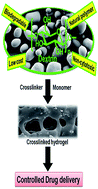
This review describes hydrogels and their classifications along with the synthesis and properties of biopolymer-dextrin based crosslinked hydrogels and their potential applications in controlled drug delivery. Modified dextrin based crosslinked hydrogels exhibit unique characteristics in terms of their mechanical properties, stimuli-responsive behaviour and drug release characteristics. Herein, first an outline is given of hydrogels and their classifications, various biopolymer based hydrogels and their synthetic procedures. Secondly, the significance of biopolymer dextrin for developing hydrogels, requirements of modification, and various dextrin based hydrogels developed in the authors’ laboratory are discussed. Finally, the importance of crosslinked hydrogels in drug delivery applications and various dextrin based hydrogels used in sustained drug delivery studies have been explored, which confirm that future clinical applications of these materials in the biomedical and pharmaceutical fields are feasible.


image file: c4ra16103c-p1.tif

Dipankar Das

Dipankar Das was born in Birbhum, West Bengal, India and completed his undergraduate studies at Burdwan University, India, in 2009. Dipankar obtained his Master of Science degree in Chemistry from Indian School of Mines, Dhanbad, India, in 2011. Currently he is pursuing his doctoral research in polymer chemistry under the supervision of Dr Sagar Pal, Associate Professor, Indian School of Mines, Dhanbad, India. His research interest is modified dextrin based crosslinked hydrogels/nanogels: development and application as matrices for controlled drug delivery.

image file: c4ra16103c-p2.tif

Sagar Pal

Dr Pal is working as an Associate Professor in the department of Applied Chemistry, Indian School of Mines, Dhanbad. He was the recipient of the DAAD FELLOWSHIP 2012. He is also a Fellow of Indian Chemical Society. He has ∼14 years of research experience in the field of polymer chemistry. His research interest is on the development of polymeric biomaterials, hybrid nanocomposites, semi-IPN hydrogels, and nanogels, and their applications in the fields of controlled drug release and wastewater treatment. Presently he has 56 international publications, 4 book chapters, 2 patents and 3 technical articles. One of his students has been awarded a Ph. D. degree, two Ph. D. students have submitted their theses and six Ph. D. students are presently working under him.


1. Introduction

In recent times, hydrogels have drawn significant attention because of their various applications in the pharmaceutical and biomedical fields.1–6 Remarkable efforts have been made to develop novel hydrogels with combined features such as: tuneable chemical and 3-dimensional physical structure,7–10 desired mechanical properties,11–14 and high water absorption ability, biodegradability and biocompatibility,7–10 all of which are indispensable for biomedical applications.15–17

In addition, biopolymers/modified biopolymer based stimuli-responsive hydrogels are widely used as drug carriers that improve drug release characteristics.5,18–20 The use of natural biopolymers over synthetic polymers for controlled drug release applications has increased markedly because of their low cost, high availability, and non-cytotoxic and biodegradable nature.21 However, they have certain shortcomings like microbial contamination, uncontrolled rates of hydration, and fall in viscosity during storage etc.22 The grafting/crosslinking of synthetic polymer chains on natural polymer backbones in the presence of external crosslinkers has been envisaged to develop hydrogels/crosslinked hydrogels which have improved characteristics, that may be applicable as sustained drug release matrices.23 Polymers synthesised from natural or synthetic sources which have hydroxyl, amine, amide, ether, and sulfonate groups in their side chains are used for developing chemically crosslinked hydrogels, and might be suitable for both hydrophilic and hydrophobic drug molecules.24

For oral administration of drugs, safe, nontoxic, biocompatible and biodegradable matrices are essential.5 But despite drug delivery requirements like improving the bioavailability of drugs, a good biocompatibility between the drug and hydrogel, the non-cytotoxic nature of the gel and sufficient drug stability in tablet formulations, chemically crosslinked hydrogels are less studied. From a drug delivery perspective, covalently crosslinked hydrogels are interesting because of their unique characteristics like reducing burst release effects and prolonged drug release behaviour.

Various modified biopolymer based hydrogels have been developed and reported. However, we for the first time developed chemically crosslinked stimulus responsive hydrogels based on modified biopolymer-dextrin and their potential applications in the field of controlled drug delivery. This review thus aims to provide an in-depth knowledge of various modified biopolymer based hydrogels and dextrin based chemically crosslinked hydrogels for controlled drug release applications. Here, we mainly focus on dextrin based chemically crosslinked hydrogels, proposed mechanisms for their synthesis, their detailed characteristics which include the surface morphology, cytoxicity studies and biodegradability, and finally their application as potential matrices for sustained release of various model drugs in vitro. The release kinetics and mechanism have also been explored.

This review is organized in the following way: we commence with a brief overview of hydrogels and their classifications, biopolymer based hydrogels, and various procedures for the development of hydrogels. Afterwards, we focus on the significance of biopolymer-dextrin for developing hydrogels, requirements of modification and various dextrin based hydrogels, which were developed in the authors’ laboratory. In the next section, we present an idea about controlled drug delivery and the importance of crosslinked hydrogels in drug delivery applications. Finally, we have explained in detail the release study of model drugs (such as ciprofloxacin and ornidazole) using synthesized modified dextrin based crosslinked hydrogels. In addition, we also explored the advantages of dextrin based hydrogels over other biopolymeric hydrogels reported in the literature.

2. Polymeric hydrogels

According to the literature, the name ‘hydrogel’ first appeared in 1894 when it was depicted as a colloidal gel of inorganic salts.25,26

In 1958, crosslinked PVA hydrogel was developed in the presence of gamma irradiation.26 In 1960, Wichterle and Lim were the first to introduce hydrogels on poly(HEMA) as water-swollen crosslinked networks for contact lense applications (Fig. 1).27 Hydrogels are hydrophilic, physically or chemically crosslinked, three-dimensional natural or synthetic polymeric networks, which are capable of imbibing large amounts of water or biological fluids.4–6,28–30 Hydrogels also have a degree of flexibility which resembles natural tissue, owing to their significant water content. Their excellent water absorption efficacy is mainly because of the presence of hydrophilic groups like –OH, –CONH–, –CONH2–, and –SO3H in the polymeric network.31 Due to the presence of these groups and domains in the network, the polymers are thus hydrated to different degrees (sometimes more than 90% wt), depending on the nature of the aqueous environment and polymer composition.32–35


image file: c4ra16103c-f1.tif
Fig. 1 Important events in hydrogel research.

Hydrogels are commonly considered as highly biocompatible, owing to their high water content and also their physiochemical similarity with the native extracellular matrix.36 In spite of their high water absorbing ability, hydrogels swell rapidly instead of dissolving in aqueous media, because of the presence of crosslinkers in the hydrogel network. These crosslinks are mainly of two types: (i) physical (entanglements or crystallites), and (ii) chemical (tie-points and junctions).37–42 The crosslinks may remain in the polymer network in the forms of covalent attachment, hydrogen bonding, van der Waals interactions or physical entanglements.43

Besides, hydrogels demonstrated phase transition with changes in external conditions such as pH,4,44 temperature,45 light,46 electric field,47 and magnetic field48 and are known as “stimuli-responsive” or “smart” gels.49–51 These smart gels exhibit numerous outstanding characteristics like defined morphology, high porosity, flexible dimensions and the ability of their network to absorb large amounts of water. These properties make them promising candidates for controlled drug release applications.52–54

There are several reports on synthetic polymer based hydrogel matrices used for controlled/sustained drug delivery applications.55–77 Lalloo et al. reported on poly(ethylene glycol) hydrogel for controlled delivery of camptothecins.55 Dadsetan et al. used oligo(poly(ethylene glycol) fumarate) hydrogel for the delivery of doxorubicin.56 Controlled delivery of recombinant hirudin based on thermo-sensitive Pluronic F127 (PF127) hydrogel was investigated by Liu et al.57 PAE-PCL-PEG-PCL-PAE pentablock copolymer hydrogel as a sustained insulin delivery system was evaluated by Huynh et al.58 Sustained release of paclitaxel by (pNIPAm-PEG-pNIPAm) hydrogel was investigated by Graaf et al.59 Delivery of ketoprofen and spironolactone drugs from PEG–PLGA–PEG triblock copolymeric hydrogel was described by Jeong et al.60 Wang et al. described the controlled release of paclitaxel from poly(lactic acid)/(methoxyl poly(ethylene glycol)/itaconic acid based hydrogel.61 Camptothecin release from PLGA–PEG–PLGA triblock copolymers hydrogel was developed by Yu et al.62 Yu et al. described controlled release of the anticancer drug doxorubicin from NPOD-PEG/α-CD hydrogels.63 Riboflavin release was studied using PVA-star PDMAEMA based hydrogel by Wu et al.64 Yu et al. also described Avastin release from crosslinked PEG hydrogel.65 Pindolol, quinine and timolol maleate release from RADA 16 peptide hydrogel was detected by Briuglia et al.66 The sustained release of dextran-fluorescent isothiocyanate (FITC) from MPEG–PCL–MPEG triblock copolymeric hydrogel was investigated by Wu et al.67 PEOz–PCL–PEOz triblock hydrogel was used as a controlled drug delivery matrix for Bevacizumab as studied by Wang et al.68 Cavalieri et al. described doxorubicin release from PVA–PMA based hydrogel.69 Wieduwild et al. investigated the release of ATIII-F drug from peptide hydrogel.70 Dexamethasone acetate release from hyper branched polyester hydrogel was studied by Zhang et al.71 The sustained release of a model protein drug (bovine serum albumin) using PEG-α-cyclodextrin based hydrogel was described by Ma et al.72 A release study of methylene blue and bovine serum albumin from NIPAm/dextran/HEMA based hydrogel was carried out by Huang et al.73 Nugent et al. used PVA/poly(acrylic acid) hydrogel for theophyline delivery.74 Poly(organophosphazene) hydrogel was used for the controlled delivery of FITC-dextran and human serum albumin by Kang et al.75 Mishra et al. reported on PEG–PVL–PEG copolymers hydrogel for sustained release of dexamethasone.76 Babu et al. described poly(HEMA-co-MMA) based hydrogel for controlled release of cisplatin.77

2.1. Classification of polymeric hydrogels

Hydrogels are broadly classified into two categories:

Permanent or chemical gel: these gels form stable covalently crosslinked networks.78 They attain an equilibrium swelling state which depends on the polymer–water interactions as well as on crosslink density.79

Reversible or physical gel: these networks are held together by molecular entanglements, ionic interactions, hydrogen bonding or hydrophobic interactions. In physically crosslinked gels, dissolution is prevented due to physical interactions between different polymer chains.78 The interactions are reversible, and can be interrupted by changing physical conditions or the application of stress.79

Hydrogels can also be classified on the basis of various features (Fig. 2)11 such as the nature of side groups, method of preparation, physical structure, mechanical and structural characteristics, and responsiveness to stimuli.1,28,29,35,37–42,80–84


image file: c4ra16103c-f2.tif
Fig. 2 Classifications of polymeric hydrogels.

However, the hydrogels mainly used in biomedical applications have been synthesized from polymers of natural or synthetic origins.1,3,85–88

2.2. Biopolymer based hydrogels

The utilization of biopolymers as materials is not new. For example, cellulose is used frequently to provide structure for plants. Chitin is used as the exoskeleton of crawfish and shrimps. Collagen is utilized for mechanical support in connective tissues, and silk in spider’s webs.89 These materials possess a great deal of significance as materials for biomedical applications owing to their good biocompatibility and low cost of disposal.90,91 Besides, the chemical structures of these materials assist in the synthesis of advanced modified materials which would be useful in multidisciplinary applications. The degradation properties of natural polymers into physiological metabolites make them potential candidates as drug delivery matrices or for regenerative medicine.

Among the biopolymers, polysaccharides have some good characteristics like non-toxicity, water solubility or high capability for swelling induced by simple chemical modifications, which make them suitable for biomedical applications.1,92,93–95

Polysaccharides are obtained from numerous sources including seaweeds, plants, fungi, bacteria, insects, crustacea, animals and even humans.96–99

Chemically, polysaccharides contain large carbohydrates which are formed by only one type of repeating monosaccharide or by two or more different monomeric units. Polysaccharides can also be classified as non-polyelectrolytes and polyelectrolytes, which are either positively or negatively charged.100 Typical examples of polysaccharides as biopolymers are chitosan, hyaluronan, agar, dextran, dextrin, cellulose, chondroitin sulphate, pullulan, sterculia gum, alginate etc. (Fig. 3).101,102 The conformation of the polysaccharide chains depends on various factors like pH, ionic strength of the medium, temperature and the concentration of certain molecules (such as lecitins).103 Polysaccharides usually respond to external stimuli, which makes them attractive as potential candidates for sustained drug release applications. In addition, some polysaccharides also have unique features such as providing nanocarriers with surface properties that control the interactions with blood, mucosa and target cells during absorption and biodistribution.104 By biomimicking the surface of eukaryotic cells, bacteria and viruses, polysaccharides can assist the recognition and binding to desirable surfaces, while escaping from opsonization and complement activation.105,106


image file: c4ra16103c-f3.tif
Fig. 3 Chemical structure of different polysaccharides.

However, polysaccharides have some limitations like microbial contamination, uncontrolled rate of hydration, and drop in viscosity on storing. Fortunately, several strategies have already been developed and reported to overcome these drawbacks by suitable chemical modification, which includes grafting/crosslinking of synthetic polymer chains onto their backbone.4,5 These modified biopolymers form a 3-dimensional, crosslinked, hydrophilic network which can absorb large amount of water when remain in contact with aqueous medium or in biological fluids without showing solubility. These materials are considered to be hydrogels or biomaterials based hydrogels.

The era of biomaterials based hydrogels started in 1960, when Wichterle and Lim first developed poly(HEMA) based copolymer for contact lense applications.27 Later, various hydrogel materials were developed and reported using different techniques. The early methods of synthesis were focused on the development and structure determination, but later, attention was shifted towards the applicability of hydrogels in various fields. The 3-dimensional polymeric networks in a swollen state interact with active compounds through covalent interactions or through H-bonding or ionic interactions. Biopolymer based hydrogels possess all the properties of their synthetic counterparts as well as being inherently renewable, abundant in nature, nontoxic, biodegradable, and relatively cheap. In addition, these hydrogels possess a high content of functional groups, which are utilized in crosslinking with additional functional crosslinkers and can be bioconjugated with cell targeting materials.107 The chemical and/or physical crosslinks are responsible for the insoluble nature of grafted/crosslinked hydrogels. It is also easy to tune the physico-chemical properties of grafted/crosslinked hydrogels, which are sensitive towards various stimuli present in our body like pH, ionic strength, temperature etc.108–111 Such hydrogels are called smart polymers. Thus, the use of these modified biopolymer based hydrogels specifically for drug delivery applications is more advantageous than that of neat polysaccharides. This is because the drug release behaviour of these hydrogels not only depends on the extent of crosslinking but also on various other factors, including their better swelling characteristics, restricted dissolution characteristics, the pH of the release media and interactions between drugs and hydrogels.4

Therefore, the distinct features of polysaccharides/biopolymers make these hydrogels effective in various applications such as biomedical applications, agricultural applications and industrial applications (Fig. 4).


image file: c4ra16103c-f4.tif
Fig. 4 Applications of polysaccharide based hydrogels in various fields.
2.2.1. Applications of polysaccharide based hydrogels in various fields.
2.2.1.1. Biomedical applications. Polysaccharide based hydrogels have been utilized in various biomedical applications because of their non-toxicity, biocompatibility, suitable performance for in vivo transplantation, capability to be fabricated into an ample range of morphologies, structural similarity to natural materials and degradation by enzymes.112

Among different biomedical applications, hydrogel based drug delivery systems have become a major area of interest and have achieved success at the industrial level. There are several reports on hydrogels including modified biopolymeric hydrogels based on agar, chitosan, dextran, carrageenan, gelatin, gellan gum, konjac glucomannan, locust bean gum, cyclodextrin, guar gum, Stereculius gum etc. for drug delivery applications.113–142

Usha Rani et al. reported on the synthesis and applications of polyacrylamide grafted agar based hydrogel as a matrix for the controlled release of 5-ASA.113 Cyclodextrin–agar hydrogels for ciprofloxacin delivery were studied by Fernandez et al.114 Kulkarni et al. synthesized pH-responsive interpenetrating network hydrogel beads of polyacrylamide grafted carrageenan (PAAm-g-CG) and sodium alginate for targeting ketoprofen to the intestine.115 Hydrogel based oil encapsulation for the controlled release of curcumin by using a ternary system of chitosan, kappa-carrageenan, and carboxymethylcellulose sodium salt was described by Nakagawa et al.116 Hezaveh et al. designed kappa-carrageenan-based hydrogel for controlled release applications.117 Gelatin–carrageenan hydrogels for quercetin delivery were designed by Varghese et al.118 Controlled delivery formulations of caffeine in alginate hydrogel beads combined with pectin, carrageenan, chitosan and psyllium were described by Cvitanovic et al.119 Kappa-carrageenan/polyvinyl alcohol crosslinked hydrogels were formulated using genipin as a natural and nontoxic crosslinker to achieve a controlled release of β-carotene.120 Sustained delivery of latanoprost by thermosensitive chitosan–gelatin-based hydrogel for controlling ocular hypertension was reported by Cheng et al.121 Chitosan–gold hybrid hydrogel and its application for drug delivery was described by Chen et al.122

Synthesis of oxidized glycerol monooleate–chitosan polymer and its hydrogel formation for sustained release of trimetazidine hydrochloride was reported by Zhang et al.123 Peng et al. described chitosan/glycerophosphate disodium thermosensitive hydrogels for the sustained delivery of venlafaxine hydrochloride.124 Genipin-crosslinked catechol–chitosan mucoadhesive hydrogels for buccal drug delivery were depicted by Xu et al.125 A magnetic chitosan hydrogel for sustained and prolonged delivery of Bacillus Calmette–Guérin in the treatment of bladder cancer was reported by Zhang et al.126 van Dijk-Wolthuis et al.127 reported dextran hydrogels, which were prepared by radical polymerization of aqueous solutions of glycidyl methacrylate-derivatized dextran (dex-MA), hydroxyethyl methacrylate-derivatized dextran (dex-HEMA), and HEMA-oligolactate-derivatized dextran (dex-lactate HEMA), using potassium peroxydisulfate and N,N,N′,N′-tetramethylethylenediamine (TEMED) as the initiating system for controlled drug release applications. The release characteristics of salmon calcitonin from dextran hydrogels for colon-specific delivery were reported by Basan et al.128 In vivo controlled release of cisplatin from biodegradable gelatin hydrogel was performed by Konishia et al.129 The in vivo anti-tumour effect of dual release of cisplatin and adriamycin from biodegradable gelatin hydrogel was studied by Konishia et al.130 Gaowa et al. reported gelatin based biodegradable hydrogel for in vivo controlled release of anti-tumour activity.131 Synthesis of polymethacrylamide-grafted-gellan gum (PMaa-g-GG) for controlled drug delivery applications was reported by Nandi et al.132 Chitosan/poly-γ-glutamic acid nanoparticles incorporated into pH-sensitive hydrogels were developed as an efficient carrier for amoxicillin delivery.133 Cyclodextrin mediated controlled release of naproxen from pH-sensitive chitosan/poly(vinyl alcohol) hydrogels for colon targeted delivery was described by Das et al.134 The use of pH-sensitive cationic guar gum/poly(acrylic acid) polyelectrolyte hydrogels for controlled drug delivery applications was described by Huang et al.135 Synthesis of acryloyl guar gum and its hydrogel materials for use in the slow release of L-DOPA and L-tyrosine was reported by Thakur et al.136 A thermoresponsive magnetic nanoparticle–aminated guar gum hydrogel system for sustained release of doxorubicin hydrochloride was described by Murali et al.137 Chen et al. explained the synthesis and properties of degradable hydrogels of konjac glucomannan grafted acrylic acid for colon-specific drug delivery of 5-ASA.138 Liu et al. described the synthesis and characteristics of pH-sensitive semi-interpenetrating polymer network hydrogels based on konjac glucomannan and poly(aspartic acid) for in vitro drug delivery of 5-fluorouracil.139 Ibuprofen release from the pH- and electro-response cellulose nanofiber/sodium alginate hybrid hydrogels was described by Zheng et al.140 Optimal response surface design of Gum tragacanth-based poly[(acrylic acid)-co-acrylamide] IPN hydrogel for the controlled release of antihypertensive drug losartan potassium was reported by Saruchi et al.141 Photo thermally enhanced drug release from κ-carrageenan hydrogels reinforced with multi-walled carbon nanotubes was described by Estrada et al.142

Wound dressing is one of the most significant biomedical applications of polysaccharide based hydrogels. The adhesive nature, antifungal/antibacterial activity and permeability to oxygen make hydrogels suitable for the treatment of wounds and burns.112 Chitosan and its derivative based hydrogels have been widely used as wound dressing materials.143 Alginate/asiaticoside based hydrogels have been studied as wound dressing materials by Sikareepaisan et al.144 Balakrishnan et al. reported periodic-alginate crosslinked hydrogel as a wound dressing agent.145 Singh et al. synthesized sterculia gum crosslinked PVA–PVP hydrogel through a radiation crosslinking method for the delivery of antimicrobial agents to wounds.146 Poly(vinyl alcohol)-alginate physically crosslinked hydrogel membranes for wound dressing applications were reported by Kamoun et al.147


2.2.1.2. Agricultural applications. Storage of water in industrial fields is challenging. Hydrogels have usually been used as water storage materials in the agricultural field.112 Cellulose based hydrogels are potential materials to absorb and retain water. Sannino and coworkers reported cellulose based hydrogels for agricultural purposes.148 A sodium alginate–glutaraldehyde crosslinked hydrogel has been investigated as a matrix for natural liquid pesticide neem seed oil.149 A starch–alginate–clay composite was used for the release of a fungicide thiram.150 Microspheres of sodium alginate/starch crosslinked with Ca2+ have been used as a controlled release matrix for the release of the pesticide chlorpurifos.151 Starch/ethylene glycol-co-methacrylic acid hydrogel was designed for the controlled release of pesticides like fluometuron, thiophanate, thiophanate methyl, and trifluralin.152
2.2.1.3. Industrial applications. Polysaccharide based hydrogels have a wide range of industrial applications. In the food industry, pectin is used as a gelling and thickening reagent. The inclusion of pectin in gel-like products achieves the desired firmness and alters the texture of the gel. For these reasons, pectin has been used in foods, cosmetics, and environmental applications to modify the release of fragrance compounds and enhance the perception of flavours.153–155

Heavy metal ion adsorption is an important application of polysaccharide based hydrogels. Psyllium husk and acrylic acid based crosslinked hydrogel was used for the removal, separation, and enrichment of hazardous metal ions from aqueous solution.156 Cellulose and its derivatives such as hydroxypropyl cellulose, cyanoethyl cellulose, hydroxyethyl cellulose, hydrazinodexy cellulose, cellulose phosphate with acrylamide and N,N′-methylene bisacrylamide based hydrogels have been studied for the sorption of Fe2+, Cu2+ and Cr2+ ions.157 A polyacrylic acid and sodium alginate based hydrogel has been used for the adsorption of Cu2+, Co2+, and Ni2+ ions.158 Hydrogels based on chitosan, maltodextrin and gum arabic have been employed as adsorbents for removing Cd2+ from aqueous solutions.159 Calcium alginate hydrogel beads containing vineyard pruning waste have been used for dye removal.160 Agarose based hydrogel has been utilized for stacking electrophoresis.161 Cellulosic hydrogels were used for chromatographic supports.162 Chitosan based hydrogels have been used as corrosion inhibitors.163 Water soluble and biodegradable pectin-grafted polyacrylamide and pectin-grafted polyacrylic acid hydrogel as corrosion inhibitors on mild steel in 3.5% NaCl media were described by Geethanjali et al.164 Polyacrylamide grafted with okra mucilage has been tested as a corrosion inhibitor for mild steel in 0.5 M H2SO4 by Banerjee et al.165 Polyacrylamide grafted guar gum (GG-g-PAM) has been tested as a potential green inhibitor of corrosion of mild steel in 1 M HCl by Roy et al.166 Polysaccharides are also used as thickeners, bioadhesives, stabilizers, probiotics, and gelling agents in the food and cosmetics industries.167–169 Radiation synthesis of eco-friendly water reducing sulfonated starch/acrylic acid hydrogel designed for the cement industry has also been reported.170

2.2.2. Classifications of polysaccharide based hydrogels. Polysaccharide based hydrogels can be classified on the basis of method of preparation, types of monomers involved and ionic charges (Fig. 5).112
image file: c4ra16103c-f5.tif
Fig. 5 Classifications of polysaccharide based hydrogels.

On the basis of method of preparation, hydrogels are mainly of two types: (i) physically crosslinked hydrogels, and (ii) chemically crosslinked hydrogels.


2.2.2.1. Physically crosslinked hydrogels. These are reversible and unstable hydrogels in which polymeric units are connected to each other by the secondary force of interactions such as hydrogen bonding, ionic or hydrophobic interactions. The stability of these hydrogels also depends on the external conditions and may change with a changing environment or by application of stress. These hydrogels have a wide range of biomedical applications because of the absence of any external crosslinkers. There are several reports on physically crosslinked hydrogels used in biomedical applications such as blends of starch/carboxymethyl cellulose, gelatine/agar and chitosan/glycerol-2-phosphate hydrogels.171–174
2.2.2.2. Chemically crosslinked hydrogels. Because of the presence of various functional groups like hydroxyl, amine and carboxylic acid groups in the polymeric backbone, it can covalently connect with the crosslinker and form chemically crosslinked hydrogels. These are irreversible in nature and stable due to the covalent attachment between the crosslinker and the polymer. Crosslinking may occur by various mechanisms such as condensation, addition and vulcanization. Because of the stable network which arises from covalent attachment, these types of hydrogels attain equilibrium swelling that depends on the polymer–solvent interactions as well as on crosslinking density. Polysaccharides like starch and its derivatives, cellulose, dextrin, dextran, chitosan, agar, alginate, guar gum, hyaluronan etc. were chemically crosslinked with crosslinkers in the presence of an initiator or radiation to form chemically crosslinked hydrogels.175,176
2.2.2.3. Homopolymer hydrogels. When a single species acts as a building block in the network and forms a three dimensional polymeric network by crosslinking, then the hydrogel is considered as a homopolymer hydrogel. The crosslinking may occur in the presence of crosslinkers or with the polymer molecules themselves. Cellulose can form a hydrogel in urea/NaOH solution in the presence of epichlorohydrin crosslinkers.177 Irradiation of polysaccharide solutions can also form hydrogels in the absence of a gelating agent. In addition, polysaccharides such as chitin, chitosan, agar, alginate, xanthan, guar gum, hyaluronan etc. can form homopolymeric hydrogels in the presence of high energy radiation.178
2.2.2.4. Copolymer hydrogels. When two or more polysaccharide units or one polysaccharide and one synthetic polymer take part in hydrogel formation (out of which, one is hydrophilic) to form a 3-dimensional polymeric network, this is considered a copolymer hydrogel. The arrangements of copolymer hydrogels may be random, block or alternate with respect to the main polymeric network. The properties of these hydrogels depend on the different combinations of polymeric units as well as on the arrangement of the polymeric network.179,180
2.2.2.5. Interpenetrating network hydrogels. Interpenetrating polymer networks (IPNs) are crosslinked polymers181 in which at least one polymer network is synthesized or crosslinked independently in the presence of the other, without any covalent bonds between them.182 Synergistic effects are observed as the properties of both of the polymers are involved, which can reduce the limitations of natural as well as synthetic polymers. On the basis of synthesis, IPNs are of two types: (a) a sequential IPN in which one network is swollen and polymerized in the presence of the other, or (b) a simultaneous IPN, in which both of the network precursors are synthesized at the same time by independent routes.

If only one component of the assembly is crosslinked leaving the other in a linear form, the system is termed as semi-IPN. The significant point during the fabrication of IPNs is the mutual miscibility of the interpenetrating polymers. Generally, polymers do not mix well with each other, resulting in the phase separation of the resultant blend.183,184 However, crosslinking provides an easy route for mixing the two components to form IPNs for a variety of applications.185


2.2.2.6. Neutral hydrogels. When neutral polysaccharides covalently crosslink with neutral polysaccharides and/or neutral monomeric units like acrylamide, N-vinyl pyrollidone, hydroxyalkyl methacrylate etc. to form three dimensional polymeric networks then the network is called a neutral hydrogel. These gels are irreversible and can absorb water without dissolution. These hydrogels demonstrate temperature sensitive swelling–deswelling behavior.186

pH sensitive N-succinyl chitosan grafted polyacrylamide hydrogel for oral insulin delivery was reported by Mukhopadhyay et al.187 Cheng et al. described the sustained delivery of latanoprost by thermosensitive chitosan–gelatin-based hydrogel for controlling ocular hypertension.188 The biodegradability and swelling capacity of kaolin based chitosan-g-PHEMA nanocomposite hydrogel was described by Pradhan et al.189 Thermoresponsive chitosan–agarose hydrogel for skin regeneration was reported by Miguel et al.190 Carboxymethyl cellulose-g-poly(2-(dimethylamino) ethyl methacrylate) hydrogel as an adsorbent for dye removal was reported by Salama et al.191 PVA/gelatin hydrogel beads and their adsorption mechanism for advanced Pb(II) removal were reported by Hui et al.192


2.2.2.7. Ionic hydrogels. Ionic hydrogels are of two types: (a) cationic, and (b) anionic. When ionic polysaccharides are covalently crosslinked with each other or synthetic/ionic polymers to form a 3-D polymeric network, then these are called ionic hydrogels.

The only naturally existing cationic polysaccharide is chitosan, which is obtained from the partial alkaline deacetylation of chitin.193,194 The amine groups are responsible for the pH-responsiveness of chitosan and also its enhanced affinity for mammalian cell components as well as its microbicide characteristics. Besides, these groups also provide affinity towards oppositely charged drugs and reactivity for crosslinking or grafting modifications.193,194

Again, condensing agents like β-glycerophosphate, citrate, calcium phosphate, or tripolyphosphate can form ionically crosslinked hydrogels.195–197

The swelling of the cationic hydrogels is dependent on the pH of the aqueous medium, which determines the extent of dissociation of the ionic chains. At an acidic pH, cationic hydrogels show excellent swelling because their chain dissociation is favoured in low pH.

Anionic polysaccharides extracted from seaweeds (alginate, agar, carrageenan) and plant cell walls (pectin) and exudates (gum arabic) have been used in the food industry as thickening or gelling agents. At basic pH, anionic hydrogels dissociate more and show significant swelling than they do in neutral solution. The sensitivity of alginate towards pH and calcium ions is mainly because of the different relative positions of the carboxylic acid groups in each block.198 Alginate also offers a large number of potential derivatives for biomedical applications.199 Agar or agar-agar is obtained from red algae or seaweed and is widely used as a component in foods and microbial cultures, because of its performance as a thickener and stabilizer. Agar is a heterogeneous mixture of agaropectin and agarose, where agaropectin is modified with acidic side-groups such as sulphate and pyruvate, and agarose has a neutral charge and possesses longer chains.200

Due to this structural conformation it can form an ionic hydrogel. The use of agar as a drug delivery matrix is still quite recent and mostly limited to physical blends with other polysaccharides.201–204 Pectin and gum arabic are branched heteropolysaccharides which have carboxylic acid groups and can form ionic hydrogels. These can be found in the cell walls of plants in the form of free acids, simple salts, esters or aminated polysaccharides. Pectin is suitable for colon delivery of drugs because of its selective digestion by the micro flora present in the colon (Bacteroides ovatus) and its resistance to the proteases and amylases of the upper gastrointestinal tract.205,206 Carrageenans or carrageenins are linear polysaccharides containing a variable density of sulphated groups. They are extracted from red seaweeds and commercialized as kappa (κ, 1 sulphate group per dimer), iota (ι, 2 sulphate groups per dimer) and lambda (λ, 3 sulphate groups per dimer). Only the first two varieties can form stable physical gels and κ-carrageenan hydrogels show pH- and temperature-responsive behaviour.201

Wang et al. developed three ionic starch-based polymers, namely a cationic starch (C-Starch), an anionic starch (A-Starch) and a zwitter ionic starch (Z-Starch) via etherification reactions for the prevention of nonspecific protein adsorption.207 Heparin is a highly sulphated glycosaminoglycan and has the highest negative charge density of any known biological molecule. It is widely used as an injectable anticoagulant. Recently it is gaining interest as a component of growth factor delivery systems.208,209 Chondroitin sulphate is a linear polysaccharide based on (1,3)-β-N-acetyl-D-galactosamine and (1,4)-β-glucuronic acid with sulphate, hydroxyl and carboxylic acid functionalities. It can be found attached to proteoglycans in connective tissue matrices with structural functionalities or as a receptor on cell or basement membranes. Mainly it has been used as a component of scaffolds for cartilage repair in tissue engineering.210 Nanocomposite hydrogels of calcium alginate and carboxymethyl-cellulose-g-poly(acrylic acid)/attapulgite for controlled release of ibuprofen have also been reported.211


2.2.2.8. Ampholytic hydrogels. When polymeric hydrogels with a 3-dimensional network contain both cationic and anionic moieties, these are considered ampholytic hydrogels. Cationic/anionic polysaccharides can covalently attach with oppositely charged synthetic polymers to form ampholytic hydrogels. At a defined pH, modified polysaccharides can also behave like ampholytic hydrogels. Zwitter ionic starch (Z-Starch) hydrogel via etherification reactions for the prevention of nonspecific protein adsorption was developed by Wang et al.212

3. General procedures for the development of polymeric hydrogels

Hydrogels open a new pathway for the advancement of various applications including tissue engineering,213,214 cell based biosensors,215 coatings,216 drug delivery,4–6 packers in oil and gas recovery217 and cell transplantation218 and so on.

They are composed of hydrophilic polymer chains, which can be either natural or synthetic.219 The structural integrity of hydrogels depends on crosslinks between the polymer chains, by chemical bonds and physical interactions. Besides, as the biomedical applications of hydrogels are increasing, so considerable efforts have been made to develop new hydrogel systems from a variety of synthetic and natural materials.220

It is essential to modify the properties of a polymer with tailor-made specifications, which are essential for target applications. There are various ways to modify the properties of polymers like blending, grafting/crosslinking, and curing. ‘Blending’ is the physical mixture of two or more polymers to get the required properties. ‘Grafting’ is a method wherein monomers are covalently bonded onto the polymer chain. ‘Chemical crosslinking’ is a method where monomers are covalently bonded onto the polymer chain in the presence of crosslinkers. In curing, the polymerization of an oligomer mixture forms a coating which adheres to the substrate by physical forces.221 A schematic representation of these polymer modifications is presented in Fig. 6.


image file: c4ra16103c-f6.tif
Fig. 6 Schematic representation of the methods for polymer modification.

Although polysaccharides have several advantages, polysaccharide itself is not sufficient for recent industrial requirements. Thus it is essential to modify biopolymers for the advancement of their applications in biomedical research, particularly in drug delivery applications. One of the easiest and most efficient ways to modify natural polysaccharides is the ‘grafting’ of synthetic polymer chains onto the polysaccharide backbone, followed by ‘chemical crosslinking’ using an external crosslinker. This modification provides chemically crosslinked hydrogels which have immense applications in the biomedical field due to their desired properties.

3.1. Blending

Polymer blends represent a very important field to assist in the development of new low-cost products with better performance in comparison with neat polymers. Blends and composites are extending the employment of polymers from renewable resource into new value-added products.222 They are significant also from the ecological and economical viewpoints. For example, municipal commingled plastic waste, composed of various immiscible polymers can be recycled by mixing in a molten state, and so it can be transformed into materials which would satisfy relevant applications.223 The properties of natural polymers can be significantly advanced by blending with synthetic polymers. The objective for developing a novel blend of two or more polymers is not to change the properties of the components drastically, but to capitalize on the maximum feasible performance of the blend. In the 1970s and 1980s, several blends of starch with various polyolefins were developed. However, these blends were not biodegradable, and thus the advantage of using a biodegradable polysaccharide was lost. Since most natural polymers are water soluble, water has been used as a solvent, dispersion medium and plasticizer for blending purposes.224
3.1.1. Melt processed blends. Arvanitoyannis et al. reported on biodegradable blends based on gelatinized starch and 1,4-trans-polyisoprene (gutta percha) by thermal pressing for food packaging or biomedical applications.225 Carvalho et al. prepared a blend of thermoplastic starch/natural rubber polymer using natural latex and cornstarch at 150 °C.226 Kokini et al. reported protein/starch blends.227 Matveev et al. studied protein/polysaccharide blends, considering inter-macromolecular hydrogen and dipole–dipole interactions.228 Warth et al. developed starch/cellulose acetate blends.229
3.1.2. Aqueous blends. Several natural polymers cannot be melt processed, either because they degrade on or before melting (softening). For this reason, aqueous blending is the preferred technology to modify the polymers.230 Pereira et al. reported on biodegradable hydrogels, based on cornstarch/cellulose acetate blends, produced by free-radical polymerization with methyl methacrylate and/or acrylic acid monomers.231 Espigares et al. developed partially biodegradable acrylic bone cements based on cornstarch/cellulose acetate blends.232 Arvanitoyannis and Biliaderis reported on aqueous blends of methyl cellulose and soluble starch, plasticized with glycerol or sugars, prepared by casting or by extrusion and hot pressing.233

Ikada et al. studied various poly(lactic acid) blends to improve their thermal properties.234 A stereocomplex was formed from enantiomeric PLAs, poly(L-lactic acid) (PLLA) and poly(D-lactide) (i.e. poly(D-lactic acid) (PDLA)) due to the strong interactions between PLLA and PDLA chains.234 Zhang et al. reported poly(3-hydroxybutyrate)/poly(lactic acid) blends prepared by casting a film from a common solvent at room temperature.235 Suyatma et al. reported on biodegradable film blends of chitosan with PLA by solution mixing and film casting.236

3.2. Grafting

Several methods (Fig. 7) have been used for the development of graft copolymeric hydrogels of different monomers on a polymeric backbone. These techniques include chemical, radiation, photochemical, plasma-induced and enzymatic grafting techniques.221
image file: c4ra16103c-f7.tif
Fig. 7 Schematic presentation of the methods of grafting.
3.2.1. Grafting initiated by chemical means. Graft copolymerization can be performed by two main processes, i.e. free radical and ionic. In addition, grafting can also be done by atom transfer radical polymerization (ATRP).
3.2.1.1. Free-radical grafting. In the free radical grafting technique, an initiator first generates radicals on a polysaccharide backbone, which further react with monomers to form the graft copolymers. Generally, the free radicals are generated by indirect or direct methods. A probable mechanism of grafting in the presence of a radical initiator is as follows:
I2 → 2I˙

I˙ + M → IM˙

I˙ + Polymer–OH → Polymer–O˙ + IH

IMM˙ + Polymer–OH → Polymer–O˙ + IMMH

Polymer–O˙ + nM → Polymer–O–Mn˙

IMM˙ + nM → IM–Mn–H

Polymer–O–Mn˙ + I˙ → Polymer–O–Mn–I

Polymer–O–Mn˙ + IMM˙ → Polymer–O–Mn+2–I

where, I and M indicate initiator and monomer.

An example of an indirect method for free radical production is a redox reaction using Mn+/H2O2 and persulphates.237–241

Fe2+ + H2O2 → Fe3+ + OH + OH˙

Fe2+ + S2O82− → Fe3+SO42− + SO4˙

Again, SO4˙ and OH˙ can also directly react with the polymer backbone to generate the free radical species.

SO4˙ + Polymer–OH → HSO4 + Polymer–O˙

OH˙ + Polymer–OH → H2O + Polymer–O˙

Misra et al. described that OH˙ is more reactive than SO42− during the grafting of vinylic monomers onto wool/cellulose.239

Similarly, organic hydro peroxides, persulphates, Fe3+, Cu2+ etc. along with a reducing agent like sodium bisulphite, thiosulphate or Ag+ are also used in free radical grafting methods.

S2O82− + HSO3 → SO42− + SO4˙ + HSO3˙

Polymer + SO4˙ → Polymer˙ + HSO4

SO4˙ + H2O → HSO4 + OH˙

HSO3˙ + H2O → H2SO3 + OH˙

Bajpai et al. reported peroxydisulphate–ascorbic acid initiated graft copolymerization using Ag as a catalyst.242 In this reaction sulphate ion radicals are produced which act as chain carriers.242 In addition, a tertiary butyl hydroperoxides–Fe2+ system can also initiate the radical polymerisation as parallel with Fenton’s reagent (Fe2+/H2O2). As a result, t-butoxy radicals are formed from one electron transfer between t-butyl-hydroperoxide (TBHP) and Fe2+. The resulting t-butoxy radical basically abstracts hydrogen from the monomer/polymer/water to generate the corresponding radical species.243

Transition metals ions like Ce4+, Cr6+, V5+ and Co3+ can also generate free radical sites on a polymeric backbone by direct oxidation. The redox potential of the metal ions is the driving force in determining the grafting efficiency. Metal ions with low oxidation potential have better grafting efficiency than those with a higher oxidation state. It is assumed that the reaction proceeds through the formation of a metal ion–polymer chelate complex as an intermediate.244–250 Sen et al. have synthesized CMS-g-PAM using ceric ammonium nitrate as the initiator.251

Ce4+ + Polymer–OH → Chelate complex

Chelate complex → Polymer–O˙ + Ce3+ + H+

Polymer–O˙ + M → Polymer–OM˙ → Polymer–OMM˙

where, M is the monomer.

MnO4 was also used for the formation of graft copolymers through radical polymerization. Here, MnO4 produces Mn3+ via Mn4+ in the presence of an acid solution. Highly reactive Mn3+ ions initiate the graft copolymerization and homopolymerization.252 Tsubokawa et al. described the polymerization of organic pigments incorporating azo groups, such as quinacridone, diketopyrrolopyrrole, anthraquinone etc., with vinyl monomers through a radical mechanism.253

Chemical pre-treatment (e.g. ozonation, diazotization and xanthation) of the polymer moiety may also produce free-radical sites, which can offer sites for grafting.254–264


3.2.1.2. Grafting through living polymerization. The ‘living polymerization’ technique offers a great potential for grafting reactions. Szwarc et al. state that living polymers can retain their ability to propagate for a long time and grow to a desired maximum size when the degree of termination or the chain transfer is negligible.265 Controlled free-radical polymerizations combine characteristics of both conventional free-radical and ionic polymerizations. In conventional free-radical polymerization, termination occurs through coupling or disproportionation reactions, which lead to a broad molecular weight distribution. Meanwhile, living polymerization provides living polymers with regulated molecular weights and low polydispersity.266–273

Controlled free-radical polymerization may be effective through ATRP. ATRP of styrene and various methacrylates has been reported, using various catalytic systems.274,275 In that method, inactive chains are capped by halogen atoms, which are reversibly transferred to metal complexes in the lower oxidation state. This creates the short-lived growing radicals and complexes in the higher oxidation state. ATRP reactions mainly proceed through the activation–deactivation dynamic equilibrium process.

image file: c4ra16103c-t1.tif
where, Mn − X is the polymeric halide and CuX/2L is Cu(I) (X = Cl/Br and L = 2,2′-bipyridine or a 4,4′-disubstituted 2,2′-bipyridine).

The copper(I) complex acts as a reversible halogen atom transfer agent between the active and inactive polymer chains (Scheme 1). The kinetically optimal ratio of ligands to copper(I) halide for this polymerization is found to be 2[thin space (1/6-em)]:[thin space (1/6-em)]1, which indicates that the coordinated copper(I) contains two bipyridine ligands.275


image file: c4ra16103c-s1.tif
Scheme 1 Schematic representation of the ATRP reaction.

Multidentate amine based ligands such as tetramethylene diamine (TMEDA), N,N,N′,N′,N′′-pentmethyl diethylene triamine (PMDETA) and (tris-2-aminoethyl) amine (TREN) are also used in the copper mediated ATRP of styrene, methyl acrylate and methyl methacrylate.276,277 Grafting from polymers including poly(vinyl chloride) (PVC), polyisobutene, polyethylene, and ethylene-co-vinyl acetate copolymer through ATRP has been reported in various works.278–282 Sonmez et al. reported acrylamide grafting by ATRP using an N-chlorosulfonamide/CuBr catalyst.283 Sakaguchi and Shimada depicted the grafting of polystyrene on a polypropylene backbone in the presence of γ-irradiation in air with 2,2,6,6-tetramethyl-1-piperidinoxyl (TEMPO).284 Janata et al. reported the design of a multifunctional ATRP macroinitiator for the preparation of graft copolymer.285 Carlmark et al. have reported surface grafting by controlled radical polymerization using 2-bromoisobutyryl bromide by reaction with the hydroxyl groups on filter paper. Then, grafting is achieved by immersing the modified paper into a reaction mixture containing methyl acrylate, Cu(I)Br, Tris 2-(dimethyl amino) ethyl amine (Me6-TREN), sacrificial initiator and ethyl acetate.286 Mishra et al. reported grafting of 4-aminoantipyrine from guar gum through graft ATRP using Cu(I)Br/2,2′-bipyridyl as the initiator.287 Wang et al. developed ethyl cellulose-graft-PDEAEMA copolymer through ATRP for drug (rifampicin) release applications.288 In this reaction, pyridine was used as a base for the first step and CuBr/bpy as the initiator.288 Huang et al. described poly(methacrylic acid)-grafted chitosan microspheres via a surface initiated ATRP technique for Cd(II) ion removal from aqueous solutions. In the first step of the reaction, 2-bromoisobutyryl bromide was used for the synthesis of the macroinitiator by TEA as a base. Then grafting of sodium methacrylate was performed with CuBr, CuBr2 and PMDTEA as the catalyst.289


3.2.1.3. Ionic grafting. In this method, grafting is performed by an ionic approach. Alkali metal suspensions in a Lewis base liquid, organometallic compounds and sodium naphthalenide are useful initiators. Alkyl aluminium (R3Al) and the halide form of the polymer (PCl) combine to form carbonium ions in the polymer chain, which is responsible for copolymerization. The reaction follows cationic and anionic polymerization mechanisms. The cationic catalyst BF3 can also be used.221

Cationic mechanism:

PCl + R3Al → P+R3AlCl

P+ + M → PM+–M → Graft copolymer

where, P+ is the cationic form of polymer and M is the monomer.

Sodium–ammonia or the methoxide of alkali metals forms the alkoxide of the polymer (PONa+), which reacts with the monomer to produce the graft copolymer.

Anionic mechanism:

P–OH + NaOR → PONa+ + ROH

PO + M → POM–M → Graft copolymer

where, M is the monomer.

3.2.2. Grafting initiated by irradiation techniques.
3.2.2.1. Free-radical grafting. The irradiation of polymers creates free radicals through homolytic fission. In the radiation technique, the presence of an initiator is not necessary, rather the medium. Thus, if irradiation is performed in open air, peroxides may be generated on the polymer backbone. The lifetime of the free radicals depends on the nature of the polymer moiety. Grafting usually takes place in three different ways: (a) pre-irradiation, (b) peroxidation or (c) the mutual irradiation technique (Scheme 2).221
image file: c4ra16103c-s2.tif
Scheme 2 Schematic representation of grafting through irradiation methods.

In the pre-irradiation grafting method, at first, the polymer backbone is irradiated in vacuum or in an inert gas to form free radicals. Then, the irradiated polymer is treated with the monomer in a liquid or vapour state, or in a suitable solvent.290–294

In the peroxidation grafting process, high-energy irradiation is applied to the polymer molecule in the presence of air or oxygen to form hydroperoxides or diperoxides. Radiation is used on the basis of the nature of the polymeric backbone and the irradiation conditions, resulting in the formation of a stable peroxy compound. After that, it is treated with the monomer at higher temperature, where the peroxides undergo decomposition to radical species. This initiates the grafting process. The advantage of this method is the formation of intermediate peroxy products that can be stored for long periods for grafting. On the other hand, in the mutual irradiation method, the polymer and the monomers are irradiated simultaneously to form free radicals and consequent addition occurs.295–301 The advantage of the pre-irradiation method is that the process is relatively free from homopolymer formation because the monomers are not irradiated. But, a certain disadvantage of the pre-irradiation method is scission of the base polymer due to its direct irradiation, which can result in the development of block copolymers.


3.2.2.2. Ionic grafting. Ionic grafting can also proceed via ion formation in the presence of high-energy irradiation. Ionic grafting may be of two different types: cationic or anionic. The polymer is irradiated to produce the polymeric ion, and afterwards reacted with the monomer to form the graft copolymer. The advantage of ionic grafting is a high reaction rate.

The cationic grafting mechanism can also proceed through the formation of a monomer radical cation, which then forms a dimer. Charge localization in the dimer occurs in such a way that the dimer radical cation then reacts with the radical formed by the irradiation of the polymer.302

Similarly in the anionic grafting mechanism, an anion controls the grafting, acting as the initiator. Kitamuru et al. and Mazzi et al. applied proton beams as ionizing radiation to prepare amidoxime type adsorbents on PE film using acrylonitrile.303,304 The hydrogen atoms which were liberated from chemically active sites of the polymer film irradiated by the ion beam reacted with carbon radicals, C[double bond, length as m-dash]C bonds, C–C bonds and crosslinking to form the graft-polymer.

3.2.3. Photochemical grafting. Because of the presence of chromophores in the polymeric chain, it can absorb light. As a result, the molecule enters an excited state and dissociates into reactive free-radicals, which initiates the grafting process. Sometimes, the absorption of light does not form free-radicals through bond rupture. Then the reaction can be promoted by the addition of photosensitizers, e.g. benzoin ethyl ether, dyes (Na-2,7-anthraquinone sulphonate or acrylated azo dye), aromatic ketones (benzophenone or xanthone) or metal ions such as UO22+. Thus the photochemical grafting process can proceed in two ways: with or without a sensitizer.305

Generally, when a sensitizer is not involved in the reaction, the generated free radicals of the polymeric backbone react with the monomer free radicals to form a graft copolymer. On the contrary, when a sensitizer is involved it forms free radicals. These free radicals can undergo diffusion so that they abstract H-atoms from the polymer. This results in the formation of radical sites, which are required for grafting.306

3.2.4. Plasma radiation induced grafting. Recently, the plasma radiation induced polymerization method has gained increasing significance. This method proceeds through a similar mechanism as with ionizing radiation.307,308 It involves electron-induced excitation, ionization and dissociation. Thus, the accelerated electrons from the plasma have adequate energy to cleave the chemical bonds in the polymeric structure. This results in the formation of polymeric radicals, which consequently initiate graft copolymerization.
3.2.5. Enzymatic grafting. Enzymes are also used in grafting technology. In this method an enzyme initiates the chemical/electrochemical grafting reaction.309 For example; tyrosinase can convert phenol into reactive o-quinone, which undergoes a subsequent non-enzymatic reaction with chitosan. Cosnier et al. reported enzymatic grafting on a poly(dicarbazole-N-hydroxysuccinimide) film.310

3.3. Methods of crosslinking

Both physical and chemical crosslinking methods (Fig. 8) have been used to prepare hydrogels. In chemically crosslinked gels, covalent bonds are present between different polymer chains or with an external crosslinking agent and polymer molecules. Whereas, in physically crosslinked gels, polymer chains are bound by physical interactions.311
image file: c4ra16103c-f8.tif
Fig. 8 Schematic representation of the methods of crosslinking.
3.3.1. Physically crosslinked hydrogels.
3.3.1.1. Crosslinking by ionic interactions. Physically crosslinked hydrogels can be developed by ionic interactions. Alginate is an example of a polymer that can be crosslinked by ionic interactions. Alginate is a polysaccharide with mannuronic and glucuronic acid residues and can be crosslinked by calcium ions.312 Crosslinking can be performed at room temperature and physiological pH. Consequently, alginate gels are usually used as matrices for encapsulation of living cells313 and for the release of proteins.314

The gels can be destabilized by extraction of the Ca-ions from the gel by a chelating agent. The ionotropic hydrogels degrade under physiological conditions. The degradation rate has been increased by incorporating hydrolysis-sensitive glycinato groups in the polymer.315 Polycations can also be crosslinked with anions. For example, chitosan-based hydrogels were obtained by crosslinking with glycerol–phosphate disodium salt.316 It was observed that below room temperature, chitosan solutions remain liquid in this salt solution, but it formed a gel when heated. With an increasing degree of deacetylation, the sol–gel transition temperature declined. Histological evaluations revealed that the gel can deliver an active protein (BP) inducing bone and cartilage formation.317

The presence of ionic groups in the polymer chains is not essential to form hydrogels by ionic interactions. For example, Watanabe et al. described that dextran forms a hydrogel in the presence of potassium ions.318 They pointed out that it forms a microstructure as the ionic radius of the potassium ion fits into the cage formed by six oxygen atoms of the glucose units of three polymer chains.318 On the other hand, dextran/potassium gel is unstable in water and consequently is less suitable for drug delivery applications. Carrageenan also contains a variable proportion of sulphate groups which can form a gel with potassium ions, but also exhibits gelation under salt-free conditions. However, gels obtained in the presence of metallic ions were considerably stronger than those under salt-free conditions.319 Besides, anionic polymers can crosslink with metallic ions to form hydrogels. Ionically crosslinked chitosan hydrogels were formed by complex formation between chitosan and dextran sulphate or polyphosphoric acid.320 Doxorubicin was encapsulated in nanoparticles of this chitosan hydrogel. These particles act as a controlled drug delivery matrix by showing non-cytoxicity and minimal burst release.


3.3.1.2. Crosslinking by crystallization. An aqueous solution of poly(vinyl alcohol) forms a gel at room temperature with low mechanical strength. In addition, aqueous solutions of PVA form highly elastic gels through a freeze-thawing process.321 The properties of the gel depend on the molecular weight of PVA, PVA-concentration, temperature and time of freezing and the number of freezing cycles. Gel formation was attributed to the development of PVA crystallites which act as physical crosslinking in the polymer network.321 The synthesized optimized gels were stable for up to 6 months at 37 °C.322 BSA loaded PVA gels were obtained by dissolving the protein in the aqueous PVA solution through freeze-thawing cycles.323

It was verified that with the addition of alginate to the PVA solution before freeze-thawing, the mechanical strength of the gel was increased drastically which was related to the release behaviour of a model drug.324


3.3.1.3. Crosslinking by amphiphilic block and graft copolymers. Amphiphilic block and graft copolymers can form hydrogels through self-assembly in water, where the hydrophobic segments of the polymers are aggregated. Amphiphilic diblock copolymers form micelles, lamellar phases, etc.325 Physically crosslinked hydrogels are usually obtained from multiblock copolymers or graft copolymers.

The biodegradability of poly(glycolic acid) and the biocompatibility of poly(ethylene glycol) make them suitable to design block copolymer based hydrogels for drug delivery applications. For example, PEG–PLGA based triblock polymers have been developed and reported.326–328 Thermo-reversible gels were formed at low concentrations in water via micelle formation. The critical gel concentration and gel-to-sol transition temperature (i.e. UCST) depend on the molecular weight as well as on the composition of the blocks. The PEG-PLGA-PEG block copolymers also exhibit LCST, which make them attractive for drug delivery applications, since the polymers remain in a liquid state at room temperature and a gel state at body temperature.327

Triblock copolymers with PEG and lactides were prepared by ring opening polymerization (ROP) for the release of BSA and fibrinogen.329–333 Contrary to BSA, fibrinogen showed an almost linear release rate. This indicates a reservoir drug delivery system and the incompatibility of the protein with the aqueous PEG phase. Kissel et al. described the synthesis and characteristics of microspheres of triblock and star-branched block copolymers for a protein delivery system.334–338

Multiblock copolymers of PEG and PLGA were prepared by polycondensation of dicarboxylated PLA and PEG.339,340 Graft copolymers of PEG and PLA were developed for protein release.341 Multiblock copolymers of PEG and poly(butylene terephthalate) were investigated by Feijen and coworkers.342 Anirudhan et al. developed a novel composite hydrogel, namely maleated cyclodextrin-grafted-silylated montmorillonite (MACD-g-MPTMS/MMT), for colon specific tetracycline hydrochloride (TCH) delivery.343 Anirudhan et al. also reported a novel pH switchable gelatin based hydrogel via a grafting method for the controlled delivery of the anti cancer drug 5-fluorouracil.344 Boruah et al. described the preparation of a biocompatible nanocomposite hydrogel based on CMC-g-PAA and organo-MMT nanoclay by using methylene bis-acrylamide (MBA) as a crosslinker and potassium persulfate (KPS) as an initiator through radical graft polymerization for the release of vitamin B12.345 Kajjari et al. described acrylamide-grafted-guar gum (pAAm-g-GG) blended with a chitosan (CS) hydrogel for ciprofloxacin delivery.346 Stimuli-responsive bacterial cellulose-g-poly-(acrylic acid) hydrogels were investigated as an oral delivery system for proteins by Ahmad et al.347


3.3.1.4. Crosslinking by hydrogen bonds. A complex was prepared using poly(acrylic acid) and poly(methacrylic acid) with poly(ethylene glycol) and the predominant hydrogen bonding interactions were mainly between the oxygen of the poly(ethylene glycol) and the carboxylic group of poly(methacrylic acid).348 While for poly(methacrylic acid), hydrophobic interactions also play a role.348 Hydrogen bonding interactions were also observed in poly(methacrylic acid-g-ethylene glycol).349,350 The hydrogen bonds are only formed when the carboxylic acid groups get protonated. This suggests that the swelling of these gels is strongly dependent on the pH.

Using the concept of hydrogen bonding of the double strands in DNA, Nagahara et al. designed a hydrogel system in which crosslinking was established by hybridization. They developed oligodeoxyribonucleotides and poly(N,N′-dimethylacrylamide-co-N-acryolyloxysuccinimide) based hydrogels at room temperature, which were dissociated at higher temperature.351


3.3.1.5. Crosslinking by protein interactions. Tirrell and Cappello first established a new class of hydrogels using protein engineering in materials chemistry research.352,353 The main benefit of these hydrogels are the sequence of peptides. This characteristic helps to control the physical and chemical properties by the proper design of the genetic code in synthetic DNA sequences.354

With the help of genetic engineering, Cappello and his co-workers developed sequential block copolymers which contain silk-like as well as elastine-like blocks. The silk-like segments were associated through hydrogen bonding to form beta strands or sheets.353,355 They undergo an irreversible sol to gel transition with time in physiological conditions owing to crystallization of the silk-like domains. The rate of gelation and subsequent drug release depends on various factors like concentration, polymer composition, and temperature. The drug release rate depends on the water content of the gels and the molecular weights of the incorporated species and follows first-order release kinetics.

Some natural proteins having α-helices (coiled coils) structure were characterized by a hydrophilic and a hydrophobic part due to the repeating unit of amino acids with different polarities. The proteins undergo conformational transition in response to temperature and pH.

Tirrell and co-workers genetically engineered a polypeptide which is called ‘leucine zipper’, which also demonstrated the above characteristics.356 The hydrogel formed by the coiled-coil interactions slowly makes a viscous solution with increasing temperature and pH (above 8.0). Kopecek and co-workers used coiled-coil forming natural and engineered proteins as crosslinkers for poly [N-(2-hydroxypropyl) methacrylamide].357,358 The proteins were connected with one end of the polymer backbone through metal complexes between histidine tags and metal-chelating ligands on the polymer.357

3.3.2. Chemically crosslinked hydrogels.
3.3.2.1. Crosslinking by radical polymerization. Chemically crosslinked gels can be synthesized by radical polymerization of low molecular weight monomers in the presence of crosslinking agents (Scheme 3).
image file: c4ra16103c-s3.tif
Scheme 3 Mechanism for the synthesis of crosslinked hydrogels through radical polymerization.

Wichterle and Lim first described a poly(2-hydroxyethyl methacrylate) based chemically crosslinked hydrogel using ethylene glycol as a crosslinker.27

Using this technique, various hydrogel systems were synthesized.359 The hydrogel’s features, mainly the swelling, can be controlled using the amount of crosslinker. Moreover, stimuli responsive materials can be obtained by the addition of e.g. methacrylic acid for pH-sensitive gels360 or N-isopropylacrylamide for temperature responsive gels.361 Additionally, by radical polymerization of mixtures of vinylic monomers, chemically crosslinked hydrogels can also be obtained.

Hovgaard et al. reported dextran crosslinked with diisocyanate hydrogel through radical polymerization for colon specific delivery of hydrocortisone.362 Edman et al. described dextran–glycidylacrylate based hydrogel synthesized by radical polymerization using N,N,N′,N′-tetramethylenediamine and ammonium peroxydisulphate as initiators and N,N′-methylene bisacrylamide as a crosslinker.363,364 Water-soluble polymers other than dextran, namely albumin,365 hydroxyethyl starch,366–368 poly-aspartamide,369–371 poly(vinyl alcohol)372 and hyaluronic acid373 were also derivatized with methacrylic groups using the process developed by Edman et al. The synthesis of methacrylated dextran using glycidylmethacrylate and catalyzed by 4-(N,N′-dimethylamino) pyridine was described by van Dijk-Wolthuis et al.374

Methacrylate groups were also introduced into dextran molecules using methacryloyl chloride,375,376 methacrylic anhydride,377 bromoacetyl bromide and sodium acrylate.378 In addition, hydrogels were also prepared by introducing methacrylic groups into mono- and di-saccharides using enzymes as catalysts.379–381 The synthesis was carried out in anhydrous pyridine and the products obtained were of high yield (75%). In contrast to chemical methods, enzymatic synthesis also results in a very good regioselectivity.

Hubbell and co-workers developed macromers which have a poly(ethylene glycol) central block, extended with oligomers of a hydroxy acid and terminated with acrylate groups. By radical polymerization of the acrylate groups of the macromer, a hydrogel was formed. Radicals were produced after an aqueous solution of the macromer was exposed to UV light in the presence of a 2,2′-dimethoxy-2-phenylacetophenone photo-initiator dissolved in N-vinylpyrrolidone solvent.382 UV-curable PEG-PLA-acrylate gels were used as matrices for protein delivery.383–385 van Dijk-Wolthuis et al. synthesized dextran derivatives by introducing hydrolytically sensitive groups to a dextran backbone. The hydrolysable groups are either a carbonate ester in dex–HEMA or a combination of carbonate ester and lactic acid groups (dex–lactate–HEMA).386

Dextran-based hydrogels were also developed via a UV-induced polymerization technique by Zhang et al. Dextran was modified with acryloyl chloride387 or allyl isocyanate.388 The dextran derivatives were dissolved in DMF with a poly(d,L-lactic acid) diacrylate macromer (PDLLAM). These gels were investigated as matrices for the release of albumin. Its release was dependent on the gel composition and occurred by a combination of diffusion and degradation of the matrix.389


3.3.2.2. Crosslinking by chemical reaction of complementary groups. Water-soluble polymers containing –OH, –COOH and –NH groups are used for the formation of hydrogels. Polymers can be covalently connected with complementary groups, like an amine-carboxylic acid or an isocyanate OH/NH reaction, or by Schiff base formation.
3.3.2.2.1. Crosslinking with aldehydes. Polymers containing hydroxyl groups like poly(vinyl alcohol) were crosslinked using glutaraldehyde.390,391 In drastic conditions like low pH, high temperature and in the presence of a quencher (methanol), they form hydrogels. In addition, amine-polymers were crosslinked with a similar reagent under mild conditions by forming Schiff bases (Scheme 4).
image file: c4ra16103c-s4.tif
Scheme 4 Mechanisms of crosslinking by the chemical reaction of complementary groups.

This was observed previously during the preparation of crosslinked proteins (e.g. albumin,392 gelatin393,394 and amine containing polysaccharides395). Crosslinking of gelatin using polyaldehydes obtained by partial oxidation of dextran was developed for wound treatment applications.396

The in vitro and in vivo biocompatibility of dextran dialdehyde crosslinked hydrogels was assessed.397 Poly(aldehyde guluronate) was used to develop hydrogels by crosslinking with adipic acid dihydrazide.398 Therein, daunomycin was covalently connected with the gel during hydrogel formation. Daunomycin was released through the hydrolysis of crosslinking linkages.399 Aleuronic acid hydrogel films were developed with adipic dihydrazide followed by crosslinking with poly(ethylene glycol)-propiondialdehyde. These hydrogels were enzymatically degradable and were used as matrices for the controlled release of antibacterial and anti-inflammatory drugs.400


3.3.2.2.2. Crosslinking by addition reactions. Water soluble polymers can be converted into hydrogels using functional crosslinking agents through addition reactions. Polysaccharides can be crosslinked with 1,6-hexamethylenediisocyanate,401 divinylsulfone,402 or 1,6-hexanedibromide.403 The properties of these hydrogels can be modified by changing the concentration of the polymer and the amount of crosslinking agent. Organic solvents are more preferable for this type of crosslinking reaction than water, because water can also react with the crosslinking agent. As the crosslinking agents are very toxic, either the gels were extracted or crosslinkers were removed for biomedical applications.402,403

Recently, Hubbell and co-workers developed a degradable crosslinked hydrogel by reaction of PEG-dithiol with PEG-acrylate for the delivery of albumin.404 Gel formation occurred at room temperature and physiological pH. The hydrogels were degraded in physiological conditions because of the hydrolysis of the ester bonds in the network within 5–25 days.404


3.3.2.2.3. Crosslinking by condensation reactions. Condensation reactions between hydroxyl or amine groups with carboxylic acids or derivatives commonly form polyesters or polyamides, which can also be utilized for the synthesis of hydrogels. Feijen and co-workers described the preparation of gelatin hydrogels using 1-ethyl-3-(3-dimethylaminopropyl) carbodiimide/N-hydroxysuccinimide (EDC/NHS) mixtures. The hydrogels were synthesized for the release of antibacterial proteins.405 Mooney et al. developed covalently crosslinked alginate and PEG-diamines using EDC. The mechanical properties were modified by the amount of PEG-diamine and the molecular weight of PEG.406

Crescezi et al. synthesized polysaccharide hydrogels using the Passerini and Ugi condensation reactions (Scheme 5).407,408


image file: c4ra16103c-s5.tif
Scheme 5 Passerini and Ugi condensation reactions.

In the Passerini condensation reaction, a carboxylic acid and an aldehyde or ketone are condensed with an isocyanide to produce α-(acryloxy) amide. On the contrary, in the Ugi condensation an amine is added to a carboxylic acid and an aldehyde or ketone mixture yielding α-(acylamino) amide. The reaction can be carried out in water under slightly acidic conditions at room temperature. Passerini condensation creates ester bonded crosslinked hydrogels, which are generally degraded at ambient temperature and pH 9.5. While gels synthesized from the Ugi condensation contained amide crosslinking and were stable under the above conditions.

Yui et al. developed PEG-hydrogel crosslinked by a hydrolyzable polyrotaxane. This hydrogel has ester linkages, which were degraded by the hydrolysis of this bond. The hydrogel was developed to be used as a scaffold for the regeneration of soft tissue.409


3.3.2.3. Crosslinking by high energy irradiation. High energy irradiation, in particular gamma and electron beams, is used to polymerize unsaturated compounds. Water-soluble polymers which have vinyl groups can form hydrogels in the presence of high energy irradiation.410 Hydrogels can also be synthesized by radiation-induced polymerization of acryloyl-proline methyl ester and a suitable crosslinker.411 In addition, high energy irradiation also produces crosslinked water-soluble polymers without additional vinyl groups. Irradiation of aqueous solutions of polymers can also form radicals on the polymer chain by homolytic scission of C–H bonds. Moreover, radiolysis of water molecules generates hydroxyl radicals, which can react with polymer chains and form a macroradical.412 These macroradicals on different chains connect with each other to form a crosslinked hydrogel by covalent bonding. Radiation induced hydrogel formation reactions are generally performed in an inert atmosphere (of nitrogen or argon), because the generated macroradicals can react with oxygen in the propagation step. In the presence of high energy irradiation, poly(vinyl alcohol),413 poly(ethylene glycol)414–416 and poly(acrylic acid)417 were crosslinked to form hydrogels. Thermosensitive hydrogel was synthesized by irradiation of aqueous solutions of poly(methyl vinyl ether).418,419

The advantage of hydrogel formation using the radiation-induced crosslinking method is based on the fact that this process can be carried out in water under mild conditions (room temperature and physiological pH). Moreover, the utilization of crosslinkers (which are normally toxic) is also avoided. Biologically active materials are generally loaded after their preparation because the radicals formed during irradiation may damage the materials.


3.3.2.4. Crosslinking using enzymes. Sperinde et al. synthesized PEG-based hydrogels using an enzyme.420 PEG was modified with glutaminyl groups (PEG-Qa).420 Then, trans-glutaminase was added to the aqueous solutions of PEG-Q and poly(lysine-co-phenylalanine) to form hydrogel.420 The enzyme catalyzed the reaction between the γ-carboxamide group of the PEG-Qa and the ε-amine group of lysine to produce an amide linkage between the polymers.420 Westhaus et al. developed a triggered hydrogel containing a mixture of Ca-loaded liposomes, fibrinogen and a Ca-dependent transglutaminase.421 This mixture formed a gel rapidly when heated to 378 °C. On heating, the liposomes were destabilized and Ca-ions were released into the surrounding fluid, simultaneously activating the enzyme.

3.4. Methods of curing

The curing process for developing advanced polymer composites is based on thermal curing, in which an oven or autoclave is widely used. According to the curing mechanism, the curing technologies can be categorized as radiation curing and thermal curing.422
3.4.1. Radiation curing. Radiation curing is based on the bond breaking of radiation sensitive polymers in the presence of high energy electromagnetic radiation such as accelerated electron beams,423–427 gamma rays and X-rays,428,429 or ultraviolet curing.430,431 Radiation curing offers some distinctive technological advantages compared to thermal curing, including improved resin stability, handling flexibility, fast curing speed, energy efficiency and so on.
3.4.2. Thermal curing. Thermal curing is the most popular curing method for polymer composites. Recently, various thermal heating processes for thermal curing have been used, including infrared, laser, microwave, hot shoe, hot gas, flame, oven, induction, ultrasonic, resistance heating, etc. Based on the heating mechanism, thermal curing can be classified as radiation heating (infrared, laser and microwave),432–437 convection and conduction heating (hot gas, flame, oven, and hot shoe),438,439 induction heating,440 ultrasonic heating,440 resistance heating441–443 and thermal additive-based heating.444–448

4. Conventional drug delivery systems (CDDS) and controlled drug delivery systems (CRDDS)

4.1. Conventional drug delivery systems

Drug delivery systems developed for oral delivery are generally conventional delivery systems. These are the systems which are designed for immediate release of drugs for rapid absorption.449,450 From Fig. 9, it is obvious that the conventional dosage does not maintain the required drug concentration in the plasma for an extended period of time. The short duration of action is because of the inability of conventional dosage forms to control temporal delivery.
image file: c4ra16103c-f9.tif
Fig. 9 A schematic diagram for conventional and controlled drug delivery. (MSC = Maximum Safe Concentration, MEC = Minimum Effective Concentration.)

The conventional dosage forms such as capsules, tablets etc. have some drawbacks such as drugs with short half-life needing frequent administration, which increases the probability of missing drug doses. This leads to poor patient compliance. The unavoidable fluctuations of the drug concentration in the blood may lead to under medication or over medication as the steady state concentration values fall or rise beyond the therapeutic range. Overdosing results in the fluctuation of drug concentrations which causes adverse effects, particularly of drugs with a small therapeutic index. In order to overcome the shortcomings of conventional drug delivery systems, controlled drug delivery systems were designed that revolutionize the drug release system and offer several therapeutic benefits.451

4.2. Controlled release drug delivery systems (CRDDS)

A controlled drug delivery system is typically designed to deliver the drug at a controlled rate. Safe and effective blood levels are maintained for a period as long as the system continues to deliver the drug. This predetermined rate of drug release is based on the desired therapeutic concentration and the drug’s pharmacokinetics. Besides, it also localizes the drug action by spatial placement to the targeted cell.450

CRDDS overcome patient compliance problems, reduce or eliminate local side effects, minimize or eliminate systemic side effects, and reduce drug accumulation with chronic dosing. They also improve efficiency in treatment, reduce fluctuations in drug level, and enhance the bioavailability of drugs. The average cost of treatment over an extended period of time may be less. They enhance therapeutic benefits and reduce side effects. The time required for health care is also reduced.450

4.2.1. Oral controlled drug delivery systems. Oral controlled drug delivery systems provide constant oral delivery of drugs at expected and reproducible kinetics for a programmed period during the course of GI transport. They also deliver the drug to a specifically targeted region in the GI tract for either a local or systemic action.452
4.2.1.1. Challenges of the oral mode of drug delivery. Among the various modes of drug delivery (Fig. 10),453–467 the oral mode is the most convenient one. However, current knowledge on the mechanisms of drug absorption, transit and microenvironment of the human gastrointestinal tract is still incomplete and poorly understood. The physiology of the gastrointestinal tract is beset with constraints like chemical degradation in the stomach, gastric emptying, intestinal motility, mucosal surface area, specific absorption sites and metabolic degradation during passage through mucosa and subsequently the liver. The animal models (e.g. beagle dogs) used during the design phase of oral controlled release products have different anatomical and physiological aspects (especially, a marked difference in transit time and pH in various parts of the gastrointestinal tract) relative to human subjects.452
image file: c4ra16103c-f10.tif
Fig. 10 Common modes of drug delivery.

The most prominent among these challenges is the harsh environment of the stomach, where enzymatic degradation of many drugs such as polypeptides (e.g. insulin) takes place. Furthermore, certain drugs like the salicylates can damage the stomach lining, leading to gastric ulcers. The only solution to these problems is to enclose the drug in a matrix that protects it during passage through the harsh environment of the stomach and should release the drug in the lower gastrointestinal tract. This gastric bypass can be achieved through the following strategies:


4.2.1.1.1. Delayed release formulation. In this case, the matrix releases the drug after a time delay. This time delay can be adjusted to cover the gastric region transit time. The matrix in this case essentially consists of crosslinked polymers. Here the extent of crosslinking controls the time delay.
4.2.1.1.2. pH triggered drug release. The pH of stomach is acidic, while that of the lower gastrointestinal tract is neutral/alkaline. This difference in pH can act as a trigger for drug release into the lower gastrointestinal tract (bypassing the gastric region). Evidently, this involves a drug delivery matrix which does not significantly release the drug in acidic pH (of stomach) and releases it at a much faster rate in a neutral/alkaline pH (of the lower gastrointestinal tract).
4.2.1.1.3. Drug release from a matrix degradable by colonic micro flora. The drug can be enclosed in a matrix that is not affected by (and can protect the drug from) the stomach environment, but undergoes biodegradation by colonic micro flora. Obviously, the drug remains protected during the gastric transit and as the matrix gets degraded in the colonic region, the enclosed drug gets released for subsequent absorption.
4.2.1.2. Classification of oral controlled drug delivery systems. The classification of oral controlled drug delivery systems is shown in Fig. 11.
image file: c4ra16103c-f11.tif
Fig. 11 Classification of oral controlled drug delivery systems.

4.2.1.2.1. Diffusion controlled systems. 4.2.1.2.1.1. Reservoir devices. Reservoir systems are hollow devices in which an inner core of the drug is surrounded by a polymer membrane. In this device, the drug core is encapsulated in a polymeric membrane. Drug diffusion through the membrane is rate limiting and controls the overall drug release rate. A saturated concentration of drug inside the reservoir is essential to maintain a constant concentration gradient across the membrane. The drug transport mechanism through the membrane is usually a solution–diffusion mechanism. Drug transport occurs first by dissolution of the drug in the membrane on one side followed by diffusion through the membrane and desorption from the other side of the membrane.452,468 The drug release rate is dependent on the type of polymer. High molecular weight compounds are difficult to deliver through the device.

4.2.1.2.1.2. Matrix devices. These consist of a drug dispersed homogeneously on a matrix surface. A matrix (or monolith) device is easy to formulate and gives a higher initial release rate than a reservoir device and can be made to release at a nearly constant rate. High molecular weight compounds are delivered through the devices.


4.2.1.2.2. Dissolution controlled systems. Sustained release oral products employ dissolution as the rate limiting step. In this system, the drug particles can be coated with materials of varying thickness or dispersed in a polymer matrix.

4.2.1.2.2.1. Matrix dissolution controlled systems. These are also called monoliths. The drug is dispersed in media such as bees wax, carnauba wax, castor oil and so on, which control drug dissolution by controlling the penetration of dissolution fluid into the matrix. This can be controlled by altering the porosity of the tablet matrix.

4.2.1.2.2.2. Encapsulation dissolution controlled systems. This method involves the coating of particles or granules of drug with slow dissolving materials. Particles, seeds or granules can be coated by microencapsulation. There are several methods for coating. A common method is to coat the seeds with the drug followed by a coat of slow dissolving materials such as carbohydrate sugar, cellulose, polyethylene glycol, polymeric materials or wax.


4.2.1.2.3. Diffusion and dissolution controlled systems. The main feature of these systems is that the drug is enclosed with a partially soluble membrane. In this system, the drugs are homogenously dispersed in a matrix. However, the drug release from these systems is either through a swelling controlled mechanism or by hydrolysis or enzymatic attack. The drug release from this type of matrix follows zero order kinetics.

5. Polymeric hydrogels as matrices for drug delivery

A smart polymeric drug delivery system must overcome all the restrictions and drawbacks of conventional therapeutic agents and should have the characteristic properties of biodegradability, biocompatibility, and non-toxicity.469 Recent advancements in polymer science have led to the development of various biopolymer based hydrogels for biomedical applications. Biopolymers are generally of two types, natural and synthetic polymers. They have a strong backbone as an efficient factor of drug delivery. For example, for colonic drug delivery systems, polymers should efficiently release the drug in the colon and should not release in the stomach or small intestine due to degradation. For instance, guar gum, HPMC, dextran etc. have proven to be best suited for colonic delivery as their enzymatic degradation occurs via the hydrolytic cleavage of glycosidic bonds.470 Consequently the individual properties of polymers are vital for their use for specific organs. Synthetic biodegradable polymers have showed an immense potential for use in drug delivery devices.

5.1. Application of various hydrogels in controlled drug delivery

Controlled drug delivery systems are considered as advanced materials which improve fundamental areas in biomedical fields of human health care. These delivery systems offer various advantages over conventional drug delivery systems. These include improved efficacy, reduced toxicity, improved patient compliance and convenience along with the release of the drug at a specific rate in a spatial manner within the body to accomplish specific therapeutic requirements. Hydrogels, among the different controlled-release systems developed so far, have particular properties such as the capability to mimic many physical properties of tissues, biocompatibility, potential responsiveness to small environmental changes (e.g., temperature, pH, and ion concentration), and the ability to store functional chemicals and nanoparticles. These characteristics make them considered as one of the potential ideal controlled release systems for the future.

Hydrogel-based drug delivery systems are generally of two types: (i) time-controlled systems and (ii) stimuli-induced release systems.471,472 The stimuli-induced release systems are also referred to as ‘stimuli-sensitive’, ‘stimuli-responsive’, ‘environment-sensitive’, ‘environment-responsive’, or ‘responsive’ hydrogel systems. Responsive hydrogel systems are designed to deliver drugs in response to a variable condition so that they can fulfil physiological requirements at an accurate time and suitable place.473 There are several reports on hydrogels including modified biopolymeric hydrogels based on agar, chitosan, dextran, carrageenan, gelatin, gellan gum, konjac glucomannan, locust bean gum, cyclodextrin, guar gum, Stereculius gum etc. for drug delivery applications.113–142

5.2. Release mechanism from hydrogel matrices

The drug release mechanisms from hydrogels are mainly classified as: (i) diffusion-controlled, (ii) swelling-controlled, or (iii) chemically-controlled. According to Fick’s first law of diffusion, diffusion-controlled release is the most applicable mechanism to explain the drug release from hydrogels.474 The drug diffusion from the hydrogel matrix is mainly dependent on the mesh sizes within the matrix of the gel,475 which is influenced by numerous factors such as the degree of crosslinking, the chemical structure of the monomers and the intensity of the external stimuli. In addition, the mechanical strength, degradability, diffusivity, and other physical properties of a hydrogel network are also dependent on its mesh size.474–476 Typical mesh sizes reported for biomedical hydrogels are in the range of 5–100 nm (in their swollen state),476,477 which are much larger than most small-molecule drugs. Therefore, diffusion of these drugs is not significantly retarded in the swollen state. For the swelling-controlled mechanism, diffusion of the drug is significantly faster than hydrogel swelling. Here swelling is considered to be the controlling factor for the release behavior.478,479

Furthermore, chemically-controlled release is determined by chemical reactions, which take place within the hydrogel matrix. The reactions include polymeric chain cleavage through hydrolytic or enzymatic degradation, or reversible/irreversible reactions. This occurs between the polymer network and the released drug. In addition, under certain circumstances, the surface or bulk erosion of hydrogels or the binding equilibrium among the drug binding moieties within the hydrogels are two different mechanisms which also control the rate of drug release.474,480

6. Dextrin, a potential biopolymer

Although developments of various biopolymeric hydrogels based on agar, chitosan, dextran, carrageenan, gelatin, gellan gum, konjac glucomannan, locust bean gum, guar gum, Stereculius gum etc. have already been reported for biomedical applications by different research groups,113–142 the development of modified dextrin based chemically crosslinked hydrogels for oral administration for controlled drug release applications were reported for the first time by our group.4–6 Dextrin is a low molecular weight saccharine polymer, produced by acid and/or enzymatic partial hydrolysis of starch or glycogen. It consists of α-(1→4)-D-glucose units of amylose and the α-(1→4) and α-(1→4,6)-D-glucose units of amylopectin with lower polymerization.481 They contain less than 5% α-(1→6) links, and therefore show minimal branching (Fig. 12).482 It has the same general formula as starch, but smaller and less complex. The degree of hydrolysis is expressed in “dextrose equivalent” (DE) which is a measure of the total reducing power.483,484 Dextrin with the same DE can exhibit different characteristics of hygroscopicity, fermentability, viscosity, sweetness, stability, gelation, solubility, and bioavailability, which may be because of the different structural features.483
image file: c4ra16103c-f12.tif
Fig. 12 Chemical structure of dextrin.

Dextrin is an inexpensive raw material, generally regarded as safe (GRAS).485 It is a widely used material with a variety of applications such as in adhesives, foods, textiles and cosmetics.481 In biomedical applications, dextrin is still relatively unfamiliar compared with other polysaccharides. It is a biocompatible and non-immunogenic material, degradable in vivo by α-amylases and its molecular weight ensures renal elimination, avoiding tissue accumulation due to repeated administration.486,487 In spite of its limited number of recent biomedical applications,488–490 the numerous advantages of dextrin ensure its use in the biomedical field. Its solubility in water, presence of hydroxyl groups, biocompatibility and degradability give immense potential for dextrin in the design and fabrication of hydrogels. The proven clinical tolerability and absorption due to degradation by amylases491,492 also provide sufficient worth to dextrin in biomedical applications.

The surface morphology of dextrin was investigated using SEM and FESEM analyses. After complete drying, dextrin was sputter-coated with gold, and the surface morphologies were analyzed by SEM (Model: S-3400N, HITACHI, Japan) and FESEM (Supra 55, Zeiss, Germany) analysis. From SEM (Fig. 13a) and FESEM (Fig. 13b) analysis, it is obvious that dextrin has a fine oval shaped granular morphology.


image file: c4ra16103c-f13.tif
Fig. 13 Images of dextrin (a) SEM (magnification 1.50 k, scale bar = 30 μm) and (b) FESEM (magnification 756×, scale bar = 10 μm).

Hreczuk-Hirst et al. and Molinos et al. described the 1H-NMR spectrum of dextrin in D2O. It was observed that the peaks between δ 4.0 and δ 3.4 ppm are assigned to protons at positions 2, 3, 4, 5 and 6, whereas the peak at δ 5.4 ppm is attributed to the glucose anomeric proton. The spectrum also showed a small peak at δ 5.3 ppm corresponding to the anomeric proton corresponding to α-1,6 linkages (<5% of the total dextrin).486,493

6.1. Various dextrin based hydrogels

Abo-Shosha et al. reported a polyacrylic acid/dextrin (PAA/D) adduct which was prepared by free radical polymerization of highly concentrated, partially neutralized acrylic acid using a Na2S2O8/Na2S2O3 redox system.494 The rheological properties of 7% aqueous solutions of these adducts, including Na-alginate (Alg) showed non-Newtonian, shear-thinning, thixotropic behavior. Within the range of shear rates studied, the apparent viscosities of adducts were also measured.494

There are also a few reports on dextrin-adducts for biomedical applications. For example, Carvalho et al. reported on the synthesis of hydrogels obtained by free radical polymerization of dextrin–VA (Scheme 6), with different degrees of substitution and monomer concentration in water.495 These hydrogels were developed as scaffold materials for bioactive molecule and cell delivery, tissue engineering and a variety of other biomedical applications.495


image file: c4ra16103c-s6.tif
Scheme 6 Dextrin–VA synthesis and formation of the hydrogel networks. (Reproduced with permission from ref. 495, copyright 2007, Elsevier Ltd.)

The hydrogels were characterized using solid state 13C CP/MAS nuclear magnetic resonance (NMR) spectral analysis. The NMR spectrum of dextrin–VA hydrogel (containing cross-linked acrylate groups) demonstrated that the carbonyl resonance was broadened and shifted from 167 ppm in dextrin–VA to 175 ppm in the hydrogel.495 The broadening reveals the heterogeneity of crosslinked VA environments and the low field shift results from the chemical changes taking place around the carbonyl groups during the crosslinking process.495

Goncalves et al. used dextrin derivatives (dextrin–VA) for the preparation of self-assembled hydrogel nanoparticles, dextrin–VA–SC16 (dexC16) by controlling the degree of substitution with hexadecanethiol (Scheme 7).496 DexC16 self-aggregates in water, giving stable (over 2 months) nanoparticles with a narrow size distribution.496 A diameter of about 20 nm was determined by DLS and AFM. The cmc value, around 0.001 g dL−1, was determined using pyrene as a fluorescent probe and confirmed by DLS.496


image file: c4ra16103c-s7.tif
Scheme 7 Schematic representation of the synthesis of dexC16. (Reproduced with permission from ref. 496, copyright 2007, American Chemical Society.)

Again, the influence of the degree of substitution on the self-assembly process of a hydrophobized dextrin polymer (dexC16) was evaluated by Goncalves et al.497 Size distribution was also evaluated by dynamic light scattering and transmission electron microscopy which showed that the particles were spherical with a diameter of about 20 nm. The size of the self assembled hydrogel nanoparticles was assessed as a function of degree of substitution with the hydrophobic chains (DSC16, number of alkyl chains per 100 dextrin glucopyranoside residues) also. Goncalves et al. pointed out that the nanoparticle size was only slightly influenced by degree of substitution with the hydrophobic chains or polymer concentration. The nanoparticles were stable in the presence of urea and at different pH values and ionic strength.497

A novel superabsorbent nanocomposite was synthesized through intercalation polymerization of partially neutralized acrylic acid, gelatinized dextrin, and an organic-montmorillonite powder using N,N′-methylene bisacrylamide as a crosslinker, Span-60 as a dispersant, and ammonium persulfate together with sodium sulphite as a type of mixed redox initiator.498 The blood compatibility of the composite was primarily measured.498

Dextrin–vinylacrylate hydrogels were prepared by radical polymerization of aqueous solutions of vinylacrylate (VA)-derivatized dextrin for the controlled release of bioactive molecules.499 The degree of acrylate substitution (DS) was determined by proton nuclear resonance spectroscopy (1H NMR) in D2O. The authors also prepared dextrin–VA/amyloglucosidase (AMG) hydrogels using different amounts of enzyme solution (diluted to 10 U mL−1 in phosphate buffer) to the dextrin–VA solution.499 SEM analysis of three dimensional hydrogel slabs revealed a highly porous structure.499 Hydrogels with lower DS (10% and 20%) exhibit irregular pores, in the size range 20–70 μm.499 It was observed that the DS altered the pore morphology up to DS 70%, thereby obtaining a material with much lower porosity. For the DS 70%, a smooth surface was observed by SEM and large pores, and consequently interconnectivity, were no longer observed.499

The protein (bovine serum albumin) diffusion coefficients of the hydrogels were analysed using lag-time analysis.499 The release nature revealed that the diffusivity depends on the crosslinking density. The release of BSA (Fig. 14) from dextrin–VA hydrogels in the presence of amyloglucosidase was mainly dependent on the diffusion and on the degradation kinetics. The protein release can be tailored from days to months by varying the degree of substitution.499


image file: c4ra16103c-f14.tif
Fig. 14 Cumulative release of BSA (2 g L−1) from dextrin–VA hydrogels, DS 20% (A), 40% (B) and 70% (C). The data points are averages of at least three repeats that deviated less than 5% of the total amount of protein in the hydrogels. (Reproduced with permission from ref. 499, copyright 2008, Elsevier.)

image file: c4ra16103c-s8.tif
Scheme 8 Schematic representation of: (a) synthesis of dextrin–VA and hydrogel networks and (b) hydrogel cross-links. (Reproduced with permission from ref. 500, copyright 2008, Elsevier)

Garcia et al. studied the structural changes of dextrin due to the grafting of vinyl acrylate (VA) (Scheme 8).500 The degree of VA substitution (DS) and polymerization (DP) were quantified up to 40% VA by FTIR intensity measurements and partial least squares (PLS)/FTIR, the latter being a faster and less error-prone method. In addition, the solid state 13C cross polarization and magic angle spinning (CP/MAS) showed a high carbon content in the hydrogels and improved PLS/NMR models were achieved for DS and DP determination.500 In addition, a correlation FTIR/NMR study suggested that ring conformations are significantly affected in the hydrogels, compared to neat dextrin.500

Wannachaiyasit et al. synthesized a dextrin–zidovudine (AZT) conjugate designed as a sustained release prodrug of AZT for parenteral administration.501 The release in vitro of free AZT and succinoylated AZT was explored in buffer solutions at pH 5.5 and 7.4 and in human plasma. AZT and succinoylated AZT release from the conjugate was 1.4% (pH 5.5), 41.7% (pH 7.4) and 78.4% (in human plasma) after 24 h as reported by Wannachaiyasit et al.501 Release was complete in human plasma after 48 h. A pharmacokinetic study in rats following intravenous administration of the conjugate showed prolonged plasma levels of AZT compared to free AZT.501 This study implies the potential of the dextrin–AZT conjugate as a new intravenous preparation of AZT.501

A new class of degradable dextrin-based hydrogels (dextrin–HEMA) was developed by Carvalho et al.502 The hydroxyethyl methacrylate ester (HEMA) hydroxyl groups were activated with N,N′-carbonyldiimidazole (CDI), followed by coupling between dextrin and the produced derivatized material which was polymerized in aqueous solution to form hydrogels.502 A comparative study of the stability of the dextrin–HEMA hydrogels and dextrin–vinyl acrylate was performed and it was observed that only the first one is effectively hydrolyzed under physiological conditions. A rheology study suggested that physical structuring is less pronounced when dextrin is modified with methacrylate side groups.502 The biocompatibility results revealed that the dextrin hydrogels have negligible cell toxicity, irrespective of the hydrogel type (HEMA and VA).502 Given the biocompatibility and the ability to tailor the release profiles, it was concluded that the hydrogel would be a promising candidate for controlled release applications.502

Treetharnmathurot et al. reported new biodegradable, polymer–protein conjugates of dextrin and trypsin with improved stability properties.503 The dextrin II–trypsin conjugate was more stable than the other conjugates and native trypsin at all temperatures between 30 and 70 °C, and also exhibited improved thermal stability in autolysis assays at 40 °C.503 The dextrin II–trypsin conjugate underlines the potential of higher molecular weight dextrin II for protein conjugation in the context of protein masking with subsequent regeneration of activity (PUMPT).503

Silva et al. carried out the comprehensive structural characterization of several commercial dextrins, which were used to produce oxidized dextrin hydrogels reticulated with adipic acid dihydrazide.481 The cytotoxicity of the crosslinking agent was evaluated and compared with that of glutaraldehyde.481

The development of hydrogel networks based on agarose (Aga) and chitosan (Ch) using oxidized dextrins as low cytotoxicity crosslinking agents was described by Gómez-Mascaraque et al.504 Spectroscopic, thermal and swelling analyses revealed the good compatibility of the components of the hydrogels, with an absence of phase separation of the two polysaccharides at Aga/Ch proportions of 50[thin space (1/6-em)]:[thin space (1/6-em)]50 and 25[thin space (1/6-em)]:[thin space (1/6-em)]75.504 Viscoelastic analysis illustrated a constant storage modulus independent of frequency, typical of gels, for all materials, and indicated that their stiffness was strongly influenced by the degree of oxidation of the crosslinker. The overall results showed that by adjusting the Aga content and varying the degree of oxidation of the crosslinker, it was possible to obtain IPN polymer networks of physical gels of Aga and chemical gels of crosslinked Ch, or that semi-IPN networks formed between the crosslinked Ch and Aga polysaccharides (Fig. 15).


image file: c4ra16103c-f15.tif
Fig. 15 The chemical structure of a chitosan network obtained by crosslinking with oxidised Dextrin. (Reproduced with permission from ref. 504, copyright 2013, Elsevier Ltd.)

Finally, biological assays explained that cell adhesion and proliferation was dependent on an array of properties of the hydrogels that should be analysed in order to choose a particular material for tissue engineering purposes.504

A novel biodegradable, non-cytotoxic, chemically crosslinked, porous hydrogel was developed in the authors’ laboratory by grafting poly(hydroxyethyl methacrylate) in the presence of an N,N′-methylene bisacrylamide (MBA) crosslinker through a free radical polymerization technique, which finds potential application as a matrix for the controlled release of colonic drugs and antibiotics.4 We also propose that this polymerization reaction is a Michael-type addition (Scheme 9). We synthesized dextrin based hydrogels by two techniques: (i) addition of N,N′-methylene bisacrylamide as a crosslinker to a radical form of dextrin grafted polymer (Reaction-I, Scheme 9), and (ii) addition of a crosslinker on a radical form of dextrin, after which the monomer was added (Reaction-II, Scheme 10). The two different types of synthetic techniques significantly affect the gel strength and accordingly the release pattern was also changed.


image file: c4ra16103c-s9.tif
Scheme 9 Probable mechanism of the formation of a dextrin based crosslinked hydrogel (Reaction-I).

image file: c4ra16103c-s10.tif
Scheme 10 Probable mechanism of the formation of a dextrin based crosslinked hydrogel (Reaction-II).

We explained in detail the synthesis and characterization of a novel hydrogel based on dextrin and poly(HEMA) and its application as a matrix for the delivery of colonic drugs as well as antibiotics (for colonic drugs, ornidazole, and for antibiotics, ciprofloxacin).4,6 We also proposed that the hydrogel was developed through the schematic steps shown in Reaction-I.4 After modification, the physical and chemical properties were also investigated in detail.

SEM analyses showed that the granular appearance of dextrin was distorted after crosslinking and the morphology of the hydrogel became porous (Fig. 16).4,6 The incorporation of poly(HEMA) and crosslinkers onto the dextrin backbone affects the crosslinking ratio as well as the structure of the hydrogel.


image file: c4ra16103c-f16.tif
Fig. 16 SEM images of (a) dextrin and (b) Dxt/p(HEMA) 5 hydrogel. (Reproduced from ref. 4, copyright 2013, Royal Society of Chemistry.)

This leads to a change in the microstructure of the dextrin/pHEMA hydrogel. Due to the presence of several interconnected pores in the hydrogel, water molecules can easily spread and hence affect the rate of swelling. It was also expected that these pores are the regions of water permeation and interaction sites of external stimuli like pH, temperature, ionic strength etc.4

The E-SEM image of c-Dxt/pHEMA 5 in its swollen state (Fig. 17), indicates that because of its excellent swelling characteristics, a large amount of water was retained in the hydrogel structure, which results in larger pore size after lyophilization.6


image file: c4ra16103c-f17.tif
Fig. 17 E-SEM image of c-Dxt/pHEMA hydrogel (reproduced with permission from ref. 6, copyright 2014, Elsevier).

The stimuli responsive behavior of the Dxt/p(HEMA) hydrogel was investigated by measuring the equilibrium swelling ratio at pH 1.2, 6.8 and 7.4 buffer solutions at 37 °C. It was observed that dextrin showed a declining trend of swelling with time which indicates its solubility in aqueous solution. The hydrogel attained its equilibrium swelling at ∼5 h (Fig. 18).4 It was also obvious that c-Dxt/pHEMA demonstrated a faster swelling rate. This observation can be explained on the basis of the presence of poly(HEMA) and MBA in the hydrogel structure, which enhanced the hydrophilicity of the network and facilitated hydration as well as expansion of the network. In addition to that, the porous morphology of the hydrogel also enhanced the diffusion of water into the hydrogel network.4 As is obvious from Fig. 18, the hydrogel showed pH dependent swelling behaviour. This may be because at acidic pH (i.e. pH 1.2), the hydrophilic groups present in the hydrogel network became protonated which hindered the formation of H-bonds with water. However, at pH 7.4, all the hydrophilic groups remain free and thus able to form more H-bonds with the media. This results in higher swelling.4


image file: c4ra16103c-f18.tif
Fig. 18 Swelling characteristic of dextrin and various hydrogels at pH 1.2 and pH 7.4 (results presented are mean ± SD, n = 3). (Reproduced from ref. 4, copyright 2013, Royal Society of Chemistry.)

The cell viability study suggested that the hydrogel is non-cytotoxic against HaCaT cell lines.4

The c-Dxt/pHEMA hydrogel was also used as an oral route administration for ciprofloxacin hydrochloride delivery.6 A biodegradation study showed that the hydrogel is biodegradable in nature. FTIR, XRD and solid state UV-VIS-NIR analyses explain the good compatibility between the drug and the hydrogel matrix.6 The viscoelastic behavior of the c-Dxt/p(HEMA) hydrogel was investigated through measurement of its rheological parameters. The non-Newtonian shear thinning behavior of the hydrogel was observed.6 The shear thinning behavior of the hydrogel was attributed to the breakdown of the crosslinked network of the p(HEMA) crosslinked to dextrin.6

Furthermore, the gel strength of c-Dxt/p(HEMA) was studied via amplitude sweep oscillatory measurement. It was observed that both G′ and G′′ are independent of frequency, which is the characteristic feature of gel structure.505 At all shear stresses, G′ was higher than G′′ (G′/G′′ = ∼2.41), suggesting the hydrogel had an elastic nature. In addition, it was also seen that a beyond critical stress value (σ = ∼1000 Pa), both G′ and G′′ declined to a lower value (Fig. 19a), which indicates the flow behavior of the hydrogel.505 c-Dxt/p(HEMA) also showed temperature dependent swelling and deswelling behaviour which suggested the reversible nature of the gel. Moreover the swelling and deswelling kinetics were also studied using the Voigt model.4,6


image file: c4ra16103c-f19.tif
Fig. 19 Variation of G′ and G′′ vs. shear stress of (a) c-Dxt/p(HEMA) hydrogel, and (b) c-Dxt/pLA hydrogel.

Thus its distinctive features, such as soft tissue like behaviour, non-cytoxicity, biodegradability, pH and temperature dependent swelling characteristics make c-Dxt/p(HEMA) hydrogel an ideal candidate for sustained drug release applications.

Our group also developed a new class of biodegradable crosslinked hydrogel, consisting of hydrophobic poly(lactic acid) (PLA) and hydrophilic dextrin (c-Dxt/pLA) in the presence of the crosslinker N,N′-methylene bisacrylamide (MBA).5 The reaction was carried out via a free radical polymerization technique as proposed in Reaction-II (Scheme 10).

By varying the reaction parameters, a series of hydrogels were prepared and the optimized grade was selected on the basis of higher % crosslinking efficiency and lower % equilibrium swelling.5

The rheological features of c-Dxt/pLA 3 hydrogel (1 wt%) were studied in PBS media (pH 7.4) after reaching the equilibrium swelling (6 h), using a Bohlin Gemini-2 rheometer (Malvern, UK). The storage modulus (G′) and loss modulus (G′′) of c-Dxt/pLA 3 were evaluated to measure the gel strength, at a constant frequency of 0.5 Hz and different shear stresses ranging from 0 to 400 Pa using parallel plate geometry, with a fixed tool gap of 500 μm. From Fig. 19b, it has been observed that at all shear stresses G′ was observed to be higher than G′′ (G′/G′′ = ∼1.21) which suggests the hydrogel has an elastic nature. It has also been observed that beyond a critical stress value, both G′ and G′′ declined sharply to a lower value, which indicates the flow behaviour of the hydrogel.505 This is also known as the yield stress (σ) of the hydrogel. It has been found that the yield stress of c-Dxt/pLA hydrogel is ∼250 Pa (Fig. 19b). The higher the yield stress value, the stronger the gel will be. However, the gel strength of c-Dxt/pLA is lower compared to c-Dxt/pHEMA (Fig. 19).

The hydrogel also showed pH responsive swelling characteristics.5 An acute oral toxicity study of a dextrin and pLA based hydrogel was carried out using five nulliporous and nonpregnant 5 weeks old female mice (Swiss albino strain).5 It was observed that there was no mortality found within the study period of 14 days after dosing, confirming the non-toxic nature of the hydrogel.5

A cell viability and proliferation study of c-Dxt/pLA hydrogel was performed using the HaCaT cell line and an MTT assay at 37 °C. Briefly, the powdered samples were made into pellets for assessing cell viability and proliferation. The pellets were sterilized using 70% alcohol and UV followed by washing with sterilized PBS at pH 7.4. HaCaT cell lines (NCCS, Pune) were cultured in a 5% CO2 atmosphere at 37 °C (Heracell 150i, Thermo, USA) in DMEM (Himedia) supplemented with 10% foetal bovine serum, 1% antibiotics, 3.7% sodium bicarbonate, and 1% L-glutamine (all Himedia). The cells were harvested from tissue culture flasks using 0.25% trypsin in 1 mM EDTA (Himedia) and plated onto pellets. To seed similar cell densities, cell suspensions was counted using a Countess (Invitrogen, USA). 104 cells were plated on both samples and the wells of a tissue culture plate (TCP). For the cell proliferation MTT [3-(4,5-dimethylthiazol-2-yl)-2,5-diphenyl tetrazolium bromide] assay, the adherent HaCaT cells on the pellets were examined after 1, 3 and 5 days. After predetermined time intervals, the culture medium was discarded, washed thoroughly with PBS and incubated with 5 mg mL−1 MTT solution (Sigma, US) at 37 °C for 4 h. The insoluble formazan crystals that formed were allowed to dissolve in dimethyl sulfoxide and absorbance was read in 96-well plates at 570 nm on a microplate reader (RMS Instruments, India).

Cell attachment, viability and proliferation indicate cellular compatibility towards a material. An MMT reduction assay was used to determine cell viability after 1, 3, and 5 days (Fig. 20). The c-Dxt/pLA 3 hydrogel as well as the tissue culture plate (TCP) maintained viable populations of HaCaT cells throughout the study period (Fig. 20a). However, compared to the 1st day of cell seeding, the hydrogel showed significantly higher viability than the TCP. Thus, the synthesized material is not toxic for HaCaT cells. The absorbance values were further converted to rate of cell proliferation using a standard curve. After 5 days, the number of cells on the c-Dxt/pLA 3 hydrogel and the TCP were 7.33 ± 0.68 × 10⁴ and 3.21 ± 0.19 × 10⁴, respectively (Fig. 20b). This may be because the hydrogel, being hydrophilic in nature, swells to form a three dimensional structure that provides more surface area for cells to attach and proliferate whereas cell proliferation is restricted on a two dimensional surface like the TCP.4 Being biodegradable and non-cytotoxic as well as possessing stimuli responsive behavior, c-Dxt/pLA hydrogel was also found to be an excellent matrix for controlled drug release.


image file: c4ra16103c-f20.tif
Fig. 20 Cell viability study of c-Dxt/pLA hydrogel using HaCaT cell line.

6.2. Drug delivery studies using synthesized crosslinked hydrogels based on modified dextrin

We have used the synthesized crosslinked hydrogels as matrices for oral route administration in controlled drug delivery. We investigated the in vitro drug delivery through tablet formulations using two different types of drugs: ornidazole – a colon specific drug and ciprofloxacin – an antibiotic. The interactions between the drugs and the hydrogels (Fig. 21 and Fig. 22) were confirmed through FTIR analysis, XRD analysis and UV-VIS-NIR studies. Excellent compatibility was observed between the drugs and the hydrogels synthesised in the authors’ laboratory.4–6 The drug release mechanism and release kinetics were determined using various mathematical models.4–6 The diffusion coefficient values of the drugs from tablet formulations were determined by Fick’s law. Finally, we also investigated the drugs’ stability for up to 3 months.4–6
image file: c4ra16103c-f21.tif
Fig. 21 Probable interaction between c-Dxt/p(HEMA) and (a) ornidazole and (b) ciprofloxacin. (Reproduced from ref. 4, copyright 2013, Royal Society of Chemistry and reproduced with permission from ref. 6, copyright 2014, Elsevier.)

image file: c4ra16103c-f22.tif
Fig. 22 Probable interaction between c-Dxt/pLA and (a) ciprofloxacin and (b) ornidazole. (Reproduced with permission from ref. 5, copyright 2013, Wiley Periodicals, Inc.)
6.2.1. Drug loading method. Some of the most commonly used techniques for drug loading are:
6.2.1.1. Solvent swelling technique. The matrix can be left to swell in a highly concentrated drug solution. Afterwards, the solvent is removed by a suitable physical treatment (e.g. evaporation).506
6.2.1.2. Supercritical fluid technique. Supercritical fluids are as dense as liquids but have viscosity as low as that of a gas. In this technique, a solution of the drug in a supercritical fluid easily and efficiently swells the matrix. The solvent (supercritical fluid) was then easily removed by decreasing the pressure (leaving the drug behind in the matrix).507,508
6.2.1.3. Direct compression method. In this method, the drug is ground and mixed with the matrix and a binder in a definite proportion, in the presence of a volatile solvent like ethanol. Afterwards, these are compressed into tablets under high pressure (2–3 tons per cm2).509–511
6.2.2. In vitro drug release study. The evaluation of the amount of drug released from solid oral dosage formulations (e.g. tablets) is an established practice in controlled drug release study. The rationale for conducting drug dissolution tests is based on the fact that the drug should be released via the gastrointestinal tract. Being a critical part of drug formulation development, all aspects of ‘in vitro’ drug dissolution studies have been extensively standardized by USP guidelines.512

In fact, dissolution testing is basically a specific form of solubility testing. However, it differs from the latter by the fact that here the measurements are taken multiple times, usually below saturation and at physiological temperature (37 °C). Meanwhile, solubility is measured at a single point i.e. at the point of saturation and usually at 20 °C, if otherwise mentioned.

In our laboratory, drug dissolution study for oral mode formulations was performed in various buffer solutions, corresponding to the pH of different regions of the gastrointestinal tract.4–6 The tests were conducted in paddle type standard USP drug dissolution rate test apparatus (Fig. 23), in 900 mL of the buffer solution maintained at the physiological temperature of 37 °C (using an isothermal bath).4–6 The spindle rotation was maintained between 50–150 rpm. Aliquots were withdrawn at equal intervals of time. Drug content was assayed and was graphically expressed as drug release profile (% cumulative drug release vs. time).


image file: c4ra16103c-f23.tif
Fig. 23 Schematic diagram of a drug dissolution test apparatus (paddle type).

6.2.2.1. Evaluation of drug release kinetics and drug release mechanism. To investigate the release kinetics and mechanism of drugs from modified dextrin based crosslinked hydrogels developed in the authors’ laboratory, the release data were analyzed using zero order,513 first order,514 Korsmeyer–Peppas,515 Higuchi,516 Hixson–Crowell,517 and Kopcha models.518 Since, no single model successfully predicts the release kinetics as well as the release mechanism of drug from a hydrogel matrix; various mathematical models were used to explain the experimental observations during drug release.
6.2.2.2. Erosion rate determination. During drug release, some tablets partially disintegrated which also affects the rate of drug release.4–6 The degree of erosion (D) was calculated using eqn (1), based on the difference between the initial dry weight of the tablet (Wi) and the dry weight of the tablet (Wd(t)) at time t, considering the initial amount of drug in the tablet (Wd) and the fraction of drug (Mt/M) release at time t,
 
image file: c4ra16103c-t2.tif(1)

6.2.2.3. Stability study. We further investigated the efficacy of the developed hydrogels as carriers for drugs like ciprofloxacin/ornidazole, mainly to find out the compatibility in accelerated conditions. The stability study was performed for up to 3 months under the influence of various environmental factors like temperature, humidity etc.519

For this process, a tablet was packed in a glass bottle and placed in a humidity chamber where the temperature was kept at 40 ± 2 °C and relative humidity (RH) was maintained at 75 ± 5% throughout the study period.

6.2.3. Release study using the developed hydrogels. For all the synthesized crosslinked hydrogels, the tablets were prepared using the direct compression method using 450 mg of matrix, 500 mg of drug and 50 mg of binder. After mixing and sieving (20 meshes), tablets of 1 g each were prepared by compression in a tablet making machine at a pressure of 2–3 tons per cm2.4–6

An in vitro ornidazole release study was performed from c-Dxt/p(HEMA) and dextrin, under a constant rotation of 60 rpm at 37 ± 0.5 °C (Fig. 24a). The amount of drug release was measured with the help of a UV-Visible spectrophotometer (Shimadzu, Japan; Model – UV 1800). It was observed that c-Dxt/p(HEMA) showed more sustained release (∼68.7% release after 18 h) characteristics (Fig. 24a) than did dextrin.4 This can be explained on the basis of the chemical structure of the hydrogel as well as the equilibrium swelling ratio. The higher swelling ratio of hydrogels creates a larger surface area for diffusion of the drug from the inside of the hydrogel to the environment.4


image file: c4ra16103c-f24.tif
Fig. 24 Drug release profiles from dextrin and c-Dxt/p(HEMA) hydrogels of (a) ornidazole, and (b) ciprofloxacin. Results presented here are ±SD (n = 3) (reproduced from ref. 4, copyright 2013, Royal Society of Chemistry and reproduced with permission from ref. 6, copyright 2014, Elsevier).

Also, the presence of poly(HEMA) as well MBA in the hydrogel structure enhanced the hydrophilicity of the network, which offers more functional groups as well as a higher surface area. This provides the release of the enclosed drug in a more sustained way.4 It was also pointed out that the interaction between the hydrogel and ornidazole is mainly a physical interaction, as shown in Fig. 21a. The in vitro release of ornidazole from the hydrogel followed a first order kinetic and non-Fickian diffusion mechanism.4 The drug stability study confirmed that ∼98% of the ornidazole remains stable in the hydrogel for up to 3 months.4 The dextrin and poly(HEMA) based hydrogel also showed excellent release characteristics for the antibiotic ciprofloxacin (33.75% release after 18 h) (Fig. 24b).6 It was also observed through various characterizations that physical interactions mainly predominate between ciprofloxacin and the hydrogel in the tablet formulation (Fig. 21b).6 In addition, c-Dxt/pHEMA demonstrated excellent potential as ciprofloxacin carrier for up to 3 months, as ∼98.5% of the drug was stable.

Considering the above characteristics of the c-Dxt/p(HEMA) hydrogel, such as its stimuli responsive behaviour, biodegradable nature, non-cytotoxic character, compatibility with drugs for up to 3 months, and most importantly the controlled release behaviour; it is obvious that this hydrogel is an excellent alternative as a dual drugs (ciprofloxacin and ornidazole) carrier.

Dextrin/poly(lactic acid) (c-Dxt/pLA) based hydrogel was also used as a controlled drug delivery matrix.5 It was obvious that c-Dxt/pLA showed excellent sustained release behaviour, which was better than that of neat dextrin (Fig. 25).5 In addition, the drug release behavior of c-Dxt/pLA was compared with hydroxypropyl methyl cellulose (HPMC), which is a common matrix for sustained release applications. It was apparent that c-Dxt/pLA demonstrated much better sustained release characteristics than HPMC for both ciprofloxacin (Fig. 25a) and ornidazole (Fig. 25b) drugs.5 Furthermore, c-Dxt/pLA also exhibited physical interactions with both the drugs (as shown in Fig. 22).5 This observation indicates that the presence of hydrophilic–hydrophobic segments and crosslinker moieties in the network structure control the swelling ratio. This helps the release of the enclosed drug in a more sustained way. Finally, the drug stability study confirmed the efficacy of c-Dxt/pLA as a proficient carrier for ciprofloxacin and ornidazole.


image file: c4ra16103c-f25.tif
Fig. 25 (a) Ciprofloxacin release studies from dextrin, c-Dxt/pLA hydrogel and HPMC, and (b) ornidazole release studies from dextrin, c-Dxt/pLA hydrogel and HPMC. Results presented here are ±SD (n = 3). (Reproduced with permission from ref. 5, copyright 2013, Wiley Periodicals, Inc.)

6.3. Comparison of dextrin based hydrogels with other hydrogels

Dextrin based hydrogels are the most promising hydrogels for drug delivery applications compared to many other polysaccharide based hydrogels. This is because:

(i) In contrast to other polysaccharides like dextran, agar, carrageenan, chitosan, gelatin, gellan gum, hydroxypropyl methyl cellulose, locust bean gum, xanthan gum, amylopectin, glycogen etc., dextrin is a cheaper material. Thus, the average cost of the treatment would be less when dextrin based hydrogels are used in drug delivery applications compared to other polysaccharide based hydrogels.

(ii) Owing to its inherent non-cytotoxicity and biodegradable nature, which are essential requirements for drug delivery applications, dextrin based hydrogels are probably suitable alternatives as drug release matrices.

(iii) Dextrin is completely soluble in water and DMSO, while chitosan is insoluble in water. Thus, compared to chitosan, dextrin is an easier alternative for modification with different substituted groups in aqueous/organic media.

(iv) Because of the presence of primary and secondary hydroxyl groups in the dextrin backbone, it is ideal for the formation of hydrogels through the free radical polymerization technique with a higher grafting and crosslinking efficiency than other polysaccharides like hyaluronan, chondroitin sulphate, alginate, chitosan etc. This dense hydrogel structure hinders the erosion phenomena during extended drug delivery.

(v) In the developed modified dextrin based drug delivery systems, it was observed that during drug release, the rate of erosion was very low. This characteristic of the modified dextrin based hydrogels makes them suitable for controlled release applications.

(vi) In spite of the development of various drug delivery systems like carboxymethyl chitosan hydrogels, hydrogels based on Sterculia gum–polyHEMA–polyacrylic acid, hydrogels derived from Sterculia gum and poly(vinyl pyrrolidone), HPMC, and ethyl cellulose for ornidazole delivery; the dextrin based hydrogels (i.e. c-Dxt/pHEMA and c-Dxt/pLA) developed in our laboratory released ornidazole as well as ciprofloxacin in more controlled way. For example, Singh et al.520 reported that ∼65% of the ornidazole drug was released from a Sterculia-cl-poly(HEMA-co-AAc) hydrogel in 5 h and Vaghani et al.521 reported that ∼100% of the ornidazole was released in 12 h from a carboxymethyl chitosan hydrogel system. Patel et al.522 studied the ornidazole release from an HPMC based coated system, which released ∼65% of the drug in 12 h and the stability of the drug loaded tablet after 2 months for 12 h is ∼74–78% and for 24 h is ∼96–98% at 40 ± 2 °C/75 ± 5% RH. On the other hand, dextrin based hydrogels like c-Dxt/pHEMA hydrogel released ∼68.7% even after 18 h in the colonic region, slower than that of earlier reports, which is the most important prerequisite, as fast release of ornidazole may lead to severe side effects. Also the drug stability when using c-Dxt/pHEMA hydrogel as a carrier for ornidazole is ∼96% for up to 3 months, which is quite promising and restricts the degradation of the drug in the stomach region.4 In addition, the c-Dxt/pLA hydrogel released ∼65.2% of the ornidazole in 18 h with ∼98% stability for up to 1 month.5

(vii) In recent years, several drug delivery systems have also been developed for ciprofloxacin release, which include an HPMC (K100M)/xanthan gum tablet formulation,523 poloxamer-graft-hyaluronic acid hydrogel,524 HPMC/poly(vinyl alcohol) microspheres,525 Sterculia-cl-poly(MAAm) hydrogel,526 carboxymethyl locust bean gum (CMLBG),527 chitosan/(PAAm-g-GG) hydrogel,528 an HPMC (K15M)/sodium alginate tablet formulation,529 and calcium phosphate/chitosan hydrogel.530 Such systems deliver the drug in different fashions. For example, Mostafavi et al. reported ∼65–90% ciprofloxacin release from HPMC (K100M), xanthan gum, NaCMC and crospovidone in 12 h,523 Cho et al. reported that ∼90% of the ciprofloxacin drug was released from poloxamer-graft-hyaluronic acid hydrogel after 20 h,524 YerriSwamy et al. reported that ∼60% of ciprofloxacin was released from HPMC/poly(vinyl alcohol) microspheres in 13 h,525 Singh et al. reported that ∼50% of ciprofloxacin was released from Sterculia-cl-poly(MAAm) hydrogel after 5 h,526 Kaity et al. reported that ∼70% of the drug was released from sodium carboxymethyl locust bean gum (CMLBG) after 12 h,527 Kajjari et al. reported that ∼50–60% of ciprofloxacin was released from different grades of chitosan/(PAAm-g-GG) hydrogel at 12 h,528 Tadros et al. reported that ∼50% ciprofloxacin was released from an HPMC (K15M)/sodium alginate based tablet formulation,529 and Nardecchia et al. reported that ∼40% of ciprofloxacin was released from calcium phosphate/chitosan hydrogel after 9 h.530 Whereas, c-Dxt/pLA released ∼50.7% of its ciprofloxacin after 18 h with ∼97.5% stability for up to 1 month.5 c-Dxt/pHEMA hydrogel released ∼33.75% of the ciprofloxacin in 18 h with ∼98.5% stability for up to 3 months.6

(viii) Again, the drug release from the various dextrin based hydrogels generally followed a first order and non-Fickian diffusion mechanism (with negligible amounts of erosion effects) which also suggests the hydrogel systems are ideal matrices for controlled drug release applications.

7. Summary

Biopolymer based hydrogels possess some unique properties, such as biodegradability, non-cytotoxicity and an excellent water absorption capability which make them superior candidates for biomedical applications. Chemically crosslinked biopolymer based hydrogels, because of their excellent gel stability, show unique characteristics for controlled release applications. Various synthetic strategies including free radical polymerization techniques were explored to develop chemically crosslinked hydrogels.

Out of several polysaccharides/biopolymers, dextrin is one of the most important polymers which can be used in the biomedical field owing to its biocompatible and non-immunogenic nature. Compared to other polysaccharides, dextrin is less studied in biomedical applications, but more recently, dextrin based chemically crosslinked hydrogels have been developed and reported for controlled release applications. However, it is still essential to finely tune the properties of dextrin based chemically crosslinked hydrogels for in vivo applications. Additionally, modified dextrin based hollow nanogels are required to be developed, which might be potential candidates for anti-cancer drug delivery. In this perspective, the development of hollow nanogels derived from chemically crosslinked dextrin based hydrogel will be investigated and studied in the near future.

Acknowledgements

The first author wishes to acknowledge University Grant Commission, New Delhi, India (Ref. no. 19-06/2011(i) EU-IV; Sr. no. 2061110303, dated: 30.11.2011) for providing financial assistance under the Junior Research Fellowship Scheme. S.P. acknowledges the financial support from the Department of Science and Technology, New Delhi, India in the form of a research grant (no. SR/FT/CS-094/2009) to carry out the reported investigation. The authors also acknowledge the kind help of Dr Santanu Dhara, School of Medical Science & Technology, Indian Institute of Technology, Kharagpur, India for providing the cell viability study and Dr Animesh Ghosh, Department of Pharmaceutical Sciences, BIT, Mesra, Ranchi, India for providing the drug stability study.

References

  1. T. Coviello, P. Matricardi, C. Marianecci and F. Alhaigue, J. Controlled Release, 2007, 119, 5–24 CrossRef CAS PubMed.
  2. L. Pescosolido, T. Piro, T. Vermonden, T. Coviello and F. Allhaique, Carbohydr. Polym., 2011, 86, 208–213 CrossRef CAS PubMed.
  3. A. S. Hoffman, Adv. Drug Delivery Rev., 2002, 54, 3–12 CrossRef CAS.
  4. D. Das, R. Das, P. Ghosh, S. Dhara, A. B. Panda and S. Pal, RSC Adv., 2013, 3, 25340 RSC.
  5. D. Das, R. Das, A. Ghosh and S. Pal, J. Appl. Polym. Sci., 2014, 131, 40039 Search PubMed.
  6. D. Das and S. Pal, Int. J. Biol. Macromol., 2015, 72, 171–178 CrossRef CAS PubMed.
  7. P. Kurian and J. P. Kennedy, J. Polym. Sci., Part A: Polym. Chem., 2002, 40, 1209–1217 CrossRef CAS.
  8. A. Finne and A. C. Albertsson, J. Polym. Sci., Part A: Polym. Chem., 2003, 41, 1296–1305 CrossRef CAS.
  9. B. V. Slaughter, S. S. Khurshid and O. Z. Fisher, Adv. Mater., 2009, 21, 3307–3329 CrossRef CAS PubMed.
  10. N. A. Peppas, J. Z. Hilt and A. Khademhosseini, Adv. Mater., 2006, 18, 1345–1360 CrossRef CAS.
  11. M. Hamidi, A. Azadi and P. Rafiei, Adv. Drug Delivery Rev., 2008, 60, 1638–1649 CrossRef CAS PubMed.
  12. E. Soussan, S. Cassel and M. Blanzat, Angew. Chem., Int. Ed., 2009, 48, 274–288 CrossRef CAS PubMed.
  13. L. Pan, Q. He, J. Liu, Y. Chen, M. Ma, L. Zhang and J. Shi, J. Am. Chem. Soc., 2012, 134, 5722–5725 CrossRef CAS PubMed.
  14. L. Tang, T. M. Fan, L. B. Borst and J. Cheng, ACS Nano, 2012, 6, 3954–3966 CrossRef CAS PubMed.
  15. J. A. Killion, L. M. Geever, D. M. Devine, J. E. Kennedy and C. L. Higginbotham, J. Mech. Behav. Biomed. Mater., 2011, 4, 1219–1227 CrossRef CAS PubMed.
  16. S. G. Lee, G. F. Brunello, S. S. Jang and D. G. Bucknall, Biomaterials, 2009, 30, 6130–6141 CrossRef CAS PubMed.
  17. H. Shin, B. D. Olsen and A. Khademhosseini, Biomaterials, 2012, 33, 3143–3152 CrossRef CAS PubMed.
  18. Y. Cao, Y. Gu, H. Ma, J. Bai, L. Liu, P. Zhao and H. He, Int. J. Biol. Macromol., 2010, 46, 245 CrossRef CAS PubMed.
  19. W. Wu, M. Aiello, T. Zhou, A. Berliner, P. Banerjee and S. Zhou, Biomaterials, 2010, 31, 3023 CrossRef CAS PubMed.
  20. T. F. Vandamme, A. Lenourry, C. Charrueau and J. C. Chaumeil, Carbohydr. Polym., 2002, 48, 219 CrossRef CAS.
  21. X. Huang, B. R. Nayak and T. L. Lowe, J. Polym. Sci., Part A: Polym. Chem., 2004, 42, 5054 CrossRef CAS.
  22. V. Vijan, S. Kaity, S. Biswas, J. Isaac and A. Ghosh, Carbohydr. Polym., 2012, 90, 496 CrossRef CAS PubMed.
  23. B. Singh and N. Sharma, Carbohydr. Polym., 2008, 74, 489 CrossRef CAS PubMed.
  24. J. K. Oh, D. I. Lee and J. M. Park, Prog. Polym. Sci., 2009, 34, 1261–1282 CrossRef CAS PubMed.
  25. J. M. Van Bemmelen, Z. Anorg.Chem., 1894, 5, S. 466 CrossRef.
  26. S. J. Buwalda, K. W. M. Boere, P. J. Dijkstra, J. Feijenc, T. Vermonden and W. E. Hennink, J. Controlled Release, 2014, 190, 254–273 CrossRef CAS PubMed.
  27. O. Wichterle and D. Lím, Nature, 1960, 185, 117–118 CrossRef.
  28. N. A. Peppas and A. G. Mikos, Preparation methods and structure of hydrogels, in Hydrogels in Medicine and Pharmacy, ed. N. A. Peppas, CRC Press, Boca Raton, FL, 1986, vol. 1, pp. 1–27 Search PubMed.
  29. L. Brannon-Peppas, Preparation and characterization of crosslinked hydrophilic networks, in Absorbent Polymer Technology, ed. L. Brannon-Peppas and R. S. Harland, Elsevier, Amsterdam, 1990, pp. 45–66 Search PubMed.
  30. J. A. Bouwstra and H. E. Jungiger, in Encyclopedia of Pharmaceutical Technology, ed. J. Swarbrick and J. C. Boylan, Marcel Dekker Inc., New York, 1st edn, 1993, vol. 7, p. 441 Search PubMed.
  31. N. A. Peppas and A. R. Khare, Adv. Drug Delivery Rev., 1993, 11, 1–35 CrossRef CAS.
  32. P. J. Flory and J. Rehner, J. Chem. Phys., 1943, 11, 521–526 CrossRef CAS PubMed.
  33. P. J. Flory, J. Chem. Phys., 1950, 18, 108–111 CrossRef CAS PubMed.
  34. P. J. Flory, Principles of Polymer Chemistry, Cornell University Press, Ithaca, NY, 1953 Search PubMed.
  35. N. A. Peppas, J. Bioact. Compat. Polym., 1991, 6, 241–246 CrossRef CAS PubMed.
  36. M. Molinos, V. Carvalho, D. M. Silva and F. M. Gama, Biomacromolecules, 2012, 13, 517–527 CrossRef CAS PubMed.
  37. N. A. Peppas and E. W. Merrill, J. Polym. Sci., Polym. Chem. Ed., 1976, 14, 441–457 CrossRef CAS.
  38. N. A. Peppas and E. W. Merrill, J. Appl. Polym. Sci., 1976, 20, 1457–1465 CrossRef CAS.
  39. N. A. Peppas, Hydrogels of poly(vinyl alcohol) and its copolymers, in Hydrogels in Medicine and Pharmacy, ed. N. A. Peppas, CRC Press, Boca Raton, FL, 1986, vol. 2, pp. 1–48 Search PubMed.
  40. S. R. Stauffer and N. A. Peppas, Polymer, 1992, 33, 3932–3936 CrossRef CAS.
  41. A. S. Hickey and N. A. Peppas, J. Membr. Sci., 1995, 107, 229–237 CrossRef CAS.
  42. N. A. Peppas and N. K. Mongia, Eur. J. Pharm. Biopharm., 1997, 43, 51–58 CrossRef CAS.
  43. K. Kamath and K. Park, Adv. Drug Delivery Rev., 1993, 11, 59–84 CrossRef CAS.
  44. J. H. Jeong, J. J. Schmidt, C. Cha and H. Kong, Soft Matter, 2010, 6, 3930–3938 RSC.
  45. D. P. Huynh, G. J. Im, S. Y. Chae, K. C. Lee and D. S. Lee, J. Controlled Release, 2009, 137, 20–24 CrossRef CAS PubMed.
  46. J. S. Im, B. C. Bai, S. J. In and Y. S. Lee, J. Colloid Interface Sci., 2010, 346, 216–221 CrossRef CAS PubMed.
  47. S. Jin, J. Gu, Y. Shi, K. Shao, X. Yu and G. Yue, Eur. Polym. J., 2013, 49, 1871–1880 CrossRef CAS PubMed.
  48. S. A. Meenach, J. Z. Hilt and K. W. Anderson, Acta Biomater., 2010, 6, 1039–1046 CrossRef CAS PubMed.
  49. P. R. Ninawe and S. J. Parulekar, Ind. Eng. Chem. Res., 2012, 51, 1741–1755 CrossRef CAS.
  50. L. E. Nita, M. T. Nistor, A. P. Chiriac and I. Neamtu, Ind. Eng. Chem. Res., 2012, 51, 7769–7776 CrossRef CAS.
  51. L. Chen, Z. Tian and Y. Du, Biomaterials, 2004, 25, 3725–3732 CrossRef CAS PubMed.
  52. N. Patil, S. G. Roy, U. Haldar and P. De, J. Phys. Chem. B, 2013, 117, 16292–16302 CrossRef CAS PubMed.
  53. J. Wang, K. P. Loh, Z. Wang, Y. Yan, Y. Zhong, Q. H. Xu and P. C. Ho, Angew. Chem., Int. Ed., 2009, 48, 6282–6285 CrossRef CAS PubMed.
  54. T. M. L. Taylor-Pashow, J. D. Rocca, R. C. Huxford and W. Lin, Chem. Commun., 2010, 46, 5832–5849 RSC.
  55. A. Lalloo, P. Chao, P. Hu, S. Stein and P. J. Sinko, J. Controlled Release, 2006, 112, 333–342 CrossRef CAS PubMed.
  56. M. Dadsetan, Z. Liu, M. Pumberger, C. V. Giraldo, T. Ruesink, L. Lu and M. J. Yaszemski, Biomaterials, 2010, 31, 8051–8062 CrossRef CAS PubMed.
  57. Y. Liu, W. L. Lu, J. C. Wang, X. Zhang, H. Zhang, X. Q. Wang, T. Y. Zhou and Q. Zhang, J. Controlled Release, 2007, 117, 387–395 CrossRef CAS PubMed.
  58. D. P. Huynh, G. J. Im, S. Y. Chae, K. C. Lee and D. S. Lee, J. Controlled Release, 2009, 137, 20–24 CrossRef CAS PubMed.
  59. A. J. de Graaf, I. I. A. Próspero dos Santos, E. H. E. Pieters, D. T. S. Rijkers, C. F. van Nostrum, T. Vermonden, R. J. Kok, W. E. Hennink and E. Mastrobattista, J. Controlled Release, 2012, 162, 582–590 CrossRef CAS PubMed.
  60. B. Jeong, Y. H. Bae and S. W. Kim, J. Controlled Release, 2000, 63, 155–163 CrossRef CAS.
  61. K. Wang, Q. Fu, X. Chen, Y. Gaoa and K. Donga, RSC Adv., 2012, 2, 7772–7780 RSC.
  62. L. Yu, G. T. Chang, H. Zhang and J. D. Ding, Int. J. Pharm., 2008, 348, 95–106 CrossRef CAS PubMed.
  63. J. Yu, W. Ha, J. Chena and Y. Shi, RSC Adv., 2014, 4, 58982–58989 RSC.
  64. W. Wu, J. Liu, S. Cao, H. Tan, J. Li, F. Xu and X. Zhanga, Int. J. Pharm., 2011, 416, 104–109 CrossRef CAS PubMed.
  65. J. Yu, X. Xu, F. Yao, Z. Luo, L. Jin, B. B. Xie, S. Shi, H. Ma, X. Y. Li and H. Chen, Int. J. Pharm., 2014, 470, 151–157 CrossRef CAS PubMed.
  66. M. L. Briuglia, A. J. Urquhart and D. A. Lamprou, Int. J. Pharm., 2014, 474, 103–111 CrossRef CAS PubMed.
  67. D. Q. Wu, T. Wang, B. Lu, X. D. Xu, S. X. Cheng, X. J. Jiang, X. Z. Zhang and R. X. Zhuo, Langmuir, 2008, 24, 10306–10312 CrossRef CAS PubMed.
  68. C. H. Wang, Y. S. Hwang, P. R. Chiang, C. R. Shen, W. H. Hong and G.-H. Hsiue, Biomacromolecules, 2012, 13, 40–48 CrossRef CAS PubMed.
  69. F. Cavalieri, E. Chiessi, R. Villa, L. Vigano, N. Zaffaroni, M. F. Telling and G. Paradossi, Biomacromolecules, 2008, 9, 1967–1973 CrossRef CAS PubMed.
  70. R. Wieduwild, W. Lin, A. Boden, K. Kretschmer and Y. Zhang, Biomacromolecules, 2014, 15, 2058–2066 CrossRef CAS PubMed.
  71. H. Zhang, A. Patel, A. K. Gaharwar, S. M. Mihaila, G. Iviglia, S. Mukundan, H. Bae, H. Yang and A. Khademhosseini, Biomacromolecules, 2013, 14, 1299–1310 CrossRef CAS PubMed.
  72. D. Ma, K. Tu and L. M. Zhang, Biomacromolecules, 2010, 11, 2204–2212 CrossRef CAS PubMed.
  73. X. Huang and T. L. Lowe, Biomacromolecules, 2005, 6, 2131–2139 CrossRef CAS PubMed.
  74. M. J. D. Nugent and C. L. Higginbotham, Eur. J. Pharm. Biopharm., 2007, 67, 377–386 CrossRef CAS PubMed.
  75. G. D. Kang, S. H. Cheon, G. Khang and S. C. Song, Eur. J. Pharm. Biopharm., 2006, 63, 340–346 CrossRef CAS PubMed.
  76. G. P. Mishra, R. Kinser, I. H. Wierzbicki, R. G. Alany and A. W. G. Alani, Eur. J. Pharm. Biopharm., 2014, 88, 397–405 CrossRef CAS PubMed.
  77. M. Babu, H. S. Yadav, A. Moin and H. G. Shivkumar, Acta Pharm. Sin. B, 2011, 1, 261–267 CrossRef CAS PubMed.
  78. W. E. Hennink and C. F. van Nostrum, Adv. Drug Delivery Rev., 2002, 54, 13–36 CrossRef CAS.
  79. J. M. Rosiak and F. Yoshii, Nucl. Instrum. Methods Phys. Res., Sect. B, 1999, 151, 56–64 CrossRef CAS.
  80. V. A. Kabanov and I. M. Papisov, Vysokomol. Soedin., Ser. A, 1979, 21, 243–281 CAS.
  81. E. A. Bekturov and L. A. Bimendina, Adv. Polym. Sci., 1981, 43, 100–147 Search PubMed.
  82. E. Tsuchida and K. Abe, Adv. Polym. Sci., 1982, 45, 1–19 CrossRef.
  83. J. Klier and N. A. Peppas, Structure and swelling behavior of poly(ethylene glycol)/poly(methacrylic acid) complexes, in Absorbent Polymer Technology, ed. L. Brannon-Peppas and R. S. Harland, Elsevier, Amsterdam, 1990, pp. 147–169 Search PubMed.
  84. C. L. Bell and N. A. Peppas, Adv. Polym. Sci., 1995, 122, 125–175 CrossRef CAS.
  85. S. Drotleff, U. Lungwitz, M. Breunig, A. Dennis, T. Blunk, J. Tessmar and A. Göpferich, Eur. J. Pharm. Biopharm., 2004, 58, 385–407 CrossRef CAS PubMed.
  86. C. C. Lin and A. T. Metters, Adv. Drug Delivery Rev., 2006, 58, 1379–1408 CrossRef CAS PubMed.
  87. K. A. Davis and K. S. Anseth, Crit. Rev. Ther. Drug Carrier Syst., 2002, 19, 385–423 CrossRef CAS.
  88. N. A. Peppas, P. Bures, W. Leobandung and H. Ichikawa, Eur. J. Pharm. Biopharm., 2000, 50, 27–46 CrossRef CAS.
  89. D. Pasqui, M. D. Cagna and R. Barbucci, Polymers, 2012, 4, 1517–1534 CrossRef PubMed.
  90. D. L. Kaplan, Introduction to polymers from renewable resources, in Biopolymers from Renewable Resources, ed. D. L. Kaplan, Springer Verlag, Berlin, Germany, 1998, pp. 1–29 Search PubMed.
  91. I. V. Yannas, Natural materials, in Biomaterials Science. An Introduction to Materials in Medicine, ed. B. D. Ratner, A. S. Hoffman, F. J. Schoen and J. E. Lemons, Academic Press California, San Diego, CA, USA, 1996, pp. 84–94 Search PubMed.
  92. S. S. Stalling, S. O. Akintoye and S. B. Nicol, Acta Biomater., 2009, 5, 1911–1918 CrossRef CAS PubMed.
  93. R. Jin, L. S. Moreira-Teixeira, A. Krouwels, P. J. Dijstra, C. A. van Blitterswilk, M. Karperien and J. Feijen, Acta Biomater., 2010, 6, 1968–1977 CrossRef CAS PubMed.
  94. R. Jin, L. S. Moreira-Teixeira, P. J. Dijstra, C. A. van Blitterswilk, M. Karperien and J. Feijen, J. Controlled Release, 2011, 152, 186–195 CrossRef CAS PubMed.
  95. R. Barbucci, R. Giardino, M. de Cagna, L. Golini and D. Pasqui, Soft Matter, 2010, 6, 3524–3532 RSC.
  96. J. Kost and R. Goldbart, Natural and modified polysaccharides, in Handbook of Biodegradable Polymers, ed. A. J. Domb, J. Kost and D. M. Wiseman, Harwood Academic Publisher, Amsterdam, 1997, pp. 275–289 Search PubMed.
  97. I. J. Colquhoun, A. J. Jay, J. Eagles, V. J. Morris, K. J. Edwards, A. M. Griffin and M. J. Gasson, Carbohydr. Res., 2001, 330, 325–333 CrossRef CAS.
  98. G. Gomez d’Ayala, M. Malinconico and P. Laurienzo, Molecules, 2008, 13, 2069–2106 CrossRef PubMed.
  99. P. Laurienzo, Mar. Drugs, 2010, 8, 2435–2465 CrossRef CAS PubMed.
  100. Z. Liu, Y. Jiao, Y. Wang, C. Zhou and Z. Zhang, Adv. Drug Delivery Rev., 2008, 60, 1650–1662 CrossRef CAS PubMed.
  101. G. F. Payne and S. R. Raghavan, Soft Matter, 2007, 3, 521–527 RSC.
  102. R. Stern and M. J. Jedrzejas, Chem. Rev., 2008, 108, 5061–5085 CrossRef CAS PubMed.
  103. A. Shrivastava, R. G. Rhodes, S. Pochiraju, D. Nakane and M. J. McBride, J. Bacteriol., 2012, 194, 3678–3688 CrossRef CAS PubMed.
  104. C. A. Lorenzo, B. B. Fernandez, A. M. Puga and A. Concheiro, Adv. Drug Delivery Rev., 2013, 65, 1148–1171 CrossRef PubMed.
  105. C. Vauthier, I. Bertholon and D. Labarre, Integrated development of glycobiologics:from discovery to applications in the design of nanoparticular drug delivery systems, in Handbook of Pharmaceutical Biotechnology, ed. S. Coxgad, Wiley-Interscience, New Jersey, 2007, pp. 125–160 Search PubMed.
  106. I. Y. Tsai, C. C. Kuo, N. Tomczyk, S. J. Stachelek, R. J. Composto and D. M. Eckmann, Soft Matter, 2011, 7, 3599–3606 RSC.
  107. J. K. Oh, D. I. Lee and J. M. Park, Prog. Polym. Sci., 2009, 34, 1261–1282 CrossRef CAS PubMed.
  108. J. O. Karlsson and P. Gatenholm, Polymer, 1997, 38, 4727–4731 CrossRef CAS.
  109. K. Pal, A. K. Banthia and D. K. Majumdar, Des. Monomers Polym., 2009, 12, 197–220 CrossRef CAS PubMed.
  110. N. A. Peppas, P. Bures, W. Leobandung and H. Ichi, Eur. J. Pharm. Biopharm., 2000, 50, 27–46 CrossRef CAS.
  111. Y. Qiu and K. Park, Adv. Drug Delivery Rev., 2001, 53, 321–339 CrossRef CAS.
  112. J. K. Bhatia, B. S. Kaith and S. Kalia, Polysaccharides hydrogels: Synthesis, Characterization, and Applications, in Polysaccharide Based Graft Copolymers, ed. S. Kalia and M. W. Sabaa, Springer-Verlag, Berlin Heidelberg, 2013, pp. 271–289 Search PubMed.
  113. G. Usha Rani, A. K. Konreddy, S. Mishra and G. Sen, Int. J. Biol. Macromol., 2014, 65, 375–382 CrossRef CAS PubMed.
  114. B. B. Fernandez, M. L. Viota, A. Concheiro and C. A. Lorenzo, Carbohydr. Polym., 2011, 85, 765–774 CrossRef PubMed.
  115. R. V. Kulkarni, R. Boppana, G. K. Mohan, S. Mutalik and N. V. Kalyane, J. Colloid Interface Sci., 2012, 367, 509–517 CrossRef CAS PubMed.
  116. K. Nakagawa, N. Sowasod, W. Tanthapanichakoon and T. Charinpanitkul, LWT--Food Sci. Technol., 2013, 54, 600–605 CrossRef CAS PubMed.
  117. H. Hezaveh and I. I. Muhamad, J. Taiwan Inst. Chem. Eng., 2013, 44, 182–191 CrossRef CAS PubMed.
  118. J. S. Varghese, N. Chellappa and N. N. Fathima, Colloids Surf., B, 2014, 11, 346–351 CrossRef PubMed.
  119. A. B. Cvitanovic, D. Komes, S. Karlovic, S. Djakovic, I. Špoljaric, G. Mršic and D. Jezek, Food Chem., 2015, 167, 378–386 CrossRef PubMed.
  120. H. Hezaveh and I. I. Muhamad, Chem. Eng. Res. Des., 2013, 91, 508–519 CrossRef CAS PubMed.
  121. Y. H. Cheng, K. H. Hung, T. Hu Tsai, C. J. Lee, R. Y. Ku, A. W. Chiu, S. H. Chiou and C. J. Liu, Acta Biomater., 2014, 10, 4360–4366 CrossRef CAS PubMed.
  122. R. Chen, Q. Chen, D. Huo, Y. Ding, Y. Hu and X. Jiang, Colloids Surf., B, 2012, 97, 132–137 CrossRef CAS PubMed.
  123. J. Zhang, M. Fu, M. Zhang, L. Xu and Y. Gao, Int. J. Pharm., 2014, 465, 32–41 CrossRef CAS PubMed.
  124. Y. Peng, J. Li, J. Li, Y. Fei, J. Dong and W. Pan, Int. J. Pharm., 2013, 441, 482–490 CrossRef CAS PubMed.
  125. J. Xu, S. Strandman, J. X. X. Zhu, J. Barralet and M. Cerruti, Biomaterials, 2015, 37, 395–404 CrossRef CAS PubMed.
  126. D. Zhang, P. Sun, P. Li, A. Xue, X. Zhang, H. Zhang and X. Jin, Biomaterials, 2013, 34, 10258–10266 CrossRef CAS PubMed.
  127. W. N. E. van Dijk-Wolthuis, J. A. M. Hoogeboom, M. J. van Steenbergen, S. K. Y. Tsang and W. E. Hennink, Macromolecules, 1997, 30, 4639–4645 CrossRef CAS.
  128. H. Basan, M. Gumusderelioglu and M. T. Orbey, Eur. J. Pharm. Biopharm., 2007, 65, 39–46 CrossRef CAS PubMed.
  129. M. Konishia, Y. Tabatab, M. Kariyaa, A. Suzukia, M. Mandaia, K. Nanbua, K. Takakuraa and S. Fujiia, J. Controlled Release, 2003, 92, 301–313 CrossRef.
  130. M. Konishia, Y. Tabatab, M. Kariyaa, H. Hosseinkhanib, A. Suzukia, K. Fukuharaa, M. Mandaia, K. Takakuraa and S. Fujii, J. Controlled Release, 2005, 103, 7–19 CrossRef PubMed.
  131. A. Gaowa, T. Horibe, M. Kohno, K. Sato, H. Harada, M. Hiraoka, Y. Tabata and K. Kawakami, J. Controlled Release, 2014, 176, 1–7 CrossRef CAS PubMed.
  132. G. Nandi, P. Patra, R. Priyadarshini, S. Kaity and L. K. Ghosh, Int. J. Biol. Macromol., 2015, 72, 965–974 CrossRef CAS PubMed.
  133. C. H. Chang, Y. H. Lin, C. L. Yeh, Y. C. Chen, S. F. Chiou, Y. M. Hsu, Y. S. Chen and C. C. Wang, Biomacromolecules, 2010, 11, 133–142 CrossRef CAS PubMed.
  134. S. Das and U. Subuddhi, Ind. Eng. Chem. Res., 2013, 52, 14192–14200 CrossRef CAS.
  135. Y. Huang, H. Yu and C. Xiao, Carbohydr. Polym., 2007, 69, 774–783 CrossRef CAS PubMed.
  136. S. Thakur, G. S. Chauhan and J. H. Ahn, Carbohydr. Polym., 2009, 76, 513–520 CrossRef CAS PubMed.
  137. R. Murali, P. Vidhya and P. Thanikaivelan, Carbohydr. Polym., 2014, 110, 440–445 CrossRef CAS PubMed.
  138. L. G. Chen, Z. L. Liu and R. X. Zhuo, Polymer, 2005, 46, 6274–6281 CrossRef CAS PubMed.
  139. C. Liu, Y. Chen and J. Chen, Carbohydr. Polym., 2010, 79, 500–506 CrossRef CAS PubMed.
  140. X. Shi, Y. Zheng, G. Wang, Q. Lina and J. Fana, RSC Adv., 2014, 4, 47056–47065 RSC.
  141. Saruchi, B. S. Kaith, R. Jindal, V. Kumar and M. S. Bhatti, RSC Adv., 2014, 4, 39822–39829 CAS.
  142. A. C. Estrada, A. L. Daniel-da-Silva and T. Trindadea, RSC Adv., 2013, 3, 10828–10836 RSC.
  143. R. Jayakumar, M. Parbaharam, P. T. S. Kumar, S. V. Nair and H. Tamura, Biotechnol. Adv., 2011, 29, 322–337 CrossRef CAS PubMed.
  144. P. Sikareepaisan, U. Ruktanonchai and P. Supaphol, Carbohydr. Polym., 2011, 83, 1457–1469 CrossRef CAS PubMed.
  145. B. Balakrishnan, M. Mohanty, P. R. Umashankar and A. Jayakrishnan, Biomaterials, 2005, 26, 6335–6342 CrossRef CAS PubMed.
  146. B. Singh and L. Pal, Int. J. Biol. Macromol., 2011, 48, 501–510 CrossRef CAS PubMed.
  147. E. A. Kamoun, E. R. S. Kenawy, T. M. Tamer, M. A. El-Meligy and M. S. M. Eldin, Arabian J. Chem., 2015, 8, 38–47 CrossRef CAS PubMed.
  148. K. Kiran and B. S. Kaith, Fibres Polym., 2010, 11, 147–152 CrossRef.
  149. F. A. Aouada, M. R. de Moura, L. Henrique and C. Mattaso, Biodegradable hydrogel as delivery vehicle for the controlled release of pesticide, in Pesticides-formulations, effects, fate, ed. M. Stoytecheva, 2011, CC BY-NC-SA Search PubMed.
  150. B. Singh, D. K. Sharma, R. Kumar and A. K. Gupta, Appl. Clay Sci., 2009, 45, 76–82 CrossRef CAS PubMed.
  151. A. Roy, J. Bajpai and A. K. Bajpai, Carbohydr. Polym., 2009, 76, 222–231 CrossRef CAS PubMed.
  152. H. L. A. El-Mohdy, E. A. Hegazy, E. M. El-Nesr and M. A. El-Wahab, J. Appl. Polym. Sci., 2011, 122, 1500–1509 CrossRef.
  153. B. M. Yapo, Carbohydr. Polym., 2011, 86, 373–385 CrossRef CAS PubMed.
  154. P. A. Markov, S. V. Popov, I. R. Nikitina, R. G. Ovodova and Y. S. Ovodov, Russ. J. Bioorg. Chem., 2011, 7, 817–821 CrossRef.
  155. A. Gregorova, N. Saha, T. Kitano and P. Saha, Carbohydr. Polym., 2015, 117, 559–568 CrossRef CAS PubMed.
  156. B. Singh, G. S. Chauhan, S. S. Bhatt and K. Kumar, Carbohydr. Polym., 2006, 64, 50–56 CrossRef CAS PubMed.
  157. G. S. Chauhan, B. S. Singh, S. Chauhan, M. Verma and S. Mahajan, Desalination, 2006, 181, 217–224 CrossRef PubMed.
  158. H. M. Nizam El-Din, M. F. Abou Taleb, A. M. Abdel Waheb and A. M. El-Naggar, Nucl. Instrum. Methods Phys. Res., Sect. B, 2008, 266, 2607–2613 CrossRef CAS PubMed.
  159. A. T. Paulinoa, L. A. Belfiore, L. T. Kubotac, E. C. Munizd and E. B. Tambourgia, Chem. Eng. J., 2011, 168, 68–76 CrossRef PubMed.
  160. X. Vecinoa, R. Devesa-Reya, J. M. Cruza and A. B. Moldesa, Carbohydr. Polym., 2015, 115, 129–138 CrossRef PubMed.
  161. F. P. Curtis, S. Nochumson and F. H. Kirkpatrick, Fmc Corporation, Jonathan Hart Morgan, Polysaccharide resolving gels and gel system for stacking electrophoresis, Publication number-CA2042381 C, 1997.
  162. W. D. Oliveira and W. G. Glasse, J. Appl. Polym. Sci., 1996, 60, 63–73 CrossRef.
  163. M. H. M. Hussein, M. F. El-Hady, H. A. H. Shehata, M. A. Hegazy and H. H. H. Hefni, J. Surfactants Deterg., 2013, 16, 233–242 CAS.
  164. R. Geethanjali, A. A. F. Sabirneeza and S. Subhashini, Indian J. Mater. Sci., 2014, 1–9, 356075,  DOI:10.1155/2014/356075.
  165. S. Banerjee, V. Srivastava and M. M. Singh, Corros. Sci., 2012, 59, 35–41 CrossRef CAS PubMed.
  166. P. Roy, P. Karfa, U. Adhikari and D. Sukul, Corros. Sci., 2014, 88, 246–253 CrossRef CAS PubMed.
  167. F. Donot, A. Fontana, J. C. Baccou and S. Schorr-Galindo, Carbohydr. Polym., 2012, 87, 951–962 CrossRef CAS PubMed.
  168. F. Freitas, V. D. Alves and M. A. M. Reis, Trends Biotechnol., 2011, 29, 388–398 CrossRef CAS PubMed.
  169. B. Nicolaus, M. Kambourova and E. O. Toksoy, Environ. Technol., 2010, 31, 1145–1158 CrossRef CAS PubMed.
  170. H. A. Abd El-Rehim, E. S. A. Hegazy and D. A. Diaa, Radiat. Phys. Chem., 2013, 85, 139–146 CrossRef CAS PubMed.
  171. J. Liu, S. Lin, L. Li and E. Liu, Int. J. Pharm., 2005, 298, 117–125 CrossRef CAS PubMed.
  172. A. K. Bajpai and J. Shrivastava, Polym. Int., 2005, 54, 1525–1536 Search PubMed.
  173. D. Gupta, C. H. Tator and M. S. Soichet, Biomaterials, 2006, 27, 2370–2379 CrossRef CAS PubMed.
  174. A. Chenite, C. Chaput, D. Wang, C. Combes, M. D. Buschmann, C. D. Hamann, J. C. Leroux, B. L. Atkinson, F. Binette and A. Selmani, Biomaterials, 2000, 21, 2155–2161 CrossRef CAS.
  175. C. Gau, M. Liu, J. Chen and X. Zhang, Polym. Degrad. Stab., 2009, 94, 1405–1410 CrossRef PubMed.
  176. H. Mittal, B. S. Kaith and R. Jindal, Chem. Sin., 2010, 1, 59–69 CAS.
  177. J. Zhou, C. Cheng, R. Zhang and L. Zhang, Macromol. Biosci., 2007, 7, 804–809 CrossRef CAS PubMed.
  178. G. O. Phillips, T. A. Du Plessis, S. Al-Assaf and P. A. Williams, Biopolymers obtained by Solid state irradiation in a unsaturated gaseous atmosphere, US Patent 6841644, 2005.
  179. S. G. Hirsch and R. J. Spontak, Polymer, 2002, 43, 123–129 CrossRef CAS.
  180. T. Mitsumata, Y. Suemitsu, K. Fujii, T. Fujii, T. Taniguchi and K. Koyama, Polymer, 2003, 44, 7103–7111 CrossRef CAS PubMed.
  181. B. Suthar, H. X. Xiao, D. Klempner and K. C. Frisch, Polym. Adv. Technol., 1996, 7, 221–233 CrossRef CAS.
  182. V. Bhardwaj, G. Harit and S. Kumar, Int. J. Drug Dev. Res., 2012, 4, 41–54 CAS.
  183. P. J. Flory, Principles of Polymer Chemistry, Cornell University Press, Ithaca, 1953 Search PubMed.
  184. O. Olabisi, L. M. Robeson and M. T. Shaw, Polymer-Polymer Miscibility, Academic Press, New York, 1979 Search PubMed.
  185. D. Myung, D. Waters, M. Wiseman, P. E. Duhamel, J. Noolandi, C. N. Ta and C. W. Frank, Polym. Adv. Technol., 2008, 19, 647–657 CrossRef CAS PubMed.
  186. J. Ostroha, M. Pong, A. Lowman and N. Dha, Biomaterials, 2004, 25, 4345–4353 CrossRef CAS PubMed.
  187. P. Mukhopadhyay, K. Sarkar, S. Bhattacharya, A. Bhattacharyya, R. Mishra and P. P. Kundu, Carbohydr. Polym., 2014, 112, 627–637 CrossRef CAS PubMed.
  188. Y. H. Cheng, K. H. Hung, T. H. Tsai, C. J. Lee, R. Y. Ku, A. Wen-hsiang Chiu, S. H. Chiou and C. J. Liu, Acta Biomater., 2014, 10, 4360–4366 CrossRef CAS PubMed.
  189. A. K. Pradhan, P. K. Rana and P. K. Sahoo, Int. J. Biol. Macromol., 2015, 74, 620–626 CrossRef CAS PubMed.
  190. S. P. Miguel, M. P. Ribeiro, H. Brancal, P. Coutinho and I. J. Correia, Carbohydr. Polym., 2014, 111, 366–373 CrossRef CAS PubMed.
  191. A. Salama, N. Shukry and M. El-Sakhawy, Int. J. Biol. Macromol., 2015, 73, 72–75 CrossRef CAS PubMed.
  192. B. Hui, Y. Zhang and L. Ye, J. Ind. Eng. Chem., 2015, 21, 868–876 CrossRef CAS PubMed.
  193. L. Illum, Pharm. Res., 1998, 15, 979–993 CrossRef.
  194. C. Alvarez-Lorenzo and A. Concheiro, Stimuli-sensitive chitosan interpenetrating networks. Current research and developments on chitin and chitosan in biomaterials science, in Current Research and Developments on Chitin and Chitosan in Biomaterials Science, ed. R. Jayakumar and M. Prabaharan, Research Signpost, Kerala (India), 2008, vol. 1, pp. 55–81 Search PubMed.
  195. J. Berger, M. Reist, J. M. Mayer, O. Felt, N. A. Peppas and R. Gurny, Eur. J. Pharm. Biopharm., 2004, 57, 19–34 CrossRef CAS.
  196. J. Berger, M. Reist, J. M. Mayer, O. Felt and R. Gurny, Eur. J. Pharm. Biopharm., 2004, 57, 35–52 CrossRef CAS.
  197. R. Barreiro-Iglesias, R. Coronilla, A. Concheiro and C. Alvarez-Lorenzo, Eur. J. Pharm. Sci., 2005, 24, 77–84 CrossRef CAS PubMed.
  198. Y. K. Lee and D. J. Mooney, Prog. Polym. Sci., 2012, 37, 106–126 CrossRef PubMed.
  199. S. N. Pawar and K. J. Edgar, Biomaterials, 2012, 33, 3279–3305 CrossRef CAS PubMed.
  200. Y. F. Pelegrín and E. Murano, Bioresour. Technol., 2005, 96, 295–302 CrossRef PubMed.
  201. H. Sjöberg, S. Persson and N. Caram-Lelham, J. Controlled Release, 1999, 59, 391–400 CrossRef.
  202. J. Liu, S. Lin, L. Li and E. Liu, Int. J. Pharm., 2005, 298, 117–125 CrossRef CAS PubMed.
  203. E. Amici, A. H. Clark, V. Normand and N. B. Johnson, Biomacromolecules, 2002, 3, 466–474 CrossRef CAS PubMed.
  204. W. J. Frith, Adv. Colloid Interface Sci., 2010, 161, 48–60 CrossRef CAS PubMed.
  205. M. K. Chourasia and S. K. Jain, Drug Delivery, 2004, 11, 129–148 CrossRef CAS PubMed.
  206. T. W. Wong, G. Colombo and F. Sonvico, AAPS PharmSciTech, 2011, 12, 201–214 CrossRef CAS PubMed.
  207. J. Wang, H. Sun, J. Li, D. Dong, Y. Zhang and F. Yao, Carbohydr. Polym., 2015, 117, 384–391 CrossRef CAS PubMed.
  208. N. Yamaguchu, L. Zhang, B. S. Chae, C. S. Palla, E. M. Furst and K. L. Kiick, J. Am. Chem. Soc., 2007, 129, 3040–3041 CrossRef PubMed.
  209. S. H. Kim and K. L. Kiick, Macromol. Rapid Commun., 2010, 31, 1231–1240 CrossRef CAS PubMed.
  210. A. D. Baldwin and K. L. Kiick, Biopolymers, 2009, 94, 128–140 CrossRef PubMed.
  211. Q. Wang, W. Wang, J. Wu and A. Wang, J. Appl. Polym. Sci., 2012, 124, 4424–4432 CAS.
  212. J. Wang, H. Sun, J. Li, D. Dong, Y. Zhang and F. Yao, Carbohydr. Polym., 2015, 117, 384–391 CrossRef CAS PubMed.
  213. A. H. Faraji, J. J. Cui, Y. Guy, L. Li, C. A. Gavigan, T. G. Strein and S. G. Weber, Langmuir, 2011, 27, 13635–13642 CrossRef CAS PubMed.
  214. D. R. Albrecht, V. L. Tsang, R. L. Sah and S. N. Bhatia, Lab Chip, 2005, 5, 111–118 RSC.
  215. D. Q. Wu, J. Wu and C. C. Chu, Soft Matter, 2013, 9, 3965–3975 RSC.
  216. Y. Nakayama, J. Y. Kim, S. Nishi, H. Ueno and T. Matsuda, J. Biomed. Mat. Res., 2001, 57, 559–566 CrossRef CAS.
  217. S. Lin, C. Cao, Q. Wang, M. Gonzalez, J. E. Dolbow and X. Zhao, Soft Matter, 2014, 10, 7519–7527 RSC.
  218. J. Elisseeff, W. McIntosh, K. Anseth, S. Riley, P. Ragan and R. Langer, J. Biomed. Mat. Res., 2000, 51, 164–171 CrossRef CAS.
  219. J. Carvalho, C. Goncalves, A. M. Gil and F. M. Gama, Eur. Polym. J., 2007, 43, 3050–3059 CrossRef CAS PubMed.
  220. J. L. Drury and D. J. Mooney, Biomaterials, 2003, 24, 4337–4351 CrossRef CAS.
  221. A. Bhattacharya and B. N. Misra, Prog. Polym. Sci., 2004, 29, 767–814 CrossRef CAS PubMed.
  222. L. Yua, K. Dean and L. Li, Prog. Polym. Sci., 2006, 31, 576–602 CrossRef PubMed.
  223. B. A. Ibrahim and K. M. Kadum, Model Appl. Sci, 2010, 4, 157–161 CAS.
  224. Y. I. Matveev, V. Y. Grinberg and V. B. Tolstoguzov, Food Hydrocolloids, 2000, 14, 425–437 CrossRef CAS.
  225. I. Arvanitoyannis, A. Nakayama and S. Aiba, Carbohydr. Polym., 1998, 37, 371–382 CrossRef CAS.
  226. A. Carvalho, A. Job, N. Alves, A. Curvelo and A. Gandini, Carbohydr. Polym., 2003, 53, 95–99 CrossRef CAS.
  227. J. L. Kokini, G. Baumann, K. Breslauer, P. K. Chedid, L. S. Herh and H. Madeka, A kinetic model for starch gelatinization and effect of starch/protein interactions on rheological properties of 98% amylopectin and amylose rich starches, in Engineering and food-physical properties and process control, ed. W. E. L. Spiess and H. Schubert, London, Elsevier Applied Science, 1990, pp. 109–121 Search PubMed.
  228. Y. I. Matveev, V. Y. Grinberg and V. B. Tolstoguzov, Food Hydrocolloid., 2000, 14, 425–437 CrossRef CAS.
  229. H. Warth, R. Mulhaupt and J. Schatzle, J. Appl. Polym. Sci., 1997, 64, 231–242 CrossRef CAS.
  230. S. Lepeniotis, B. I. Feuer and J. M. Bronk, Chemom. Intell. Lab. Syst., 1998, 44, 293–306 CrossRef CAS.
  231. C. S. Pereira, A. M. Cunha, R. L. Reis, B. Vazquez and J. San Roman, J. Mater. Sci.: Mater. Med., 1998, 12, 825–833 CrossRef.
  232. I. Espigares, C. Elvira, J. F. Mano, B. Vázquez, J. San Roman and R. L. Reis, Biomaterials, 2002, 23, 1883–1895 CrossRef CAS.
  233. I. Arvanitoyannis and C. G. Biliaderis, Carbohydr. Polym., 1999, 38, 47–58 CrossRef CAS.
  234. Y. Ikada, K. Jamshidi, H. Tsuji and S. H. Hyon, Macromolecules, 1987, 20, 904–906 CrossRef CAS.
  235. L. Zhang, C. Xiong and X. Deng, Polymer, 1996, 37, 235–241 CrossRef CAS.
  236. N. E. Suyatma, A. Copinet, L. Tighzert and V. Coma, J. Polym. Environ., 2004, 12, 1–6 CrossRef CAS.
  237. M. A. Pepenzhik, A. D. Virnik and Z. A. Rogovin, Vysokomol. Soedin., Ser. B, 1969, 11, 245–250 Search PubMed.
  238. B. N. Misra, R. Dogra and I. K. Mehta, J. Polym. Sci., Polym. Chem. Ed., 1980, 18, 749–752 CrossRef CAS.
  239. B. N. Mishra, I. K. Mehta and R. C. Khetrapal, J. Polym. Sci., Polym. Chem. Ed., 1984, 22, 2767–2775 CrossRef.
  240. K. V. H. Prasanth and R. N. Tharanathan, Carbohydr. Polym., 2003, 54, 43–51 CrossRef.
  241. W. Xie, P. Xu, W. Wang and Q. Liu, Carbohydr. Polym., 2002, 50, 35–40 CrossRef CAS.
  242. U. D. N. Bajpai, A. Jain and S. Ray, J. Appl. Polym. Sci., 1990, 39, 2187–2204 CrossRef CAS.
  243. B. N. Misra and D. S. Sood, Graft copolymerization of vinyl monomers onto wool by use of TBHP–FAS system as redox initiators, in Physicochemical aspects of polymer surfaces, ed. K. L. Mittal, Plenum Press, New York, 1981, pp. 881–891 Search PubMed.
  244. G. Mino, S. Kaizerman and E. Rasmussen, J. Polym. Sci., 1958, 31, 242–247 CrossRef.
  245. B. N. Misra, I. Kaur and R. Dogra, J. Appl. Polym. Sci., 1979, 24, 1595–1602 CrossRef CAS.
  246. J. Zhang, Z. Yuan, Y. Yuan, J. Shen and S. Lin, Colloids Surf., B, 2003, 30, 249–257 CrossRef CAS.
  247. C. L. Hsueh, Y. J. Peng, C. C. Wang and C. Y Chen, J. Membr. Sci., 2003, 219, 1–13 CrossRef CAS.
  248. T. L. Han, R. N. Kumar, H. D. Rozman and M. A. Md Noor, Carbohydr. Polym., 2003, 54, 509–516 CrossRef CAS PubMed.
  249. J. Zhang, Y. Youling, W. K. Kehua, J. Shen and S. Lin, Colloids Surf., B, 2003, 30, 249–257 CrossRef CAS.
  250. J. Zhang, Y. Youling, J. Shen and S. Lin, Eur. Polym. J., 2003, 39, 847–850 CrossRef CAS.
  251. G. Sen, R. Kumar, S. Ghosh and S. Pal, Carbohydr. Polym., 2009, 77, 822–831 CrossRef CAS PubMed.
  252. S. Moharana, S. B. Mishra and S. S. Tripathy, J. Appl. Polym. Sci., 1991, 40, 345–357 CrossRef.
  253. N. Tsubokawa, M. Kobayashi and T. Ogasawara, Prog. Org. Coat., 1999, 36, 39–44 CrossRef CAS.
  254. H. Kubota and Y. Ogiwara, J. Appl. Polym. Sci., 1980, 25, 683–689 CrossRef CAS.
  255. H. Kubota and Y. Ogiwara, J. Appl. Polym. Sci., 1978, 22, 3363–3370 CrossRef CAS.
  256. H. Kubota and Y. Ogiwara, J. Appl. Polym. Sci., 1979, 23, 241–247 CrossRef CAS.
  257. Y. Ogiwara, T. Umasaka and H. Kubota, J. Appl. Polym. Sci., 1979, 23, 837–845 CrossRef CAS.
  258. B. V. Kokta, J. L. Valade and C. Daneault, Transactions, 1981, 7, 5–10 Search PubMed.
  259. C. I. Simonescu and C. Dumitrice, J. Polym. Sci., 1972, 37, 187–203 Search PubMed.
  260. A. Hebeish, E. M. A. Bary, A. Waly and M. S. Bedawy, Angew. Makromol. Chem., 1980, 86, 47–63 CrossRef CAS.
  261. Advances in polymer science (translated from the Russian), ed. Z. A. Rogovin, Halsted Press, New York, 1974, p. 331 Search PubMed.
  262. V. Hornof, B. V. Kokta and J. L. Valade, J. Appl. Polym. Sci., 1975, 19, 545–556 CrossRef CAS.
  263. V. Hornof, B. V. Kokta and J. L. Valade, J. Appl. Polym. Sci., 1976, 20, 1543–1554 CrossRef CAS.
  264. Y. Yun, J. Zhang, F. Di, J. Yuan, J. Zhou, J. Shen and S. Lin, Colloids Surf., B, 2003, 29, 247–256 CrossRef.
  265. M. Szwarc, J. Polym. Sci., Part A: Polym. Chem., 1998, 36, 1X–XV Search PubMed.
  266. K. E. Russell, Prog. Polym. Sci., 2002, 27, 1007–1038 CrossRef CAS.
  267. U. M. Stehling, E. E. Malmstrom, R. M. Waymouth and C. J. Hawker, Macromolecules, 1998, 31, 4396–4398 CrossRef CAS.
  268. V. Percec and B. Barboiu, Macromolecules, 1995, 28, 7970–7972 CrossRef CAS.
  269. J. S. Wang and K. Matyjaszewski, J. Am. Chem. Soc., 1995, 117, 5614–5615 CrossRef CAS.
  270. K. Matyjaszewski and J. Xia, Chem. Rev., 2001, 101, 2921–2990 CrossRef CAS PubMed.
  271. K. Matyjaszewski, Chem.–Eur. J., 1999, 5, 3095–3102 CrossRef CAS.
  272. V. Coessens, T. Pintauer and K. Matyjaszewski, Prog. Polym. Sci., 2001, 26, 337–377 CrossRef CAS.
  273. M. Kato, M. Kamigaito, M. Sawamoto and T. Higashimura, Macromolecules, 1995, 28, 1721–1723 CrossRef CAS.
  274. J. S. Wang and K. Matyjaszewski, Macromolecules, 1995, 28, 7901–7910 CrossRef CAS.
  275. K. Matyjaszewski, T. E. Patten and J. Xia, J. Am. Chem. Soc., 1997, 119, 674–680 CrossRef CAS.
  276. J. Xia and K. Matyjaszewski, Macromolecules, 1997, 30, 7697–7700 CrossRef CAS.
  277. J. Xia, S. G. Gaynor and K. Matyjaszewski, Macromolecules, 1998, 31, 5958–5959 CrossRef CAS.
  278. H. G. Borner and K. Matyjaszewski, Macromol. Symp., 2002, 177, 1–15 CrossRef CAS.
  279. H. J. Paik, S. G. Gaynor and K. Matyjaszewski, Macromol. Rapid Commun., 1998, 19, 47–52 CrossRef CAS.
  280. S. C. Hong, T. Pakula and K. Matyjaszewski, Macromol. Chem. Phys., 2001, 202, 3392–3402 CrossRef CAS.
  281. K. Matyjaszewski, M. Teodorescu, P. J. Miller and M. L. Peterson, J. Polym. Sci., Part A: Polym. Chem., 2000, 38, 2440–2448 CrossRef CAS.
  282. F. G. Garcia, M. R. Pinto and B. G. Soares, Eur. Polym. J., 2002, 38, 759–769 CrossRef CAS.
  283. H. B. Sonmez, B. F. Senkal, D. C. Sherrington and N. Bleak, React. Funct. Polym., 2003, 55, 1–8 CrossRef CAS.
  284. M. Sakaguchi and S. Shimada, Macromolecules, 2001, 34, 2089–2094 CrossRef.
  285. M. Janata, B. Masar, L. Toman, P. Vlcek, P. Policka, J. Brus and P. Holler, React. Funct. Polym., 2001, 50, 67–75 CrossRef.
  286. A. Carlmark and E. Malmstroem, J. Am. Chem. Soc., 2002, 124, 900–901 CrossRef CAS PubMed.
  287. V. Mishra and R. Kumar, Carbohydr. Polym., 2011, 86, 296–303 CrossRef CAS PubMed.
  288. D. Wang, J. Tan, H. Kang, L. Ma and X. Jin, Carbohydr. Polym., 2011, 84, 195–202 CrossRef CAS PubMed.
  289. L. Huang, S. Yuan, L. Lv, G. Tan, B. Ling and S. O. Pehkonen, J. Colloid Interface Sci., 2013, 405, 171–182 CrossRef CAS PubMed.
  290. I. E. Uflyand, I. A. Ilchenko, V. N. Sheinker and V. S. Savostyanov, React. Funct. Polym., 1992, 17, 289–296 CrossRef CAS.
  291. A. Bhattacharya, A. Das and A. De, Indian J. Chem. Technol., 1998, 5, 135–138 CAS.
  292. J. Chen, H. Iwata, Y. Maekawa, M. Yoshida and N. Tsubokawa, Radiat. Phys. Chem., 2003, 67, 397–401 CrossRef CAS.
  293. P. Marmey, M. C. Porte and C. Baquey, Nucl. Instrum. Methods Phys. Res., Sect. B, 2003, 208, 429–433 CrossRef CAS.
  294. T. Yamaki, M. Asano, Y. Maekawa, Y. Morita, T. Suwa, J. Chen, N. Tsubokawa, K. Kobayashi, H. Kubota and M. Yoshida, Radiat. Phys. Chem., 2003, 67, 403–407 CrossRef CAS.
  295. I. Kaur, R. Barsola, A. Gupta and B. N. Misra, J. Appl. Polym. Sci., 1994, 54, 1131–1139 CrossRef CAS.
  296. I. Kaur, B. N. Misra, M. S. Chauhan, S. Chauhan and A. Gupta, J. Appl. Polym. Sci., 1996, 59, 389–397 CrossRef CAS.
  297. I. Kaur, B. N. Misra, R. Barsola and K. Singla, J. Appl. Polym. Sci., 1993, 47, 1165–1174 CrossRef CAS.
  298. S. Basu, A. Bhattacharya, P. C. Mondal and S. N. Bhattacharyya, J. Polym. Sci., Part A: Polym. Chem., 1994, 32, 2251–2255 CrossRef CAS.
  299. S. Aich, A. Bhattacharya and S. Basu, Radiat. Phys. Chem., 1997, 50, 347–354 CrossRef CAS.
  300. S. Aich, T. Sengupta, A. Bhattacharya and S. Basu, J. Polym. Sci., Part A: Polym. Chem., 1999, 37, 3910–3915 CrossRef CAS.
  301. S. N. Bhattacharyya, D. Maldas and V. K. Pandey, J. Polym. Sci., 1986, 24, 2507–2515 CrossRef CAS.
  302. S. N. Bhattacharyya and D. Maldas, Prog. Polym. Sci., 1983, 10, 171–270 CrossRef.
  303. A. Kitamura, S. Hamamoto, A. Taniike, Y. Ohtani, N. Kubota and Y. Furuyama, Radiat. Phys. Chem., 2004, 69, 171–178 CrossRef CAS.
  304. R. Mazzei, D. Tadey, E. Smolko and C. Rocco, Nucl. Instrum. Methods Phys. Res., Sect. B, 2003, 208, 411–415 CrossRef CAS.
  305. H. Kubota, I. G. Suka, S. Kuroda and T. Kondo, Eur. Polym. J., 2001, 37, 1367–1372 CrossRef CAS.
  306. S. N. Bhattacharyya and D. Maldas, Prog. Polym. Sci., 1983, 10, 171–270 CrossRef.
  307. A. Wenzel, H. Yamgishita, D. Kitamoto, A. Endo, K. Haraya, T. Nakane, N. Hanai, H. Matsuda, H. Kamuswetz and D. Paul, J. Membr. Sci., 2000, 179, 69–77 CrossRef CAS.
  308. T. Yamaguchi, S. Yamahara, S. Nakao and S. Kimura, J. Membr. Sci., 1994, 95, 39–49 CrossRef CAS.
  309. T. Chen, G. Kumar, M. T. Harries, P. J. Smith and G. F. Payne, Biotechnol. Bioeng., 2000, 70, 564–573 CrossRef CAS.
  310. S. Cosnier, D. Fologen, S. Szunerits and R. S. Marks, Electrochem. Commun., 2000, 2, 827–831 CrossRef CAS.
  311. W. E. Hennink and C. F. van Nostrum, Adv. Drug Delivery Rev., 2002, 54, 13–36 CrossRef CAS.
  312. P. Gacesa, Carbohydr. Polym., 1988, 8, 161–182 CrossRef CAS.
  313. M. F. A. Goosen, G. M. O’Shea, H. M. Gharapetian, S. Chou and A. M. Sun, Biotechnol. Bioeng., 1985, 27, 146–150 CrossRef CAS PubMed.
  314. W. R. Gombotz and S. F. Wee, Adv. Drug Delivery Rev., 1998, 31, 267–285 CrossRef CAS.
  315. A. K. Andrianov, L. G. Payne, K. B. Visscher, H. R. Allcock and R. Langer, J. Appl. Polym. Sci., 1994, 53, 1573–1578 CrossRef CAS.
  316. A. Chenite, C. Chaput, D. Wang, C. Combes, M. D. Buschmann, C. D. Hoemann, J. C. Leroux, B. L. Atkinson, F. Binette and A. Selmani, Biomaterials, 2000, 21, 2155–2161 CrossRef CAS.
  317. T. Watanabe, A. Ohtsuka, N. Murase, P. Barth and K. Gersonde, Magn. Reson. Med., 1996, 35, 697–705 CrossRef CAS.
  318. K. S. Hossain, K. Miyanaga, H. Maeda and N. Nemoto, Biomacromolecules, 2001, 2, 442–449 CrossRef CAS PubMed.
  319. K. S. Hossain, K. Miyanaga, H. Maeda and N. Nemoto, Biomacromolecules, 2001, 2, 442–449 CrossRef CAS PubMed.
  320. K. A. Janes, M. P. Fresneau, A. Marazuela, A. Fabra and M. J. Alonso, J. Controlled Release, 2001, 73, 255–267 CrossRef CAS.
  321. F. Yokoyama, I. Masada, K. Shimamura, T. Ikawa and K. Monobe, Colloid Polym. Sci., 1986, 264, 595–601 CAS.
  322. C. M. Hassan and N. A. Peppas, Macromolecules, 2000, 33, 2472–2479 CrossRef CAS.
  323. N. A. Peppas and J. E. Scott, J. Controlled Release, 1992, 18, 95–100 CrossRef CAS.
  324. A. Takamura, F. Ishii and H. Hidaka, J. Controlled Release, 1992, 20, 21–27 CrossRef CAS.
  325. S. Forster and M. Antonietti, Adv. Mater., 1998, 10, 195–217 CrossRef.
  326. D. S. Lee, B. Jeong, Y. H. Bae and S. W. Kim, Proc. Int. Symp. Controlled Release Bioact. Mater., 1996, 23, 228–229 Search PubMed.
  327. B. Jeong, Y. H. Bae and S. W. Kim, Macromolecules, 1999, 32, 7064–7069 CrossRef CAS.
  328. B. Jeong, D. S. Lee, J. I. Shon, Y. H. Bae and S. W. Kim, J. Polym. Sci., Part A: Polym. Chem., 1999, 37, 751–760 CrossRef CAS.
  329. I. Rashkov, N. Manolova, S. Li, J. Espartero and M. Vert, Macromolecules, 1996, 29, 50–56 CrossRef CAS.
  330. S. Li, I. Rashkov, J. Espartero, N. Manolova and M. Vert, Macromolecules, 1996, 29, 57–62 CrossRef CAS.
  331. D. S. G. Hu and H. J. Liu, J. Appl. Polym. Sci., 1994, 51, 473–482 CrossRef CAS.
  332. H. Kricheldorf and J. Meier-Haack, Makromol. Chem., 1993, 194, 715–725 CrossRef CAS.
  333. Y. Li and T. Kissel, J. Controlled Release, 1993, 27, 247–257 CrossRef CAS.
  334. C. Witt, K. Mader and T. Kissel, Biomaterials, 2000, 21, 931–938 CrossRef CAS.
  335. C. Witt and T. Kissel, Eur. J. Pharm. Biopharm., 2001, 51, 171–181 CrossRef CAS.
  336. K. Pistel, B. Bittner, H. Koll, G. Winter and T. Kissel, J. Controlled Release, 1999, 59, 309–325 CrossRef CAS.
  337. B. Bittner, C. Witt, K. Mader and T. Kissel, J. Controlled Release, 1999, 60, 297–309 CrossRef CAS.
  338. Y. Li, C. Volland and T. Kissel, J. Controlled Release, 1994, 32, 121–128 CrossRef.
  339. K. M. Huh and Y. H. Bae, Polymer, 1999, 40, 6147–6155 CrossRef CAS.
  340. Y. H. Bae, K. M. Huh, Y. Kim and K. H. Park, J. Controlled Release, 2000, 64, 3–13 CrossRef CAS.
  341. B. Jeong, M. R. Kibbey, J. C. Birnbaum, Y. Y. Won and A. Gutowska, Macromolecules, 2000, 33, 8317–8322 CrossRef CAS.
  342. J. M. Bezemer, R. Radersma, D. W. Grijpma, P. J. Dijkstra, J. Feijen and C. A. van Blitterswijk, J. Controlled Release, 2000, 64, 179–192 CrossRef CAS.
  343. T. S. Anirudhan, S. Sandeep and P. L. Divya, RSC Adv., 2012, 2, 9555–9564 RSC.
  344. T. S. Anirudhan and A. M. Mohana, RSC Adv., 2014, 4, 12109–12118 RSC.
  345. M. Boruah, P. Gogoi, A. K. Manhar, M. Khannam, M. Mandal and S. K. Dolui, RSC Adv., 2014, 4, 43865–43873 RSC.
  346. P. B. Kajjari, L. S. Manjeshwar and T. M. Aminabhavi, Ind. Eng. Chem. Res., 2011, 50, 13280–13287 CrossRef CAS.
  347. N. Ahmad, M. C. I. M. Amin, S. M. Mahali and I. Ismail, Mol. Pharmaceutics, 2014, 11, 4130–4142 CrossRef CAS PubMed.
  348. D. Eagland, N. J. Crowther and C. J. Butler, Eur. Polym. J., 1994, 30, 767–773 CrossRef CAS.
  349. C. L. Bell and N. A. Peppas, J. Controlled Release, 1996, 39, 201–207 CrossRef CAS.
  350. A. M. Mathur, K. F. Hammonds, J. Klier and A. B. Scanton, J. Controlled Release, 1998, 54, 177–184 CrossRef CAS.
  351. S. Nagahara and T. Matsuda, Polym. Gels Networks, 1996, 4, 111–127 CrossRef CAS.
  352. K. P. McGrath, M. J. Fournier, T. L. Mason and D. A. Tirrell, J. Am. Chem. Soc., 1992, 114, 727–733 CrossRef CAS.
  353. J. Cappello, J. Crissman, M. Dorman, M. Mikolajczak, G. Textor, M. Marquet and F. Ferrari, Biotechnol. Prog., 1990, 6, 198–202 CrossRef CAS PubMed.
  354. E. Yoshikawa, M. J. Fournier, T. Mason and D. A. Tirrell, Macromolecules, 1994, 27, 5471–5475 CrossRef CAS.
  355. J. Cappello, J. W. Crissman, M. Crissman, F. A. Ferrari, G. Textor, O. Wallis, J. R. Whitledge, X. Zhou, D. Burman, L. Aukerman and E. R. Stedronsky, J. Controlled Release, 1998, 53, 105–117 CrossRef CAS.
  356. W. Petka, J. Harden, K. McGrath, D. Wirtz and D. A. Tirrell, Science, 1998, 281, 389–392 CrossRef CAS.
  357. C. Wang, R. J. Stewart and J. Kopecek, Nature, 1999, 397, 417–420 CrossRef CAS PubMed.
  358. A. Tang, C. Wang, R. J. Stewart and J. Kopecek, J. Controlled Release, 2001, 72, 57–70 CrossRef CAS.
  359. R. S. Langer and N. A. Peppas, Biomaterials, 1981, 2, 201–214 CrossRef CAS.
  360. R. Bettini, P. Colombo and N. A. Peppas, J. Controlled Release, 1995, 37, 105–111 CrossRef CAS.
  361. H. Cicek and A. Tuncel, J. Polym. Sci., Part A: Polym. Chem., 1998, 36, 543–552 CrossRef CAS.
  362. L. Hovgaard and H. Brondsted, J. Controlled Release, 1995, 36, 159–166 CrossRef CAS.
  363. P. Edman, B. Ekman and I. Sjoholm, J. Pharm. Sci., 1980, 69, 838–842 CrossRef CAS.
  364. I. P. Sjoholm and P. Edman, The use of biocompatible microparticles as carriers of enzymes and drugs in vivo, in Microspheres and Drug Therapy. Pharmaceutical, Immunological and Medical Aspects, ed. S. S. Davies, L. Illum, J. G. Vie and E. Tomlinson, Elsevier, Amsterdam, 1984, pp. 245–262 Search PubMed.
  365. K. Park, Biomaterials, 1998, 9, 435–441 CrossRef.
  366. P. Artursson, P. Edman, T. Laakso and I. Sjoholm, J. Pharm. Sci., 1984, 73, 1507–1513 CrossRef CAS.
  367. J. Heller, S. H. Pangburn and K. V. Roskos, Biomaterials, 1990, 11, 345–350 CrossRef CAS.
  368. C. Sturesson and L. D. Wikingsson, J. Controlled Release, 2000, 68, 441–550 CrossRef CAS.
  369. G. Giammona, V. Tomarchio, G. Pitarresi and G. Cavallaro, Polymer, 1997, 38, 3315–3323 CrossRef CAS.
  370. G. Giammona, G. Pitarresi, G. Cavallaro, S. Buscemi and F. Saiano, Biochim. Biophys. Acta, 1999, 1428, 29–38 CrossRef CAS.
  371. G. Giammona, G. Pitarresi, G. Cavallaro and G. Spadaro, J. Biomed. Sci. Polym. Ed., 1999, 10, 969–987 CrossRef CAS PubMed.
  372. P. Martens and K. S. Anseth, Polymer, 2000, 41, 7715–7722 CrossRef CAS.
  373. Y. Jin, J. Yamanaka, S. Sato, I. Miyata, C. Yomota and M. Yonese, J. Controlled Release, 2001, 73, 173–181 CrossRef CAS.
  374. W. N. E. van Dijk-Wolthuis, O. Franssen, H. Talsma, M. J. van Steenbergen, J. J. Kettenes-van den Bosch and W. E. Hennink, Macromolecules, 1995, 28, 6317–6322 CrossRef CAS.
  375. E. Marsano, E. Bianchi, S. Gagliardi and F. Ghioni, Polymer, 2000, 41, 533–538 CrossRef CAS.
  376. L. K. Huang, R. C. Metha and P. P. DeLucca, Pharm. Res., 1997, 14, 475–482 CrossRef CAS.
  377. S. H. Kim and C. C. Chu, J. Biomed. Mater. Res., 2000, 49, 517–527 CrossRef CAS.
  378. S. H. Kim, C. Y. Won and C. C. Chu, Carbohydr. Polym., 1999, 40, 183–190 CrossRef CAS.
  379. N. S. Patil, J. S. Dordick and D. G. Rethwisch, Biomaterials, 1996, 17, 2343–2350 CrossRef CAS.
  380. B. D. Martin, R. J. Linhardt and J. S. Dordick, Biomaterials, 1998, 19, 69–76 CrossRef CAS.
  381. N. S. Patil, Y. Li, D. G. Rethwisch and J. S. Dordick, J. Polym. Sci., Part A: Polym. Chem., 1997, 35, 2221–2229 CrossRef CAS.
  382. A. S. Sawhney, C. P. Pathak and J. A. Hubbell, Macromolecules, 1993, 26, 581–587 CrossRef CAS.
  383. J. L. West and J. A. Hubbell, React. Polym., 1995, 25, 139–147 CrossRef CAS.
  384. J. A. Hubbell, J. Controlled Release, 1996, 39, 305–313 CrossRef CAS.
  385. S. Lu and K. S. Anseth, Macromolecules, 2000, 33, 2509–2515 CrossRef CAS.
  386. W. N. E. van Dijk-Wolthuis, S. K. Y. Tsang, J. J. Kettenes-vanden Bosch and W. E. Hennink, Polymer, 1997, 38, 6235–6242 CrossRef CAS.
  387. Y. Zhang, C. Y. Won and C. C. Chu, J. Polym. Sci., Part A: Polym. Chem., 1999, 37, 4554–4569 CrossRef CAS.
  388. Y. Zhang, C. Y. Won and C. C. Chu, J. Polym. Sci., Part A: Polym. Chem., 2000, 38, 2392–2404 CrossRef CAS.
  389. Y. Zhang and C. C. Chu, J. Biomed. Mater. Res., 2000, 54, 1–11 CrossRef.
  390. N. A. Peppas and R. E. Benner, Biomaterials, 1980, 1, 158–162 CrossRef CAS.
  391. W. S. Dai and T. A. Barbari, J. Membr. Sci., 1999, 156, 67–79 CrossRef CAS.
  392. N. Willmott, H. M. H. Kamel, J. Cummings, J. F. B. Stuart and A. T. Florence, Adriamycin-loaded albumin microspheres: lung entrapment and fate in the rat, in Microspheres and Drug Therapy. Pharmaceutical, Immunological and Medical Aspects, ed. S. S. Davies, L. Illum, J. G. Vie and E. Tomlinson, Elsevier, Amsterdam, 1984, pp. 189–205 Search PubMed.
  393. Y. Tabata and Y. Ikada, Pharm. Res., 1989, 6, 422–427 CrossRef CAS.
  394. M. Yamamoto, Y. Tabata, L. Hong, S. Miyamoto, N. Hashimoto and Y. Ikada, J. Controlled Release, 2000, 64, 133–142 CrossRef CAS.
  395. S. R. Jameela and A. Jayakrishnan, Biomaterials, 1995, 16, 769–775 CrossRef CAS.
  396. J. P. Draye, B. Delaey, A. van de Voorde, A. van den Bulcke, B. Bogdanov and E. Schacht, Biomaterials, 1998, 19, 99–107 CrossRef.
  397. J. P. Draye, B. Delaey, A. van de Voorde, A. van den Bulcke, B. de Reu and E. Schacht, Biomaterials, 1998, 19, 1677–1687 CrossRef CAS.
  398. K. Y. Lee, K. H. Bouhadir and D. J. Mooney, Macromolecules, 2000, 33, 97–101 CrossRef CAS.
  399. K. H. Bouhadir, G. M. Kruger, K. Y. Lee and D. J. Mooney, J. Pharm. Sci., 2000, 89, 910–919 CrossRef CAS.
  400. Y. Luo, R. K. Kirker and G. D. Prestwich, J. Controlled Release, 2000, 69, 169–184 CrossRef CAS.
  401. H. Brondsted, L. Hovgaard and L. Simonsen, STP. Pharma Sci., 1995, 5, 65–69 Search PubMed.
  402. S. H. Gehrke, L. H. Uhden and J. F. McBride, J. Controlled Release, 1998, 55, 21–33 CrossRef CAS.
  403. T. Coviello, M. Grassi, G. Rambone, E. Santucci, M. Carafa, E. Murtas, F. M. Riccieri and F. Alhaique, J. Controlled Release, 1999, 60, 367–378 CrossRef CAS.
  404. D. L. Elbert, M. P. Luthof, A. B. Pratt, S. Halstenberg and J. A. Hubbell, Proc. Int. Symp. Controlled Release Bioact. Mater., 2001, 28, 987–988 Search PubMed.
  405. A. J. Kuijpers, P. B. van Wachum, M. J. A. van Luyn, G. H. M. Engbers, J. Krijsveld, S. A. J. Zaat, J. Dankert and J. Feijen, J. Controlled Release, 2000, 67, 323–336 CrossRef CAS.
  406. P. Eiselt, K. Y. Lee and D. J. Mooney, Macromolecules, 1999, 32, 5561–5566 CrossRef CAS.
  407. A. E. J. de Nooy, G. Masci and V. Crescenzi, Macromolecules, 1999, 32, 1318–1320 CrossRef CAS.
  408. A. E. J. de Nooy, D. Capitani, G. Masci and V. Crescenzi, Biomacromolecules, 2000, 1, 259–267 CrossRef CAS.
  409. T. Ichi, J. Watanabe, T. Ooya and N. Yui, Biomacromolecules, 2001, 2, 204–210 CrossRef CAS PubMed.
  410. G. Giammona, G. Pitarresi, G. Cavallaro and G. Spadaro, J. Biomed. Sci. Polym. Ed., 1999, 10, 969–987 CrossRef CAS PubMed.
  411. P. Caliceti, S. Salmaso, A. Lante, M. Yoshida, R. Katakai, F. Martellini, L. H. I. Mei and M. Carenza, J. Controlled Release, 2001, 75, 173–181 CrossRef CAS.
  412. N. A. Peppas and A. G. Mikos, Preparation methods and structureof hydrogels, in Hydrogels in Medicine and Pharmacy, ed. N. A. Peppas, CRC Press, Boca Raton, FL, 1986, vol. I, ch. 1 Search PubMed.
  413. N. A. Peppas and E. W. Merrill, J. Appl. Polym. Sci., 1977, 21, 1763–1770 CrossRef CAS.
  414. P. Kofinas, V. Athanasssiou and E. W. Merrill, Biomaterials, 1996, 17, 1547–1550 CrossRef CAS.
  415. E. W. Merrill, K. A. Dennison and C. Sung, Biomaterials, 1993, 14, 1117–1126 CrossRef CAS.
  416. J. L. Stringer and N. A. Peppas, J. Controlled Release, 1996, 42, 195–202 CrossRef CAS.
  417. E. Jabbari and S. Nozari, Eur. Polym. J., 2000, 36, 2685–2692 CrossRef CAS.
  418. R. Moerkerke, F. Meeussen, R. Koningsveld, H. Berghmans, W. Mondelaers, E. Schacht, K. Dusek and K. Solc, Macromolecules, 1998, 31, 2223–2229 CrossRef CAS.
  419. K. F. Arndt, T. Schmidt and R. Reichelt, Polymer, 2001, 42, 6785–6791 CrossRef CAS.
  420. J. J. Sperinde and L. G. Griffith, Macromolecules, 1997, 30, 5255–5264 CrossRef CAS.
  421. E. Westhaus and P. B. Messersmith, Biomaterials, 2001, 22, 453–462 CrossRef CAS.
  422. D. Abliz, Y. Duan, L. Steuernagel, L. Xie, D. Li and G. Ziegmann, Polym. Polym. Compos., 2013, 21, 341–348 CAS.
  423. A. Singh, Nucl. Instrum. Methods Phys. Res., Sect. B, 2001, 185, 50–57 CrossRef CAS.
  424. C. Decker, Macromol. Rapid Commun., 2002, 23, 1067–1093 CrossRef CAS.
  425. D. Bezier, P. Perilleux and Y. Grenie, Radiat. Phys. Chem., 1996, 48, 171–177 CrossRef.
  426. C. Saunders, V. Lopata, J. Barnard and T. Stepanik, Radiat. Phys. Chem., 2000, 57, 441–445 CrossRef CAS.
  427. A. J. Berejka and C. Eberle, Radiat. Phys. Chem., 2002, 63, 551–556 CrossRef CAS.
  428. C. Dispenza, S. Alessi and G. Spadaro, Adv. Polym. Technol., 2008, 27, 163–171 CrossRef CAS.
  429. A. J. Berejka, M. R. Cleland, R. A. Galloway and O. Gregoire, Nucl. Instrum. Methods Phys. Res., Sect. B, 2005, 241, 847–849 CrossRef CAS PubMed.
  430. A. Endruweit, M. S. Johnson and A. C. Long, Polym. Compos., 2006, 27, 119–128 CrossRef CAS.
  431. Q. Yuan, M. B. Yang and Y. W. Mai, Adv. Compos. Lett., 2000, 9, 341–346 Search PubMed.
  432. R. Schledjewski, Plast., Rubber Compos., 2009, 38, 379–386 CrossRef CAS PubMed.
  433. V. Tartrattanakul and D. Jaroendee, J. Appl. Polym. Sci., 2006, 102, 1059–1070 CrossRef.
  434. A. M. Visco, L. Calabrese, P. Cianciafara, L. Bonaccorsi and E. Proverbio, J. Mater. Eng. Perform., 2007, 16, 792–799 CrossRef CAS.
  435. E. T. Thostenson and T. Chou, Polym. Compos., 2001, 22, 197–212 CrossRef CAS.
  436. F. Boey, I. Gosling and S. W. Lye, J. Mater. Process. Technol., 1992, 29, 311–319 CrossRef.
  437. F. Boey and C. Y. Yue, J. Mater. Sci. Lett., 1991, 10, 1333–1334 CrossRef CAS.
  438. R. Pitchumani, J. W. Gillespie Jr and M. A. Lamontia, J. Compos. Mater., 1997, 31, 244–275 CrossRef CAS PubMed.
  439. J. Tierney and J. W. Gillespie, J. Compos. Mater., 2006, 40, 1487–1506 CrossRef CAS PubMed.
  440. S. Sharma and I. Luzinov, J. Compos. Mater., 2011, 45, 2217–2224 CrossRef CAS PubMed.
  441. C. Ageorges and L. Ye, Composites, Part A, 2001, 32, 1603–1612 CrossRef.
  442. Z. S. Colak, F. O. Sonmez and V. Kalenderoglu, J. Compos. Mater., 2002, 36, 721–744 CrossRef PubMed.
  443. D. Stavrov and H. Bersee, Composites, Part A, 2005, 36, 39–54 CrossRef PubMed.
  444. L. Ye, Y. Lu, Z. Q. Su and G. Meng, Compos. Sci. Technol., 2005, 65, 1436–1446 CrossRef PubMed.
  445. R. F. Gibson, Compos. Struct., 2010, 92, 2793–2810 CrossRef PubMed.
  446. L. Y. Sun, R. F. Gibson, F. Gordaninejad and J. Suhr, Compos. Sci. Technol., 2009, 69, 2392–2409 CrossRef CAS PubMed.
  447. J. Baur and E. Silverman, MRS Bull., 2007, 32, 328–334 CrossRef CAS.
  448. F. H. Gojny, M. Wichmann, B. Fiedler and K. Schulte, Compos. Sci. Technol., 2005, 65, 2300–2313 CrossRef CAS PubMed.
  449. J. R. Robinson and V. H. L. Lee, Controlled Drug Delivery: Fundamentals and Applications, Marcel Dekker, Inc, New York, 2nd edn, 1987 Search PubMed.
  450. R. Narasimharao, M. R. Anusha, N. R. Swetha, P. Divyasagar and K. Keerthana, Int. J. Pharm Bio Sci., 2011, 2, 1118–1133 Search PubMed.
  451. Y. W. Chien, Novel drug delivery systems, Marcel Dekker, Inc, New York, 2nd edn, 1992 Search PubMed.
  452. B. Vora, A. J. Khopade, V. V. D. Jain, Shelly and N. K. Jain, Indian Drugs, 1996, 33, 365–373 Search PubMed.
  453. X. Xu, Y. Shen, W. Wang, C. Sun, C. Li, Y. Xiong and J. Tu, Eur. J. Pharm. Biopharm., 2014, 88, 998–1004 CrossRef CAS PubMed.
  454. J. Wu, W. Wei, L. Y. Wang, Z. G. Su and G. H. Ma, Biomaterials, 2007, 28, 2220–2232 CrossRef CAS PubMed.
  455. J. Kim and A. Chauhan, Int. J. Pharm., 2008, 353, 205–222 CAS.
  456. I. P. Kaur, A. K. Mitra and D. Aggarwal, J. Drug Delivery Sci. Technol., 2007, 17, 33–41 CrossRef CAS.
  457. E. L. Nissenbaum, W. Khan, R. P. Pawar, R. Tabakman, E. Naftali, I. Winkler, O. Kaufman, L. Klapper and A. J. Domb, Eur. J. Pharm. Biopharm., 2012, 82, 85–93 CrossRef PubMed.
  458. J. Y. Leea, K. S. Kim, Y. M. Kang, E. S. Kim, S. J. Hwang, H. B. Lee, B. H. Min, J. H. Kim and M. S. Kima, Int. J. Pharm., 2010, 392, 51–56 CrossRef PubMed.
  459. N. H. Bhanji, G. Chouinard and H. C. Margolese, Eur. Neuropsychopharmacol., 2004, 14, 87–92 CrossRef CAS.
  460. J. Xu, S. Strandman, J. X. X. Zhu, J. Barralet and M. Cerruti, Biomaterials, 2015, 37, 395–404 CrossRef CAS PubMed.
  461. K. Ertopcu, C. Nayki, P. Ulug, U. Nayki, E. Gultekin, A. Donmez and Y. Yildirim, Int. J. Gynecol. Obstet., 2015, 128, 10–13 CrossRef PubMed.
  462. H. Shiohira, M. Fujii, N. Koizumi, M. Kondoh and Y. Watanabe, Int. J. Pharm., 2009, 379, 119–124 CrossRef CAS PubMed.
  463. Y. Yuan, C. Ying, Z. Li, Z. H. ping, G. Y. Sha, Z. Bo, H. Xia, Z. Ling, W. Xiao-hui and C. Li, Int. J. Pharm., 2012, 430, 114–119 CrossRef CAS PubMed.
  464. L. Wei-Ze, Z. Ning, Z. Y. Qiang, Y. Li-Bin, W. Xiao-Ning, H. Bao-Hua, K. Peng and Z. Chun-Feng, Eur. J. Pharm. Sci., 2012, 47, 162–169 CrossRef PubMed.
  465. A. Abruzzo, F. Bigucci, T. Cerchiara, B. Saladini, M. C. Gallucci, F. Cruciani, B. Vitali and B. Luppi, Carbohydr. Polym., 2013, 91, 651–658 CrossRef CAS PubMed.
  466. S. Mazzitelli, C. Pagano, D. Giusepponi, C. Nastruzzi and L. Perioli, Int. J. Pharm., 2013, 454, 47–57 CrossRef CAS PubMed.
  467. H. J. Lim, E. C. Cho, J. A. Lee and J. Kim, Colloids Surf., A, 2012, 402, 80–87 CrossRef CAS PubMed.
  468. T. Chandy and C. P. Sharma, J. Appl. Polym. Sci., 1992, 44, 2145–2156 CrossRef CAS.
  469. J. H. Kim, J. Y. Kim, Y. M. Lee and K. Y. Kim, J. Appl. Polym. Sci., 1992, 14, 1823–1828 CrossRef.
  470. A. Vashist, A. Vashist, Y. K. Gupta and S. Ahmad, J. Mater. Chem. B, 2014, 2, 147–166 RSC.
  471. C. M. Ji, H. N. Xu and W. Wu, J. Biomater. Appl., 2009, 23, 311–329 CrossRef CAS PubMed.
  472. C. C. Lin and A. T. Metters, Adv. Drug Delivery Rev., 2006, 58, 1379–1408 CrossRef CAS PubMed.
  473. A. Kikuchi and T. Okano, Adv. Drug Delivery Rev., 2002, 54, 53–77 CrossRef CAS.
  474. B. Amsden, Macromolecules, 1998, 31, 8382–8395 CrossRef CAS.
  475. T. Canal and N. A. Peppas, J. Biomed. Mater. Res., 1989, 23, 1183–1193 CrossRef CAS PubMed.
  476. M. N. Mason, A. T. Metters, C. N. Bowman and K. S. Anseth, Macromolecules, 2001, 34, 4630–4635 CrossRef CAS.
  477. G. M. Cruise, D. S. Scharp and J. A. Hubbell, Biomaterials, 1998, 19, 1287–1294 CrossRef CAS.
  478. J. Siepmann and N. A. Peppas, Adv. Drug Delivery Rev., 2001, 48, 139–157 CrossRef CAS.
  479. R. Bettini, P. Colombo, G. Massimo, P. L. Catellani and T. Vitali, Eur. J. Pharm. Sci., 1994, 2, 213–219 CrossRef CAS.
  480. D. G. Kanjickal and S. T. Lopina, Crit. Rev. Ther. Drug Carrier Syst., 2004, 21, 345–386 CrossRef CAS.
  481. D. M. Silva, C. Nunes, I. Pereira, A. S. P. Moreira, M. R. M. Domingues, M. A. Coimbra and F. M. Gamaa, Carbohydr. Polym., 2014, 114, 458–466 CrossRef CAS PubMed.
  482. D. H. Hirst, D. Chicco, L. German and R. Duncan, Int. J. Pharm., 2001, 230, 57–66 CrossRef.
  483. I. S. Chronakis, Food Sci. Nutr., 1998, 38, 599–637 CAS.
  484. D. R. White, P. Hudson and J. T. Adamson, J. Chromatogr. A, 2003, 997, 79–85 CrossRef CAS.
  485. K. Alvani, X. Qi and R. F. Tester, Starch - Stärke, 2011, 63, 424–431 CrossRef CAS.
  486. M. Molinos, V. Carvalho, D. M. Silva and F. M. Gama, Biomacromolecules, 2012, 13, 517–527 CrossRef CAS PubMed.
  487. S. Moreira, R. M. G. da Costa, L. Guardao, F. Gartner, M. Vilanova and M. Gama, J. Bioact. Compat. Polym., 2010, 25, 141–153 CrossRef CAS PubMed.
  488. E. Peers and R. Gokal, Artif. Organs, 1998, 22, 8–12 CrossRef CAS.
  489. Y. Takatori, S. Akagi, H. Sugiyama, J. Inoue, S. Kojo, H. Morinaga, K. Nakao, J. Wada and H. Makino, Clin. J. Am. Soc. Nephrol., 2011, 6, 1337–1344 CrossRef CAS PubMed.
  490. A. DeBusk and T. Alleman, Method for preparing medical dressings, US2006/0018955 A1, DeRoyal Industries, Inc, Powell, TN, 2006.
  491. J. E. Frampton and G. L. Plosker, Drugs, 2003, 63, 2079–2105 CrossRef CAS PubMed.
  492. D. H. Hirst, D. Chicco, L. German and R. Duncan, Int. J. Pharm., 2001, 230, 57–66 CrossRef.
  493. D. Hreczuk-Hirst, D. Chicco, L. German and R. Duncan, Int. J. Pharm., 2001, 230, 57–66 CrossRef CAS.
  494. M. H. Abo-Shosha, N. A. Ibrahim, E. Allam, M. R. El-Zairy and E. M. El-Zairy, J. Appl. Polym. Sci., 2006, 101, 4290–4300 CrossRef CAS.
  495. J. Carvalho, C. Goncalves, A. M. Gil and F. M. Gama, Eur. Polym. J., 2007, 43, 3050–3059 CrossRef CAS PubMed.
  496. C. Goncüalves, J. A. Martins and F. M. Gama, Biomacromolecules, 2007, 8, 392–398 CrossRef PubMed.
  497. C. Goncalves and F. M. Gama, Eur. Polym. J., 2008, 44, 3529–3534 CrossRef CAS PubMed.
  498. X. Ding, L. Li, P. Liu, J. Zhang, N. Zhou, S. Lu, S. Wei and J. Shen, Polym. Compos., 2009, 30, 976–981 CrossRef CAS.
  499. J. Carvalho, M. A. Coimbra and F. M. Gama, Carbohydr. Polym., 2009, 75, 322–327 CrossRef CAS PubMed.
  500. H. Garcia, A. S. Barros, C. Gonçalves, F. M. Gama and A. M. Gil, Eur. Polym. J., 2008, 44, 2318–2329 CrossRef CAS PubMed.
  501. S. Wannachaiyasit, P. Chanvorachote and U. Nimmannit, AAPS PharmSciTech, 2008, 9, 840–850 CrossRef CAS PubMed.
  502. J. Carvalho, S. Moreira, J. Maia and F. M. Gama, J. Biomed. Mater. Res., 2010, 93, 389–399 Search PubMed.
  503. B. Treetharnmathurot, L. Dieudonné, E. L. Ferguson, D. Schmaljohann, R. Duncan and R. Wiwattanapatapee, Int. J. Pharm., 2009, 373, 68–76 CrossRef CAS PubMed.
  504. L. G. Gómez-Mascaraque, J. A. Méndez, M. F. Gutiérrez, B. Vázquez and J. S. Román, Acta Biomater., 2014, 10, 798–811 CrossRef PubMed.
  505. A. Pal and J. K. Dey, Langmuir, 2011, 27, 3401–3408 CrossRef CAS PubMed.
  506. Controlled Release Systems: Fabrication Technology, ed. D. S. Hsieh, CRC Press, Inc., Boca Raton, Florida, 1987, vol. I Search PubMed.
  507. R. Ghaderi, P. Artusson and J. Carlfors, Eur. J. Pharm. Sci., 2000, 10, 1–9 CrossRef CAS.
  508. R. Falk, T. W. Randolph, J. D. Meyer, R. M. Kelly and M. C. Manning, J. Controlled Release, 1997, 44, 77–85 CrossRef CAS.
  509. S. Geresh, G. Y. Gdalevsky, I. Gilboa, J. Voorspoels, J. P. Remon and J. Kost, J. Controlled Release, 2004, 94, 391–399 CrossRef CAS PubMed.
  510. S. Pal, G. Sen, S. Mishra, R. K. Dey and U. Jha, J. Appl. Polym. Sci., 2008, 110, 392–400 CrossRef CAS.
  511. G. Sen and S. Pal, Int. J. Biol. Macromol., 2009, 45, 48–55 CrossRef CAS PubMed.
  512. USP, General chapter <711> Dissolution, USP 27-The United states pharmacopeial convention, Inc., Rockville, MD, 2003, p. 2303.
  513. M. Donbrow and Y. Samuelov, J. Pharm. Pharmacol., 1980, 32, 463–470 CrossRef CAS PubMed.
  514. M. Gibaldi and S. Feldman, J. Pharm. Sci., 1967, 56, 1238–1242 CrossRef CAS.
  515. R. W. Korsmeyer, R. Gurny, E. Doelker, P. Buri and N. A. Peppas, Int. J. Pharm., 1983, 15, 25–35 CrossRef CAS.
  516. T. Higuchi, J. Pharm. Sci., 1961, 50, 874–875 CrossRef CAS.
  517. A. W. Hixson and J. H. Crowell, Ind. Eng. Chem., 1931, 23, 923–931 CrossRef CAS.
  518. M. Kopcha, N. G. Lordi and K. J. Toja, J. Pharm. Pharmacol., 1991, 43, 382–387 CrossRef CAS PubMed.
  519. WHO, Technical Report Series, no. 953, Annex. 2, 2009.
  520. B. Singh and M. Vashistha, Nucl. Instrum. Methods Phys. Res., Sect. B, 2008, 266, 2009–2020 CrossRef CAS PubMed.
  521. S. S. Vaghani, M. M. Patel and C. S. Satish, Carbohydr. Res., 2012, 347, 76–82 CrossRef CAS PubMed.
  522. P. Patel, A. Roy, V. Kumar SM and M. Kulkarni, Int. J. Drug Dev. Res., 2011, 3, 52–61 CAS.
  523. A. Mostafavi, J. Emami, J. Varshosaz, N. M. Davies and M. Rezazadeh, Int. J. Pharm., 2011, 409, 128–136 CrossRef CAS PubMed.
  524. K. Y. Cho, T. W. Chung, B. C. Kim, M. K. Kim, J. H. Lee, W. R. Wee and C. S. Cho, Int. J. Pharm., 2003, 260, 83–91 CrossRef CAS.
  525. B. YerriSwamy, C. V. Prasad, C. L. N. Reddy, B. Mallikarjuna, K. C. Rao and M. C. S. Subha, Cellulose, 2011, 18, 349–357 CrossRef CAS.
  526. B. Singh and N. Sharma, Biomacromolecules, 2009, 10, 2515–2532 CrossRef CAS PubMed.
  527. S. Kaity and A. Ghosh, Ind. Eng. Chem. Res., 2013, 52, 10033–10045 CrossRef CAS.
  528. P. B. Kajjari, L. S. Manjeshwar and T. M. Aminabhavi, Ind. Eng. Chem. Res., 2011, 50, 13280–13287 CrossRef CAS.
  529. M. I. Tadros, Eur. J. Pharm. Biopharm., 2010, 74, 332–339 CrossRef CAS PubMed.
  530. S. Nardecchia, M. C. Gutierrez, M. C. Serrano, M. Dentini, A. Barbetta, M. L. Ferrer and F. del Monte, Langmuir, 2012, 28, 15937–15946 CrossRef CAS PubMed.

This journal is © The Royal Society of Chemistry 2015