Simple electrochemical synthesis of ultra-long silver telluride nanotubes

Kee-Ryung Parka, Seil Kima, Nosang V. Myungb, Sung-Oong Kang*a and Yong-Ho Choa*a
aDepartment of Fusion Chemical Engineering, Hanyang University, Ansan 426-791, Republic of Korea. E-mail: choa15@hanyang.ac.kr
bDepartment of Chemical and Environmental Engineering, University of California-Riverside, Riverside, CA 92521, USA

Received 3rd December 2014 , Accepted 19th March 2015

First published on 20th March 2015


Abstract

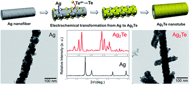
A simple synthetic route for ultra-long silver telluride (Ag2Te) nanotubes is presented. Silver (Ag) nanofibers electro-spun on a plastic substrate could be electrochemically transformed into Ag2Te nanotubes. The electrochemical transformation proceeded through the electrodeposition of tellurium (Te) on the surface of Ag nanofibers, which induces the Ag atoms to diffuse into the layers of Te, and thus leads to a structural and morphological change from nanofibers to hollow nanotubes.

Silver telluride (Ag2Te) is an intriguing phase-change material with a mixed ionic-electronic conductivity in a solid, which exhibits a structural phase transition at 150 °C from a low-temperature monoclinic phase (β-Ag2Te) of a narrow bandgap semiconductor to a high-temperature cubic phase (α-Ag2Te) of a superionic conductor.1,2 The semiconducting and magnetic properties of Ag2Te have been found to include large magnetoresistance, high carrier mobility and low thermal conductivity.3,4 In particular, the monoclinic β-Ag2Te with low thermal conductivity has been concerned as one of promising thermoelectric materials and also employed to be a second-step host material chemically transformed into a series of metal tellurides.4,5 In addition, recent theoretical and experimental studies investigated a topological insulating phenomenon in the β-Ag2Te nanostructures, which consist of a fully filled insulating bulk state and a highly anisotropic massless Dirac cone of two-dimensional surface states from the periodic quantum interference of electron phase.6,7

For enhancement in thermoelectric efficiency of the metal telluride nanostructures, researchers have predicted that one-dimensional (1D) telluride nanostructures, including nanofibers, nanotubes and nanowire-based composites, would be advantageous over their bulk counterparts mainly due to the reduction of lattice thermal conductivity based on the stronger phonon-surface scattering.8,9 Various synthetic routes have been developed to prepare the 1D telluride nanostructures. Yang et al. reported a tellurium-templated chemical bath reaction for a series of metal tellurides with the composition modulation, closely related to the resultant thermoelectric properties.4,10 Electrochemical growth in anodic alumina nanopores yielded single-crystalline Ag2Te nanowire arrays with a narrow diameter distribution.11 In addition, different types of solvothermal methods synthesized a bunch of Ag2Te nanowires based on the topotactic transformation and the composite-hydroxide-mediated redox reaction, respectively.12,13 It has been generally believed that the hollow 1D nanostructures with ultra-long lengths would be beneficial to enhance the thermoelectric properties stemming from the stronger phonon scattering and the coverage of the wider temperature gradients.8,9 However, the synthetic approaches mentioned above have been mostly effective in producing the specific morphology of nanowires with relatively short length up to tens of micrometers. To this end, we previously reported a two-step synthetic method for the ultra-long 1D telluride nanostructures from the hollow nanofibers to the hierarchical nanotubes using a combination of electro-spinning process for the sacrificial nickel (Ni) nanofibers and the following galvanic displacement of Ni nanofibers.14–17

In this work, we report the simpler and more facile electrochemical synthesis of ultra-long Ag2Te hollow nanotubes, which could be directly transformed from the electro-spun Ag nanofibers without the conventional step of the sacrificial Ni nanofibers. The electrodeposition of tellurium (Te) on the surface of Ag nanofibers induced the chemical transformation into the Ag2Te nanotubes with the ultra-long lengths spanning over the substrate. Structural and microscopic study demonstrated the morphological and structural changes during the electrochemical transformation. We believe that this synthetic approach would open an effective way to produce a wide range of 1D metal telluride nanostructures.

Detailed experiments from the electro-spinning of Ag nanofibers to the transformation into the Ag2Te nanotubes are described in the ESI. Silver nitrate/polyvinylpyrrolidon (AgNO3/PVP) composites were first electro-spun and thermally reduced to the Ag nanofiber on a plastic substrate (polyimide) in a reducing condition. Structural transition and morphological change between the AgNO3/PVP and the Ag nanofibers could be confirmed by scanning electron microscopy (SEM) and X-ray diffraction (XRD). As shown in Fig. 1a and b, the diameters of electro-spun AgNO3/PVP nanofibers (125 ± 25 nm) decreases to 85 ± 15 nm in the Ag nanofibers due to evaporation of PVP binder and formation of Ag nanoparticles: the diameters and a standard deviation were counted from SEM images. The thermal reduction of AgNO3/PVP composite leads to a structural change into the Ag phase from the AgNO3 crystallites, as verified in XRD patterns of samples (Fig. 1c). All diffraction peaks in the XRD pattern of Ag nanofibers are indexed to the cubic Ag (JCPDS no. 01-07-4613).


image file: c4ra15688a-f1.tif
Fig. 1 SEM images of (a) AgNO3/PVP nanofibers and (b) Ag nanofibers. (c) XRD pattern indicates the structural change from the AgNO3 crystallites to the Ag phase induced by the thermal reduction.

Microstructure and crystallinity of the Ag nanofibers were observed using transmission electron microscopy (TEM). A bright field TEM image in Fig. 2a visualizes the bumpy surface of Ag nanofiber made of multigrains of Ag nanoparticles, which are nucleated and reduced from AgNO3 crystallites in a PVP polymer matrix. The randomly oriented crystal lattices visualized in a high-resolution TEM (HR-TEM) micrograph show the interplanar spacing of 0.238 nm, corresponding to (111) planes of cubic Ag structure. A ring pattern of selected area electron diffraction (SAED) is displayed in the inset of Fig. 2b. The XRD, HR-TEM and SAED results indicates the polycrystalline nature of as-prepared Ag nanofiber.


image file: c4ra15688a-f2.tif
Fig. 2 (a) TEM image of a typical Ag nanofiber and (b) HR-TEM image of lattice fringes randomly oriented in the Ag nanofiber.

The growth mechanism on the specific morphology of Ag2Te hollow nanofibers can be explained by a well-known Kirkendall effect or topotactic transformation. First, the Kirkendall effect may influence the growth of hollow nanostructure based on a mutual diffusion process with different diffusion coefficients of Ag and Te atoms.18,19 During the reaction, the so-called ‘Kirkendall voids’ are created from the condensation of excess vacancies at the original interface and within the faster diffusion side. For the formation of hollow nanostructure, the Ag nanofibers are coated by the electrodeposited Te layers and the diffusion Ag and Te atoms occur in opposite directions at the different diffusion rates. In order to confirm the Kirkendall effect, we characterized the phases of sample collected at an early stage of reaction using XRD (Fig. S1 in the ESI). Once the Kirkendall effect determines the growth of hollow nanofibers, the product at the early stage should consist of three phases: unreacted Ag inside wall, Ag2Te phase in the mid of nanotubes and the undiffused Te outer wall. However, the XRD result of the sample reacted for 10 m indicates that the product is composed of only two phases of the unreacted Ag and Ag2Te. As a result, the phases identified in this early-staged sample may rule out the Kirkendall effect on the growth of hollow nanofibers because the growth of hollow nanostructures affected by the Kirkendall effect generally follow the growth steps from the early-staged core/shell structure, through the movement of interface between a diffusion couple and to the final hollow morphology.

Instead, the growth mechanism of topotactic transformation appears to be more plausible. Due to the topotactic lattice matching between hexagonal t-Te and metal telluride crystal structures, such a structural conversion system has been reported to be thermodynamically favoured, and thus derive the self-templated transformation of Te nanowires into metal telluride nanowires.4,5 Here, we should note that the specific morphology of metal telluride nanowires, not hollow nanofibers, were chemically transformed from the Te nanowires in a conventional chemical bath reaction.5 This structural and morphological preservation in the chemical transformation from Te nanowires to Ag2Te nanowires may indicate the diffusion of Ag atoms into the layer structure of Te nanowires. In a given contrary case of Te diffusion into Ag structure, the chemical transformation is expected to result in Ag2Te hollow nanofibers from Te nanowires. As a result, in our electrochemical scheme, it is expectable that Te4+ ions in the electrochemical solution reduce to Te on the surface of Ag nanofibers (a working electrode) by applying a negative potential. The Te atoms reduced from Te4+ ions are envisaged to nucleate and grow on the surfaces of Ag nanofibers in the forms of thin layers or porous sheath.15 In parallel with the deposition of Te layers, the Ag atoms from the nanofibers diffuse into the layers of Te and chemically transform into the monoclinic Ag2Te structure. Through this electrochemical reaction, the core Ag nanofibers are sourced and exhausted for the chemical transformation and finally produce the hollow nanotubes. A schematic for the electrochemical transformation through the electrodeposition of Te is depicted in Fig. 3. Cyclic voltammetry was used to monitor the cathodic reduction of Te4+ ions on the working electrode of Ag nanofibers. A scan in an electrochemical solution containing 1 M HNO3 and 10 mM TeO2 indicated that the Te4+ ions reduce at a potential of −0.05 V (Fig. S2 in the ESI).


image file: c4ra15688a-f3.tif
Fig. 3 A schematic of electrochemical transformation from the Ag nanofiber into the Ag2Te nanotubes.

The electrochemical transformation from the Ag nanofibers to the Ag2Te nanotubes resulted in the changes in morphologies and crystal structure. The structure identification of Ag2Te nanotubes using XRD demonstrates the structure transformation from the cubic Ag into the monoclinic Ag2Te, which was electrodeposited for 30 m with an applied potential of −0.05 V (Fig. 4a). The XRD pattern of Ag2Te nanotubes exhibits the diffraction peaks that can be indexed to the crystal phase of monoclinic Ag2Te (JCPDS no. 34-0142, a = 8.16 Å, b = 8.93 Å, c = 8.01 Å) without by-product (e.g., Ag7Te4) or Ag phase. The different reaction periods longer and shorter than 30 m produced the Te-rich Ag4.53Te3 phase and the unreacted Ag phase in the final products, respectively. The structural transformation should be accompanied with the morphological changes in the Ag2Te nanotubes. Being discernible in SEM image (Fig. 4b), the diameters of nanotubes enlarge to 143 ± 21 nm in comparison with those of Ag nanofibers (85 ± 15 nm). The ultra-long morphology of Ag2Te nanotubes in lengths is depicted in Fig. S3 (see the ESI), which is inherited from the electro-spun Ag nanofibers. The low-magnification SEM image (Fig. S3) indicates that the nanotubes were fabricated to span over the substrate with the lengths longer than at least 200 μm and a large aspect ratio higher than 1400. The diameters of as-prepared nanotubes are slightly larger than those of hydrothermally rolled-up lead telluride (PbTe) nanotubes but considerably smaller than those of hydrothermally prepared Ag2Te nanotubes.20,21 However, these hydrothermally produced metal telluride nanotubes were reported to have relatively short lengths up to several micrometers and wide size distributions. In comparison with our previous work on bismuth telluride (Bi2Te3) nanotubes prepared through a combinational route of electrospinning of sacrificial Ni metal and its galvanic displacement,15 the diameters and size distributions of nanotubes fabricated in this work could be notably reduced and improved.


image file: c4ra15688a-f4.tif
Fig. 4 (a) XRD and (B) SEM identification of the Ag2Te nanotubes.

The electrochemical transformation into the nanotubes was further investigated by a TEM technique. Fig. 5a displays a representative TEM image of Ag2Te nanotubes, which are straight in length with rough surfaces. Differences in brightness and contrast apparently visualize the hollow morphology of nanotubes. Chemical composition of nanotubes was studied using energy-dispersive X-ray spectroscopy (EDS). The high-angle annular dark-field scanning TEM (HAADF-STEM) and EDS mapping results indicate the homogeneous distribution of Ag and Te elements within the nanotubes, which signifies the complete electrochemical transformation into the Ag2Te nanotubes (Fig. 5b–e). EDS measurement to check the stoichiometry of nanotubes presents the stoichiometric ratio of 2[thin space (1/6-em)]:[thin space (1/6-em)]1 between the elements of Ag and Te (Fig. 5f). EDS peaks for Cu originates from the TEM grid. The morphological transformation into nanotubes could be also evidenced by the cross-sectional SEM observation (Fig. S4 in the ESI).


image file: c4ra15688a-f5.tif
Fig. 5 TEM study on the Ag2Te nanotubes. (a) TEM image, (b) HAADF-STEM image and (c–e) EDS mapping results of the Ag2Te nanotubes. (f) EDS pattern for the stoichiometry of product.

The complete electrochemical transformation into the Ag2Te nanotubes could be further confirmed through a cross-sectional TEM study. As recognized in the cross-sectional TEM image (Fig. 6a), the nanotube has an apparent hollow morphology. The HR-TEM image (obtained from the area marked in a red circle in Fig. 6a) shows the randomly oriented polycrystalline grains with the interplanar spacing of 0.21, 0.23 and 0.28 nm that correspond to the (200), (212) and (312) planes in monoclinic Ag2Te (Fig. 6b).


image file: c4ra15688a-f6.tif
Fig. 6 (a) A cross-sectional TEM image of nanotube and (b) HR-TEM image representing the crystal lattices of polycrystalline grains in the nanotube.

In summary, we presented a facile and high-throughput synthetic route to produce the ultra-long Ag2Te nanotubes. The structural and microscopic characterization demonstrated that the electro-spun Ag nanofibers are readily and completely transformed into the Ag2Te nanotubes via the post-electrodeposition of Te. This synthetic approach is believed to significantly contribute to direct production of various 1D metal telluride nanostructures. Furthermore, the Ag2Te nanotubes prepared in this work could be a useful host material for the topotactic transformation into a wide range of metal telluride nanotubes, which have great applications from sensors to thermoelectrics.

Acknowledgements

This work was supported by the Industrial Strategic Technology Development Program (10045177, Development of Resistive Ceramic Thin Film using Solution Process and Low Temperature Thin Film Vacuum Getter) and (no. 20112010100100) funded by the MOTIE (Ministry of Trade, Industry, and Energy), Republic of Korea.

Notes and references

  1. R. Dalven and R. Gill, J. Appl. Phys., 1967, 38, 753 CrossRef CAS PubMed .
  2. R. Chen, D. Xu, G. Guo and L. Gui, J. Mater. Chem., 2002, 12, 2435 RSC .
  3. R. Xu, A. Husmann, T. F. Rosenbaum, M. L. Saboungi, J. E. Enderby and P. B. Littlewood, Nature, 1997, 390, 57 CrossRef CAS PubMed .
  4. H. Yang, J.-H. Bahk, T. Day, A. M. S. Mohammed, B. Min, G. Jeffrey Snyder, A. Shakouri and Y. Wu, Nano Lett., 2014, 14, 5398 CrossRef CAS PubMed .
  5. G. D. Moon, S. Ko, Y. Xia and U. Jeong, ACS Nano, 2010, 4, 2307 CrossRef CAS PubMed .
  6. A. Sulaev, P. Ren, B. Xia, Q. H. Lin, T. Yu, C. Qiu, S.-Y. Zhang, M.-Y. Han, Z. P. Li, W. G. Zhu, Q. Wu, Y. P. Feng, L. Shen, S.-Q. Shen and L. Wang, AIP Adv., 2013, 3, 032123 CrossRef PubMed .
  7. S. Lee, J. In, Y. Yoo, Y. Jo, Y. C. Park, H.-J. Kim, H. C. Koo, J. Kim, B. Kim and K. L. Wang, Nano Lett., 2012, 12, 4194 CrossRef CAS PubMed .
  8. R. Yang and G. Chen, Nano Lett., 2005, 5, 1111 CrossRef CAS PubMed .
  9. H. Fang and Y. Wu, J. Mater. Chem. A, 2014, 2, 6004 CAS .
  10. H. Yang, S. W. Finefrock, J. D. Albarracin Caballero and Y. Wu, J. Am. Chem. Soc., 2014, 136, 10242 CrossRef CAS PubMed .
  11. R. Chen, D. Xu, G. Guo and L. Gui, J. Mater. Chem., 2002, 12, 2435 RSC .
  12. F. Xiao, Q. Wang, L. Wnag, J. Pei and N. Zhou, J. Solid State Chem., 2010, 183, 2382 CrossRef CAS PubMed .
  13. F. Li, C. Hu, Y. Ziong, B. Wan, W. Yan and M. Zhang, J. Phys. Chem. C, 2008, 112, 16130 CAS .
  14. F. Xiao, B. Yoo, K. H. Lee and N. V. Myung, J. Am. Chem. Soc., 2007, 129, 10068 CrossRef CAS PubMed .
  15. K.-J. Lee, H. Song, Y.-I. Lee, H. Jung, M. Zhang, Y.-H. Choa and N. V. Myung, Chem. Commun., 2011, 47, 9107 RSC .
  16. H. Park, H. Jung, M. Zhang, C. H. Chang, N. Ndifor-Angwafor, Y.-H. Choa and N. V. Myung, Nanoscale, 2013, 5, 3058 RSC .
  17. C. M. Hangarter, Y.-I. Lee, S. C. Hernandez, Y.-H. Choa and N. V. Myung, Angew. Chem., Int. Ed., 2010, 49, 7081 CrossRef CAS PubMed .
  18. A. D. Smigelskas and E. O. Kirkendall, Trans. AIME, 1947, 171, 130 Search PubMed .
  19. Y. Yin, R. M. Rioux, C. K. Erdonmez, S. Hughes, G. A. Somorjai and A. Paul Alivisatos, Science, 2001, 304, 711 CrossRef PubMed .
  20. L. Zhang, J. C. Yu, M. Mo, L. Wu, K. W. Kwong and Q. Li, Small, 2005, 1, 349 CrossRef CAS PubMed .
  21. A. Qin, Y. Fang, P. Tao, J. Zhang and C. Su, Inorg. Chem., 2007, 46, 7403 CrossRef CAS PubMed .

Footnote

Electronic supplementary information (ESI) available: Experimental and supplementary characterizations. See DOI: 10.1039/c4ra15688a

This journal is © The Royal Society of Chemistry 2015