DOI:
10.1039/C4RA14905J
(Paper)
RSC Adv., 2015,
5, 18041-18050
Abietane diterpenoid of Salvia sahendica Boiss and Buhse potently inhibits MCF-7 breast carcinoma cells by suppression of the PI3K/AKT pathway†
Received
19th November 2014
, Accepted 22nd January 2015
First published on 22nd January 2015
Abstract
In the current study, we report on the bioactive compounds isolated from the roots of Salvia sahendica Boiss and Buhse using bioassay-guided procedures and their biological effects against MCF-7 breast carcinoma cells. In comparison with other solvents, the hexane-based extraction resulted in the most potent anti-cancer activity, and hence it was subjected to more phytochemical fractionation analyses using vacuum liquid chromatography (VLC), reversed-phase high pressure liquid chromatography (HPLC) and NMR spectroscopy. The biological impacts of the isolated pure compounds were evaluated using MTT, DAPI and acridine orange/ethidium bromide staining (AO/EB) assays. Cell cycle analysis was performed to assess the sub-G1 population of the cells treated with the extracted compounds, while the FITC-labeled annexin V assay was used to study the apoptosis profile. The gene expression profile of the treated cells was studied by quantitative PCR, looking at key genes (Caspase 9, Bax, Akt and Bcl-2) involved in apoptosis. Ketoethiopinone (1) and ortho-diacetate aethiopinone (2) compounds were identified using 1H and 13C-NMR. Compounds 1 and 2 showed profound inhibitory impact on the treated MCF-7 cells with IC50 values of 8.6 and 14.2 μg mL−1 at 48 h, respectively. DAPI and AO/EB assays resulted in significant alterations in the nucleus through chromatin remodeling in the treated cells which somewhat impacts the integrity of the cell membrane. An annexin V flow cytometry assay revealed that the cells treated with compound 2 resulted in early and late apoptosis (∼30%). Gene expression profiling demonstrated significant (p < 0.05) changes in the expression of Bcl-2, Caspase 9, Bax and Akt in the cell treated with compound 2 with profound impact on the Bax and Akt pathways. Taken all together, we propose ortho-diacetate aethiopinone as a new class of anticancer agents with great translational potential for clinical uses against solid tumors.
1. Introduction
Pursuant to the report of World Health Organization (WHO), breast cancer is the most common life-threatening malignancy in women, which caused about 13.7% of cancer deaths worldwide in 2008.1
Most patients with breast cancer require chemotherapy after initial surgery and radiotherapy modalities. Although chemotherapy with potent anticancer agents inhibits the cancerous cells proliferation and growth, population of untouched cancerous cells remain, resulting in recurrence of the disease. Furthermore, these cells often show resistance to chemotherapies via various mechanisms, which demands administration of different anticancer agents. In fact, the majority of patients need multiple lines of therapy or alteration in the treatment protocol because of the occurrence of such resistance to the chemotherapeutic agents. Resistance to the currently used chemotherapeutics in combating the breast cancer has highlighted the demands for novel anticancer agents, perhaps with minimal side effects yet maximal effectiveness against malignant cells.
Of various classes of anticancer agents, natural products such as sesquiterpenes,2 steroids, polysaccharides, flavonoids, terpenoids and alkaloids have been the main source for development of a number of clinically important anticancer agents such as vincristine, vinblastine and paclitaxel.
Salvia genus is the most common member of the Labiatae (Lamiaceae) family. It features conspicuously in the pharmacopoeias of different countries from the Far East to Europe. Different Salvia species have been used in a number of medical applications such as aromatization. Salvia species, especially Salvia miltiorrhiza, are considered as a source of anticancer compounds.3
Of a large number of Salvia species dispensed worldwide, almost 20 species are endemic of Iran.4 Salvia sahendica Boiss and Buhse is a known medicinal species of the Iran's Azerbaijan flour that the species names is given from its origin mountain; “Sahand”. It has been traditionally used as antifungal and antibacterial herbal medicine, in addition to its application for management of dyspepsia.5 Furthermore, various extracts of S. sahendica were found to impose anti-proliferative effect on the human melanoma and pancreatic cancer.3
Few studies have been reported upon phytochemical constituents extracted from different part of S. sahendica. For example, the extraction of sesquiterpene methylester, sclareol and salvigenin from the aerial parts has recently been reported.6 Further, Jassbi et al. and Fronza et al. reported on the extraction of abietane diterpenoids (ferruginol and sahandinone) from the root parts of the plant.3,7 Beside, sahandinone, prionitin and horminon have been detected in the root of S. sahendica.7 Some other important compounds have also been isolated from the root of S. sahandica, including: (a) sesterterpene 8α-hydroxy-13-hydroperoxylabd-14,17-dien-19,16;23,6α-diolide, (b) salvileucolide-6,23-lactone, (c) norsesterterpene 17,18,19,20-tetranor-13-epi-manoyloxide-14-en-16-oic acid-23,6α olide, (d) norambreinolide-18,6α-olide, and (e) 8α-acetoxy-13,14,15,16-tetranorlabdan-12-oic acid-18,6α-olide.8
All these studies have highlighted the importance of S. sahandica as a source for some key compounds, however little is known about their biological activities in malignancies. Here in the current study, for the first time, we report on a bioassay-guided isolation and characterization of bioactive compounds of S. sahandica that imposed remarkable inhibitory effects on the human breast cancer cells.
2. Experimental
2.1. Material
MCF-7 cell line was purchased from Pasteur Institute (Tehran, Iran). RPMI Medium 1640, FBS, streptomycin and penicillin were provided from Gibco Invitrogen Corporation (Gibco, Invitrogen, UK). Pipettes, tissue culture flasks, 96 well plates, trypan-blue, and MTT were from Sigma-Aldrich (Sigma-Aldrich Co., Ltd., UK). RNX plus lysis buffer was purchased from CinnaGen (CinnaGen, Tehran, Iran). For the cDNA synthesis, Reverta-L reagent kit was used (Inter LabService, Russia). Hot Taq EvaGreen® qPCR Mix was used for the real time PCR (SinaClon Co., Tehran, Iran). DMSO and DAPI were from Merck (Darmstadt, Germany) and diethylpyrocarbonate (DEPC), Triton-X100 was purchased from Sigma-Aldrich Chemical Co. (Poole, UK). Annexin V-FITC Kit, propidium iodide (PI), acridine orange and ethidium bromide were obtained from eBioscience (CA, San diego, USA). All other materials that are not mentioned were from Stratagene (La Jolla, CA, USA) or Fermentas Life Science (Burlington, Canada).
2.2. Plant material
Root parts of Salvia sahendica Boiss and Buhse, were gathered from the mountains of Tabriz-Basminj road, Iran in spring, 2012. The plant was identified by Professor Hossein Nazemiyeh, Head of the Herbarium at Tabriz University of Medical Sciences (TUOMS) and a voucher specimen (Tbz-FPh 736) representing the collection was deposited in the Herbarium at TUOMS, Tabriz, Iran. The plant root parts were dried at room temperature while it was protected from direct sunlight. Then, they were comminuted and kept in the closed containers at 2–8 °C.
2.3. Preparation of the extractions
Air dried powdered plant (200 g) were consecutively extracted by soxhlet using organic solvents including n-hexane (Hex), dichloromethane (DCM) and methanol. All the extraction solvents were evaporated in vacuo by rotary evaporator at ambient temperature. Anti-proliferate properties of the extracts were evaluated using MTT cytotoxicity assay. The hexane-based extraction showed the highest cytotoxic effect, and hence was subjected to further fractionation using VLC.
2.4. Compounds isolation and identification
The Hex extract (3 g) was fractionated using VLC on a stationary phase of Merck Silica gel 60 GF254, eluting with a gradient admixture of organic solvents including: Hex
:
acetone (98
:
2, 96
:
4, 92
:
8, 90
:
10; 200 mL each), Hex
:
acetone (80
:
20, 60
:
40, 40
:
60, 20
:
80, 0
:
100; 400 mL each), and finally acetone
:
methanol (60
:
40, 50
:
50, 40
:
60, 30
:
70, 20
:
80, 10
:
90, 0
:
100; 300 mL each). The vacuum chromatography was repeated for 3 times to get enough amount of each fraction. The solvents of fractions were then removed under the circumstance of low pressure at 40 °C. The yielded fractions were subjected to MTT assay, and the fractions with a dominant anti-proliferate activity (eluted by 92–8%, 80–20%; Hex
:
acetone) were further evaluated and fractionated using appropriate HPLC procedures.
2.5. Preparative HPLC
The fractions obtained by VLC were screened towards their cytotoxic impacts on the cultivated cells. Then, the designated fractions with the highest cytotoxic impacts were further isolated by preparative HPLC eluted with a linear gradient of acetonitrile (ACN)/water and monitored using a photo-diode-array detector at a range of 190 to 400 nm. For purification of 92
:
8 (Hex
:
acetone) fraction the most suitable HPLC program was set as system A, i.e. mobile phase: 0–50 min, ACN from 70 to 90% in H2O; 50–55 min, 90% ACN in H2O; 55–56 min, ACN from 90 to 70% in H2O; 56–62 min ACN 70% in H2O, flow rate 20 mL min−1. For 80
:
20 (Hex
:
acetone) fraction system B was developed as follows: mobile phase: 0–30 min, ACN from 60 to 70% in H2O; 30–35 min, 70% ACN in H2O; 35–45 min, ACN from 70 to 90% in H2O; 45–50 min ACN 90% in H2O; 50–51 min, ACN from 90 to 60% in H2O; 51–60 min, ACN 60% in H2O, flow rate 20 mL min−1. Then, the solvents of eluted fractions were removed by the rotary evaporator in vacuo. All the collected sub-fractions were monitored on TLC plates and the similar compounds were integrated. Once again, the cytotoxicity effects of the fractions were evaluated by MTT assay and the most potent anti-proliferate fractions were selected for the chemical structure determination and further biological investigations.
2.6. Determining the chemical structures
The structure of purified compounds were elucidated by UV-visible, 1H-NMR and 13C-NMR spectroscopy techniques. For 1H-NMR and 13C-NMR spectroscopy the sufficient amount of yielded compounds were dissolved in deuterated chloroform.
2.7. Cell culture and treatments
MCF-7 cells were cultured in Roswell Park Memorial Institute medium (RPMI) 1640 medium, containing 1% penicillin/streptomycin and 10% FBS in a humidified incubator (5% CO2–95% air atmosphere) at 37 °C. Various concentrations of the compounds ranging from 5–100 μg mL−1 were prepared in RPMI containing DMSO as co-solvent (not more than 0.3%) and 10% FBS. Subsequently, prior to treatments the serial dilutions were sterilized by filtration methods using 0.22 μm syringe filter (JET BIOFIL, Interlab Ltd, New Zealand).
2.8. Cell viability
MTT cytotoxicity assay is frequently used to measure the cell proliferation/viability and the mitochondrial activity. Mitochondrial NAD(P)H-dependent cellular oxidoreductase enzymes may reflect the number of viable cells present. These enzymes are capable of reducing the tetrazolium dye MTT 3-(4,5-dimethylthiazol-2-yl)-2,5-diphenyltetrazolium bromide to its DMSO soluble formazan, which has a purple color. Thus, UV absorbance of the respective color may effectively show the extent of the viable cells. The MTT assay was performed for the cytotoxicity evaluation of the extracts, fractions and pure compounds in MCF-7 cells as reported previously.9,10 In this study, cells were cultivated at a density of 3.0 × 10⁴ cells per cm2 in 96-well plates and incubated at 37 °C in a humidified atmosphere and allowed to attach overnight. At 70% confluency, the medium was substituted with a designated amount of the selected compound (as 200 μL of 5–100 mg mL−1) and the cells were incubated over different time periods (i.e., 24, 48 and 72 h). After designated incubation period, 20 μL per well of MTT solution in PBS (5 mg mL−1, pH 7.4) was added and the cells were incubated at 37 °C for 4 h in dark. Thereafter, the media/MTT mixture was replaced by 200 μL of DMSO containing 25 μL Sorenson's glycine buffer (0.1 M NaCl, 0.1 M glycine, pH 10.5). The absorbance of dissolved formazan crystals was determined spectrophotometrically at a wavelength of 570 nm using a Biotek microplate reader (BioTek Instruments, Friedrichshall, Germany).
2.9. Cell morphology and nuclear staining
2.9.1. Morphological assessment. After incubation with compounds for 24 h, the cells were monitored for any morphological alternations using Olympus IX81 fluorescence microscope (Olympus optical Co., Ltd. Tokyo, Japan) equipped with XM10 monochrome camera (Olympus, Hamburg, Germany).
2.9.2. DAPI staining. For the nuclei condensation and fragmentation studies, the treated and untreated cells after 24 h incubation were fixed in 4% paraformaldehyde for 2 h, washed with PBS and then stained by DAPI.11,12 After washing with PBS, the cells were permeabilized using 0.1% Triton-X-100 for 5 min. Afterword, the cells were exposed to DAPI (final concentration 0.2 mg mL−1) in darkness for 5 min. Finally, using fluorescent microscopy, the morphology of the cells were investigated for possible changes in the pattern of nucleus and the remodeling of chromatin.
2.9.3. Acridine orange and ethidium bromide staining. Apoptosis occurrence was further verified, morphologically after staining the cells with acridine orange and ethidium bromide (AO/EB) by fluorescence microscopy as described previously.13 Briefly, after 24 h incubation of the MCF-7 with different compounds, treated cells, were rinsed with PBS and exposed to the 50 μL of acridine orange/ethidium bromide solution (100 μg mL−1 of acridine orange and 100 μg mL−1 of ethidium bromide in PBS). Microscopic analyses were performed subsequent staining followed by a brief washing.
2.10. Apoptosis detection and quantification
2.10.1. Cell cycle analysis. Cell cycle analysis was performed to assess the sub-G1 population of the cells treated with 20 μg mL−1 of the compounds for 24 h. Briefly, the cells were harvested with trypsin, centrifuged and washed (3×) with PBS. The cells were then resuspended and fixed with 1.0 mL ice cold ethanol (70%), and the samples were stored at 4 °C for 30 min. Then they were washed with PBS (3×) by centrifugation at 850 × g. To avoid the inadvertent staining of ds RNA and also to solely stain DNA, the cells were treated with 50 μL ribonuclease A at 37 °C for 30 min. Next, the samples were washed and stained with propidium iodide at the final concentration of 5 μg mL−1 dissolved in PBS. Flow cytometry analysis was carried out for 10
000 events per sample through FL2-A band-pass filter (propidium iodide) using Becton Dickinson (BD) fluorescence-activated cell sorting (FACS) flow cytometer, FACScalibar (San Jose, CA, USA). To analyze the fluorescence of the cell population(s), we used the freely available WinMDI 2.8 software (http://facs.scripps.edu/).
2.10.2. Annexin V detection of apoptosis. To find out the stage of the apoptosis/necrosis in the treated cells, the Annexin V flow cytometry analysis was performed as described previously.14,15 It should be stated that the annexin V is a phospholipid-binding protein with high affinity for phosphatidylserine, which translocate from the inner sheet to the external cell surface concurrent with early apoptosis event. In this study, annexin V-fluorescein isothiocyanate (FITC) apoptosis detection kit (BD science) was used following the manufacturer's protocol. Briefly, the treated cells were detached by gentle trypsinization and a total of 1.5 × 10⁶ cells were washed (2×) with 1× binding buffer. Then, the cells were resuspended in 100 μL binding buffer containing 5 μL annexin V. Subsequent to incubation in the dark at room temperature for 20 min, 5 μL PI were added to the samples, which were analyzed in comparison with the untreated cells as negative control using BD FACScalibur flow cytometer (San Jose, CA, USA) and WinMDI 2.8 software.
2.11. Quantitative PCR
The cultivated cells treated with compound 2 (60 μg mL−1 for 24 h) were further subjected to the gene expression profiling. Total RNA was extracted by RNXplus lysis buffer according to the manufacturers' protocol.16 The quantity and quality of the isolated RNA was evaluated using a NanoDrop® ND-1000 Spectrophotometer (Nano-Drop Technologies, Wilmington, DE, USA) and RNA gel electrophoresis. The reverse transcription reaction was carried out according to manufacturer's protocol. Briefly, 10 μL of RNA-samples (1 μg μl−1) were added to the appropriate test tube containing 10 μL of ready-to-use reagent mix incubated at 37 °C for 30 min using an Astec thermal cycler PC-818 (Astec, Fukuoka, Japan).
The qPCR reactions were carried out in a total volume of 20 μL using Bio-Rad iQ5 multicolor thermal cycler (Bio-Rad, Inc., Hercules, CA, USA). Each well contained: 1 μL primer (10 pmol μL−1 each primer) (Table 1), 1 μL cDNA, 4 μL of 5× HotTaq EvaGreen qPCR Mix and 16 μL DNAse/RNAse free DEPC treated water. The thermal cycling conditions for the real time PCR were as following: 94 °C for 10 min, 40 cycles of 95 °C for 15 s, 55–61 °C for 1 min, and 72 °C for 30 s.
Table 1 Primer's sequence for the genes studied
| Primer |
Primer sequence |
Gene bank accession no. |
Annealing (Tm) |
| 18S rRNA |
F: 5′-CGATGCGGCGGCGTTATTC-3′ |
NR_003286.1 |
61 |
| R: 5′-TCTGTCAATCCTGTCCGTGTCC-3′ |
| Bcl-2 |
F: 5′-CATCAGGAAGGCTAGAGTTACC-3′ |
NM_000633.2 |
56 |
| R: 5′-CAGACATTCGGAGACCACAC-3′ |
| Caspase 9 |
F: 5′-TGCTGCGTGGTGGTCATTCTC-3′ |
NM_001229.2 |
62 |
| R: 5′-CCGACACAGGGCATCCATCTG-3′ |
| Akt |
F: 5′-CGCAGTGCCAGCTGATGAAG-3′ |
NM_005163.2 |
62 |
| R: 5′-GTCCATCTCCTCCTCCTCCTG-3′ |
| Bax |
F: 5′-AAGCTGAGCGAGTGTCTCAAGCGC-3′ |
NR_027882 |
53 |
| R: 5′-TCCCGCCACAAAGATGGTCACG-3′ |
Analyses of the results were performed by the Pfaffl technique and the closure times (CTs) were normalized to the expression of 18S rRNA as a housekeeping gene.17 All reactions were accomplished as triplicates with internal negative controls.
2.12. Statistical analysis
Data obtained from the assays were analyzed by either Student's t-test or One-Way ANOVA using SPSS 11.0 software (Statistical Package for the Social Sciences 11.0) followed by a post-hoc multiple comparison analysis. A p value less than 0.05 was considered for the statistical significance. Data presented in this study are replicative of 3–4 experiments.
3. Results and discussion
3.1. Determining the chemical structures
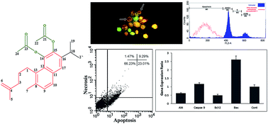
This study was planned to evaluate the bioactive compounds isolated from the root of S. sahendica. To pursue this aim, a bioassay-guided isolation and fractionation platform was recruited. The purified compounds were characterized by UV/vis, and lH- and 13C-NMR spectroscopies, and also compared with previously reported structures. As shown in Fig. 1, the phytochemical analyses of the compounds led to the isolation of two abietane diterpene compounds (ketoethiopinone (1) and ortho-diacetate aethiopinone (2)) which showed anti-proliferative properties.
 |
| | Fig. 1 Structures of the isolated compounds from the n-hexane extract of S. sahendica roots. | |
Ketoethiopinone (1) is a known abietane diterpene that has been recognized and elucidated from the roots of Salvia aethiopis18 and Salvia argentea.19 To the best of our knowledge, this is the first report on ketoethiopinone existence in the roots of S. sahendica. Using preparative HPLC separation method, an amorphous red residue was obtained after evaporation of the excess solvents in vacuo. The compound (1) displayed λmax (online) at 244, and 337 nm which were in well-consensus with the presence of an orthoquinone moiety.18 In the first evaluation, according to the 13C-NMR results together with the number of detected carbon, a diterpene structure has been proposed for (1). Occurrence of three characteristic peaks at δ 200.72, 184.42 and 182.64 proposed three carbonyl groups in the structure of (1). Also, existence of a typical peak at δ 110.64 in C-NMR and δ 4.89 (2H, bs, H6) in 1H-NMR suggested one exocyclic double bond. 1H-NMR spectrum of (1) also showed the presence of one aromatic methyl group at δ 2.23 (3H, s, H7), an isopropyl group with signals at δ 1.11 (6H, d, J = 6.74, H19–20) and a methine septet at δ 2.98 (H, m, H18).
ortho-Diacetate aethiopinone (2) has previously been reported by Boya et al.18 However, there is no sufficient evidence in relation with compound (2) elucidation from the other sources. To the best of our knowledge, our study is the first report on the presence of ortho-diacetate aethiopinone in the root of S. sahendica. The 1H-NMR spectrum of (2) preserved the same pattern of aromatic signals observed for compound (1). The remaining signals were the same as ketoethiopinone, with some exception upon two more aliphatic methyl groups that appeared as singlet peaks in δ 2.33 (3H, s, H24) and 2.31 (3H, s, H22). Also, a prominent peak corresponding the carbonyl group in the 13C-NMR spectrum of (1) was not observed in the spectrum of (2). Further, in compound (2), two carbonyl groups were observed as characteristic peaks in δ 181.18 (C21), 180.31 (C23), which seemed to be shifted towards the low magnetic field as compared to (1). Beside, as compared to the 13C-NMR data of (1), there were two more aromatic carbon signals in δ 134.15 and 146.91 in compound (2), respectively corresponding the (C14) and (C15).
3.2. Chromatographic and spectroscopic data
3.2.1. Ketoethiopinone (1). Red amorphous solid; Rt: 7.30 min (purified by the system A chromatography); 1H-NMR (CHCl3-d4, δ/ppm, J/Hz): 7.06 (1H, d, J = 8.3, H10), 6.97 (1H, d, J = 8.5, H9), 6.89 (H, s, H17), 4.89 (2H, bs, H6), 2.91–2.98 (H, m, H18), 2.59–2.70 (2H, m, H2), 2.23 (3H, s, H7), 2.02 (3H, s, H5), 1.80–1.86 (2H, m, H3), 1.11 (6H, d, J = 6.74, H19–20). 13C-NMR (CHCl3-d4, δ/ppm, J/Hz): 200.72 (C1), 184.42 (C14), 182.64 (C15), 149.75 (C4), 148.17 (C9), 140.00 (C8), 138.22 (C16), 136.26 (C13), 134.56 (C11), 129.10 (C10), 125.51 (C17), 110.64 (C6), 44.77 (C3), 44.68 (C2), 24.94 (C18), 20.48 (C5), 20.34 (C7), 18.71 (C19–C20).
3.2.2. ortho-Diacetate aethiopinone (2). Red gum; Rt: 9.25 min (purified by the system B chromatography); 1H-NMR (CHCl3-d4, δ/ppm, J/Hz): 7.3 (1H, d, J = 7.64, H10), 7.05 (1H, s, H17), 6.99 (1H, dd, J = 7.59, 1.3, H9), 4.67 (2H, br s, H6), 2.91–3.01 (3H, m, Hl, H18), 2.33 (3H, s, H24), 2.31 (3H, s, H22), 2.08–2.20 (2H, m, H3), 1.66–1.77 (2H, m, H2), 1.58 (3H, s, H5), 1.11 (6H, d, J = 6.8, H19, H20). 13C-NMR (CHCl3-d4, δ/ppm, J/Hz): 181.18 (C21), 180.31 (C23), 147.47 (C4), 146.91 (C15), 144.43 (C13), 143.25 (C16), 139.05 (C8), 135.56 (C10), 134.15 (C14), 131.14 (C11), 126.90 (C17), 122.61 (C12), 118.16 (C9), 108.97 (C6) 37.23 (C3), 29.30 (C1), 25.65 (C2), 25.40 (C18), 24.60 (C19–C20), 21.36 (C5), 21.25 (C22), 18.77 (C7), 16.44 (C24).
3.3. Cytotoxic effects on MCF-7 cells
The cytotoxic effects of the compounds on MCF-7 cells were evaluated by MTT assay. As shown in Fig. 2, compounds (1) and (2) were able to induce cytotoxicity in the treated cells in a time- and dose-dependent manner, which respectively resulted in the IC50 values of ∼8.6 and 14.2 μg mL−1 at 48 h (Table 2). Furthermore, the light microscopic visualization illustrated that the treated cells displayed distinct morphologic alterations in comparison with the normal untreated cells in the appearance and the number of cells (Fig. 3).
 |
| | Fig. 2 In vitro cytotoxicity of (A) ketoethiopinone (1), (B) ortho-diacetate aethipinone (2) extracted from S. sahendica and (C) doxorubicin (Dox) in MCF-7 cells. Data represent cell viability of cells exposed to the demonstrated concentration (5–100 μg mL−1) for 24, 48 and 72 h. Significant differences in cell viability were observed after different times with 10 μg mL−1 of ketoethiopinone (1) and with 10 and 20 μg mL−1 of ortho-diacetate aethipinone (2). | |
Table 2 IC50 values for ketoethiopinone (1) and ortho-diacetate aethiopinone (2)
| Exposure time |
Ketoethiopinone (1) |
ortho-Diacetate aethiopinone (2) |
Doxorubicin |
| 24 h |
10 μg mL−1 |
21 μg mL−1 |
70 μg mL−1 |
| 48 h |
8.6 μg mL−1 |
14.2 μg mL−1 |
42 μg mL−1 |
| 72 h |
5.9 μg mL−1 |
7.4 μg mL−1 |
38 μg mL−1 |
 |
| | Fig. 3 Light microscopy and DAPI staining for nuclei condensation and fragmentation assessment (magnification 200×): (A and B) untreated control MCF-7 displaying normal epithelial morphology, (C and D) 24 h after exposure to 20 μg mL−1 of ketoethiopinone (1), (E and F) 24 h after exposure to 20 μg mL−1 of ortho-diacetate aethiopinone (2), and (G and H) 24 h after exposure to the 5% DMSO as a positive control. Control cells possess normal nuclear morphology, whereas apoptotic cells showed clear morphological changes such as nuclear fragmentation and chromatin condensation (represented by white arrows). | |
3.4. DAPI staining assay and AO/EB staining assay
Due to the direct interaction of plant-derived cytotoxic compounds with the cellular compartments, we expected to see some inadvertent biological alterations such as chromatin remodeling and detrimental impacts in cell membrane and nucleus of the treated cells. There are a number of investigations which have used DAPI staining and AO/EB assays to study the cellular impacts of natural products or synthetic compounds.20,21 In this investigation to reveal the cytotoxicity of the compounds, DAPI staining and AO/EB assays were utilized to assess possible remodeling of chromatin and nuclear fragmentation. Throughout these techniques, a significant nuclear fragmentation and chromatin condensation were observed in the MCF-7 cells treated with the compounds (1) and (2).
Fig. 3 represents the fluorescence microscopy micrographs of the DAPI-stained cells after exposure to 20 μg mL−1 of (1) and (2), as well as 5% DMSO (positive control).22,23 It seems that the apoptotic cells are principally detected in the positive control, as well as compounds (1) and (2) treated cells. All treatments caused a statistically significant nucleus fragmentation and condensation in the chromatin and DNA, nevertheless their morphology was not altered in the untreated control cells.24
We also surveyed the viability of the treated cells by staining the cells with the fluorochromes AO/EB (Fig. 4). Live cells are not permeable to EB, yet permeable to AO. Hence, in the viable cells the interaction of AO dye with the DNA can produce green nuclear fluorescence. As shown in Fig. 4, the apoptotic cells revealed yellow chromatin in fragmented and condensed nucleus, however the necrotic cells appeared to have red nucleus, indicative of the interaction of EB dye with DNA in damaged cells. Treatment with (1) and (2) compounds (20 μg mL−1) for 24 h appeared to increase the percentage of nonviable cells. Compound (2) considerably increased the number of apoptotic cells in the MCF-7 cells (plane B). However, in the case of compound (1), markedly higher level of necrosis was observed as compared to the untreated control cells.
 |
| | Fig. 4 Apoptotic morphological variations of MCF-7 cells identified with AO/EB staining and observed under fluorescence microscope (magnification 200×): (UT) untreated control MCF-7 displaying normal epithelial morphology, (A) 24 h after exposure to 20 μg mL−1 of ketoethiopinone (1), (B) 24 h after exposure to 20 μg mL−1 of ortho-diacetate aethiopinone (2), (C) 24 h after exposure to the 5% DMSO as a positive control. The viable cell possess unchanged green nuclear, apoptotic cells have bright green-orange areas of fragmented or condensed chromatin in the nuclear, and the necrotic cells have uniform bright red nuclear. White empty arrows show the apoptotic cells and white filled arrows indicate the necrotic cells. | |
3.5. Cell cycle analysis
Cell cycle arrest analysis also displayed the interaction of the compounds with DNA. Any cleavage in the chromosome at the inter-nucleosomal sites might lead to the activation of proteins that contributed in the regulation of the checkpoints in the cell cycle. It should be noted that the cell cycle arrest has already been reported as the main biochemical signs of the apoptotic cell death.25 In order to test whether the isolated compounds can cause cell cycle arrest, we used propidium iodide (PI) for the staining of double strand DNA whose levels are elevated in G0/G1 and G2/M. We followed sub-G0 population of the cells representing the fragmented ds DNA and condensed chromatin as the sign for occurrence of apoptosis. We compared the effects of (1) and (2) compounds in the treated cells in comparison with the untreated cells. Both compounds (1) and (2) appeared to exhibit similar patterns of cell cycle arrest. The effects of the compounds on the cell cycle modulation are shown in Fig. 5. The compound (1) exhibited a higher toxicity in cell viability assay, however the count of cells with fragmented DNA was slightly lower than that of the compound (2). Therefore, we postulate that the compounds could induce pyknosis and karyopyknosis, which are the irreversible chromatin condensation and ds DNA strand breakages in the nucleus undergoing apoptosis26 or necrosis.27 The cells treated with compound (2) seems to be associated with a sharp sub-G1 apoptotic peak (Fig. 5C), which may confer compound (2) to be an apoptosis promoting entity in the cells. Similarly, the compound (1) treated cells were found to cause a sub-G1 peak in the MCF-7 cells, even though the cell population count appears to be subordinated in association with a marked reduction of the sub-G1 peak.
 |
| | Fig. 5 Cell cycle analysis of MCF-7 cells treated with (A) 5% DMSO as positive control, (B) 20 μg mL−1 of ketoethiopinone (1), (C) 20 μg mL−1 of ortho-diacetate aethiopinone (2) for 24 h, analyzed by FACS flow cytometry for the distribution of cells in different phases of cell cycle. | |
3.6. Apoptosis detection using annexin V staining
Specific staining, using annexin V-FITC/PI flow cytometry, was performed to differentiate the necrotic cells form the apoptotic cells. It should be pointed out that annexin V is a cellular protein with a high affinity for phosphatidylserine (Ptd-L-Ser) in the presence of calcium ion, which identifies the alteration of Ptd-L-Ser on the outer leaflet of the plasma membrane as an early distinctive of apoptotic cells when labeled with a fluorescent probe.28,29 In our experiments, the FITC-labeled annexin V flow cytometry analyses confirmed the occurrence of apoptosis stages in the MCF-7 cells treated with compound (2) (Fig. 6). These results revealed that more than 30% of (2)-treated cells underwent the apoptotic stage, while 2% of the cells underwent the necrotic stage after 24 h (Fig. 6). The results obtained from FACScalibur disclosed that the compound (2) could cause cell death, in large part because of the activation of apoptosis pathway(s). We also speculate that compound (2) may have pro-apoptotic properties in the treated dosage, and cell death prompted by the compound may be associated with the activation of apoptosis pathways similar to previous reports.30 These data enabled us to distinguish the apoptotic cells from the necrotic and/or living cells.31
 |
| | Fig. 6 FITC-labeled Annexin V flow cytometric detection of apoptosis (A) MCF-7 cells treated with 20 μg mL−1 of ortho-diacetate aethiopinone (2) for 24 h, (B) untreated MCF-7 cells, and (C) MCF-7 cells exposed to the 5% DMSO as a positive control for 24 h. Considerable late stages of apoptosis were detected in the cells treated with ortho-diacetate aethiopinone compared to untreated cells (p < 0.05). | |
3.7. Quantitative real-time PCR
Finally, we looked at various gene expression profiles known to be involved in apoptosis, in which the death signals are afforded after a chemical treatment in the cells directing to liberation of mitochondrial factors such as small mitochondria-derived activator of caspases (SMACs) into the cytosol.32,33 The intracellular apoptotic pathway can be regulated with the help of intracellular signals, which puts forward the cell in programmed death. An alteration in the permeability of the mitochondrial membrane can obligate the apoptotic proteins to release into the cell. It seems that some pores known as the mitochondrial outer membrane permeabilization pores (MACs) can control the permeability of the membrane to the apoptotic proteins. Proteins belonging to the Bcl-2 family can control the MACs.34 The activated Bax protein (Bcl-2-associated X protein) dimerizes in the mitochondrial membrane. This dimerization stimulates the MAC pore development, causing apoptotic leakage of proteins into the cytosol. In contrast, the proteins Bcl-2 and Mcl-1 can inhibit the MAC creation, suppressing the release of apoptotic proteins into the cell.35 The SMACs can bind to the inhibitors of apoptosis (IAP), which activates caspases in the cell.36 Caspases are enzymes that can damage intracellular proteins, which finally leads to the entire cell death. In this study, we studied the expression of some of these genes (Bcl-2, Akt, Caspase 9 and Bax) and found a significant regulation in the gene expression profile of the treated cells with compound (2) after 24 h. As illustrated in Fig. 7, the expression of Caspase 9 gene was not significantly changed by the compound (2). Moreover, the expression of Akt and Bcl-2 genes were significantly down-regulated in comparison with the untreated MCF-7 cells.
 |
| | Fig. 7 Gene expression ratios of Akt, Caspase 9, Bcl-2 and Bax in the treated cells with ortho-diacetate aethiopinone, untreated control MCF-7 cells. *Represent significant differences between defined group (p < 0.05). | |
However, there was a significant up-regulation in the Bax gene (Akt's downstream gene) in the cells treated with the compound (2) after 24 h, which was not amazing due to the down-regulation of Akt. We speculate that the initiation of apoptosis in the treated cells by ortho-diacetate aethiopinone may be through PI3K/AKT pathway that is a known pathway involves in breast cancer.37
4. Conclusion
Taken all, the current study showed that the two abietane diterpene extracted from S. sahendica inhibited the growth of MCF-7 cells in a time and dose-dependent manner and persuaded cytotoxicity via inducing apoptosis and necrosis. It was found that ketoethiopinone (1) and ortho-diacetate aethiopinone (2) are able to inhibit the proliferation of the MCF-7 cells by stimulating apoptosis via DNA and chromatin fragmentation. We also showed the incidence of early/late stages of apoptosis within MCF-7 cells treated with compound (2) by FITC-labeled annexin V flow cytometry and nuclear staining assays. Furthermore, using the cell cycle arrest and DNA fragmentation assays, significant fragmentation of DNA were found in the treated cells with compound (2). Technically, significant decreases in Akt, Bcl-2 expressions and an increase in Bax expression may lead us towards possible involvement of the PI3K/AKT pathway in the modulation of MCF-7 cells proliferation by the compound (2). In conclusion, all the data presented pinpointed that ortho-diacetate aethiopinone is able to elicit profound cytotoxic impacts in the MCF-7 cells. We envision this compound as potential candidate for further translational/clinical studies that may provide a novel chemotherapy agent to tackle the breast cancer and perhaps other types of solid tumors.
Disclosure of interest
The authors declare no conflicts of interest concerning this article.
Abbreviations
| NMR | Nuclear magnetic resonance |
| MCF-7 | Michigan cancer foundation-7 |
| DMSO | Dimethyl sulfoxide |
| MTT | 3-[4,5-Dimethylthiazol-2-yl]-2,5-diphenyltetrazolium bromide |
| DAPI | 4′,6-Diamidino-2-phenylindole |
| PBS | Phosphate buffered saline |
| FBS | Fetal bovine serum |
| RT-PCR | Reverse transcription and real time polymerase chain reactions |
| MMLV-rt | Moloney murine leukemia virus reverse-transcriptase |
Acknowledgements
This work was supported by the Research Center for Pharmaceutical Nanotechnology (RCPN) at Tabriz University of Medical Sciences (grant no.: 90011, which is a part of PhD thesis no.: 90/011/101/1).
References
- D. S. Alberts, K. S. Griffith, G. E. Goodman, T. S. Herman and E. Murray, Cancer Chemother. Pharmacol., 1980, 5, 11–15 CrossRef CAS.
- M. Eskandani, J. Abdolalizadeh, H. Hamishehkar, H. Nazemiyeh and J. Barar, Fitoterapia, 2015, 101, 1–11 CrossRef CAS PubMed.
- M. Fronza, R. Murillo, S. Ślusarczyk, M. Adams, M. Hamburger, B. Heinzmann, S. Laufer and I. Merfort, Bioorg. Med. Chem., 2011, 19, 4876–4881 CrossRef CAS PubMed.
- M. Alikhani, A. Rezwani, E. Rakhshani and S. Madani, J. Entomol. Res., 2010, 2, 7–16 Search PubMed.
- F. Lotfipour, M. Samiee and H. Nazemiyeh, Pharmaceut. Sci., 2007, 1, 3 Search PubMed.
- F. M. Moghaddam, B. Zaynizadeh and P. Rüedi, Phytochemistry, 1995, 39, 715–716 CrossRef CAS.
- A. R. Jassbi, M. Mehrdad, F. Eghtesadi, S. N. Ebrahimi and I. T. Baldwin, Chem. Biodiversity, 2006, 3, 916–922 CAS.
- F. M. Moghaddam, M. M. Farimani, M. Seirafi, S. Taheri, H. R. Khavasi, J. Sendker, P. Proksch, V. Wray and R. Edrada, J. Nat. Prod., 2010, 73, 1601–1605 CrossRef CAS PubMed.
- M. Heidari Majd, D. Asgari, J. Barar, H. Valizadeh, V. Kafil, G. Coukos and Y. Omidi, J. Drug Targeting, 2013, 1–13 Search PubMed.
- M. Eskandani and H. Nazemiyeh, Eur. J. Pharm. Sci., 2014, 59, 49–57 CrossRef CAS PubMed.
- S. Vandghanooni, A. Forouharmehr, M. Eskandani, A. Barzegari, V. Kafil, S. Kashanian and J. Ezzati Nazhad Dolatabadi, DNA Cell Biol., 2013, 32, 98–103 CrossRef CAS PubMed.
- M. Eskandani, H. Hamishehkar and J. Ezzati Nazhad Dolatabadi, Food Chem., 2014, 153, 315–320 CrossRef CAS PubMed.
- M. Deepaa, T. Sureshkumara, P. K. Satheeshkumara and S. Priya, Chem.–Biol. Interact., 2012, 200, 38–44 CrossRef PubMed.
- V. Kafil and Y. Omidi, BioImpacts, 2011, 1, 23–30 CAS.
- M. Eskandani, H. Hamishehkar and J. Ezzati Nazhad Dolatabadi, DNA Cell Biol., 2013, 32, 498–503 CrossRef CAS PubMed.
- S. Vandghanooni, M. Eskandani, V. Montazeri, M. Halimi, E. Babaei and M. A. Feizi, J. Cancer Res. Ther., 2011, 7, 325–330 CrossRef PubMed.
- M. W. Pfaffl, Nucleic Acids Res., 2001, 29, e45 CrossRef CAS PubMed.
- M. T. Boya and S. Valverde, Phytochemistry, 1981, 20, 1367–1368 CrossRef CAS.
- A. Michavila, M. C. De La Torre and B. Rodríguez, Phytochemistry, 1986, 25, 1935–1937 CrossRef CAS.
- S. Kandekar, R. Preet, M. Kashyap, M. U. Renu Prasad, P. Mohapatra, D. Das, S. R. Satapathy, S. Siddharth, V. Jain, M. Choudhuri, C. N. Kundu, S. K. Guchhait and P. V. Bharatam, ChemMedChem, 2013, 8, 1873–1884 CrossRef CAS PubMed.
- T. W. Tan, H. R. Tsai, H. F. Lu, H. L. Lin, M. F. Tsou, Y. T. Lin, H. Y. Tsai, Y. F. Chen and J. G. Chung, Anticancer Res., 2006, 26, 4361–4371 CAS.
- C. Yuan, J. Gao, J. Guo, L. Bai, C. Marshall, Z. Cai, L. Wang and M. Xiao, PLoS One, 2014, 9, e107447 Search PubMed.
- J. L. Hanslick, K. Lau, K. K. Noguchi, J. W. Olney, C. F. Zorumski, S. Mennerick and N. B. Farber, Neurobiol. Dis., 2009, 34, 1–10 CrossRef CAS PubMed.
- F. Madeo, E. Fröhlich and K.-U. Fröhlich, J. Cell Biol., 1997, 139, 729–734 CrossRef CAS.
- G. I. Evan and K. H. Vousden, Nature, 2001, 411, 342–348 CrossRef CAS PubMed.
- G. Kroemer, L. Galluzzi, P. Vandenabeele, J. Abrams, E. S. Alnemri, E. Baehrecke, M. Blagosklonny, W. El-Deiry, P. Golstein and D. Green, Cell Death Differ., 2008, 16, 3–11 CrossRef PubMed.
- Z. Darzynkiewicz, G. Juan, X. Li, W. Gorczyca, T. Murakami and F. Traganos, Cytometry, 1997, 27, 1–20 CrossRef CAS.
- H. Hamishehkar, S. Khani, S. Kashanian, J. Ezzati Nazhad Dolatabadi and M. Eskandani, Drug Chem. Toxicol., 2014, 37, 241–246 CrossRef CAS PubMed.
- M. Eskandani, E. Dadizadeh, H. Hamishehkar, H. Nazemiyeh and J. Barar, BioImpacts, 2014, 4, 191–198 CrossRef.
- S. B. Bratton and G. S. Salvesen, J. Cell Sci., 2010, 123, 3209–3214 CrossRef CAS PubMed.
- K. S. Ravichandran, Immunity, 2011, 35, 445–455 CrossRef CAS PubMed.
- M. J. Koziol and J. B. Gurdon, Biochem. Res. Int., 2012, 2012, 1–9 CrossRef PubMed.
- M. MacFarlane, W. Merrison, S. B. Bratton and G. M. Cohen, J. Biol. Chem., 2002, 277, 36611–36616 CrossRef CAS PubMed.
- L. M. Dejean, S. Martinez-Caballero, S. Manon and K. W. Kinnally, Biochim. Biophys. Acta, Mol. Basis Dis., 2006, 1762, 191–201 CrossRef CAS PubMed.
- F. Llambi and D. R. Green, Curr. Opin. Genet. Dev., 2011, 21, 12–20 CrossRef CAS PubMed.
- S. W. Fesik and Y. Shi, Science, 2001, 294, 1477–1478 CrossRef CAS PubMed.
- M. Osaki, M. a. Oshimura and H. Ito, Apoptosis, 2004, 9, 667–676 CrossRef CAS.
Footnotes |
| † Electronic supplementary information (ESI) available. See DOI: 10.1039/c4ra14905j |
| ‡ These authors contributed equally to this work and should be considered as first authors. |
|
| This journal is © The Royal Society of Chemistry 2015 |
Click here to see how this site uses Cookies. View our privacy policy here.