An imidazo-phenanthroline scaffold enables both chromogenic Fe(II) and fluorogenic Zn(II) detection

Aykut Yoldasa and Fatih Algi*b
aLaboratory of Organic Materials (LOM), Canakkale Onsekiz Mart University, TR-17100 Canakkale, Turkey
bDepartment of Biotechnology and Molecular Biology & ASUBTAM BioNanoTech Lab., Aksaray University, TR-68100 Aksaray, Turkey. E-mail: falgi@aksaray.edu.tr

Received 9th November 2014 , Accepted 22nd December 2014

First published on 22nd December 2014


Abstract

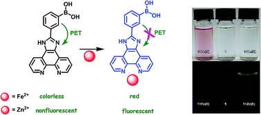
A novel, simple and efficient dual channel probe built on an imidazo-phenanthroline scaffold with a boronic acid unit, viz. 3-(1H-imidazo[4,5-f][1,10]phenanthrolin-2-yl)phenylboronic acid, is disclosed. It is found that this novel probe induces chromogenic and fluorogenic responses to Fe(II) and Zn(II) ions, respectively. To our best knowledge, this is one of the rare examples of dual channel responsive probes that can be used for visual detection of Fe(II) and turn-on fluorogenic detection of Zn(II) ions, simultaneously.


1. Introduction

Functional organic materials that allow selective and sensitive detection of target metal ions have attracted considerable attention, since metal ions play deleterious and/or essential roles in biological and environmental processes.1–8 Among these ions, iron is the most abundant transition metal ion, which is naturally present in (II) and (III) oxidation states. It plays vital roles in biological processes such as DNA synthesis, oxidative processes of living tissues, oxygen transport and storage, and mitochondrial electron transfer.9–13 Also, iron is highly necessary for energy metabolism and enzyme activities. However, either iron deficiency or overload can lead to human disorders and diseases: anemia, diabetes, heart diseases, siderosis, organ damage (liver, kidney etc.), toxicity or even death.14–24 Furthermore, the discrimination of Fe(II) from Fe(III) might be important, since they represent one of the most important redox couples.25–32 For that reason, the detection and, if possible, discrimination of Fe(II) from Fe(III) by using simple and efficient methods might be a challenge. In this context, colorimetric iron probes,33–35 which are capable of giving prominent signals that can easily be visualized by the naked eye without resorting to any instrumentation, offer a facile and prominent strategy.

On the other hand, zinc is the second most abundant transition metal in humans after iron. It is also essential to life as it plays highly crucial roles in many biological processes; regulation of apoptosis, signal transmission, enzyme function and gene expression.36 It is also reported that zinc is associated in a number of pathological processes, such as diabetes,37 Alzheimer's disease,38 and epilepsy.39 Therefore, the design of novel molecular sensors, which can recognize zinc selectively among the other metal ions is a challenging task. Fortunately, however, fluorescence spectroscopy offers a highly efficient way of detection of Zn(II) ions. Thus, many examples of fluorescent Zn(II) sensors have been reported to date in the literature.40–59 However, some of them have disadvantages concerning tedious multistep synthesis, selectivity, sensitivity, and/or interference of other ions especially in aqueous buffer solution. For that reason, design and synthesis of novel and simple yet efficient fluorescent Zn(II) sensors would be welcome.

One of the most prominent phenomena in the design of fluorescent molecular sensors is photoinduced electron transfer (PET).60–63 In principle, PET might occur in two different directions.63 In the first case, PET takes place from an excited-state fluorophore to an acceptor unit, which also acts as receptor. This process is termed as oxidative PET.64–66 In the second case, however, PET takes place from a donor unit to the excited-state fluorophore, which is known as reductive PET. Both of these processes are accompanied by quenching of the fluorophore emission.

In the context of PET based fluorescent probes, we have recently reported that cation-mediated oxidative PET could be modulated in a very rigid arrangement.67 We have designed a novel compound which is based on a 1,10-phenanthroline scaffold with cofacial BODIPY units attached orthogonally as the receptor and fluorophore units, respectively. This design afforded a turn-off fluorescent Cd(II) probe. Furthermore, we noted that a simple modification could allow us tailoring the selectivity of the probe from Cd(II) to Zn(II).68 It is important to note that those analytes, Cd(II) and Zn(II), are in the same group of the periodic table and they show very similar properties.

In the present work, we envisaged that an imidazo-phenanthroline motif with phenyl boronic acid unit could be used to create a simple, selective and viable fluorescent Zn(II) sensor and chromogenic Fe(II) probe. Herein we wish to present our research concerning the design, synthesis and properties of a novel material 1, viz. 3-(1H-imidazo[4,5-f][1,10]phenanthrolin-2-yl)phenylboronic acid. It is found that 1 induces chromogenic response to Fe(II) ions by turning from a colorless state to red color in 0.1 M HEPES buffer containing 0.1% CH3OH (v/v) (pH = 7.2) solution. It should be noted that 1 can clearly discriminate Fe(II) from Fe(III) in aqueous media. Furthermore, it is noteworthy that 1 can be utilized as a turn-on fluorescent Zn(II) sensor in aqueous buffer solution (0.1 M HEPES buffer containing 0.1% CH3OH (v/v), pH = 7.2). Importantly, this simple fluorescent sensor is highly selective and it gives fast response to Zn(II) ions. The fluorescence response of 1 is based on cation-mediated inhibition of reductive PET that results in enhancement of fluorescence emission up to 21-fold in aqueous buffer solution (0.1 M HEPES buffer containing 0.1% CH3OH (v/v), pH = 7.2). To our best knowledge, this is one of the rare examples of dual channel responsive probes, which can be used for visual detection of Fe(II) and turn on fluorogenic detection of Zn(II) ions, simultaneously.39,69,70

2. Experimental section

General

All chemicals were purchased from Sigma Aldrich Chemicals or Merck Company and used as received unless otherwise noted. FTIR spectra were recorded on Thermo Scientific Nicolet iS5 FT-IR Spectrometer with iD5-ATR apparatus. 1H (400 MHz) and 13C (100 MHz) NMR spectra were recorded on a Bruker Ultrashield 400 NMR Spectrometer. Matrix-assisted laser desorbtion/ionization (MALDI TOF) were recorded on Bruker microflex LT MALDI TOF MS system. High resolution mass spectra (HRMS) were recorded on Waters SYNAPT MS system (TOF MS ES). UV/Vis and fluorescence measurements were recorded on Varian Cary 50 and Varian Cary Eclipse spectrophotometers, respectively. Fluorescence and UV/Vis spectra were taken at room temperature. Melting points were determined on a Schorrp MPM-H2 model apparatus and are uncorrected. Column chromatography was performed on silica gel (60–200 mesh) from Merck Company. TLC was carried out on Merck 0.2 mm silica gel 60 F254 analytical aluminum plates. The synthesis of 3 (ref. 71) was carried out according to a published procedure with slight modifications. Metal solutions were freshly prepared as 1.0 × 10⁻³ M stocks from the corresponding perchlorate salts [with the exceptions of Au3+ (prepared from AuCl3) and Pt2+ (prepared from K2PtCl4)]. Compound 1 (1 mmol) was dissolved in CH3OH (10 mL). Appropriate amount of this solution was diluted in 0.1 M HEPES buffer (3 mL) at pH 7.2 at room temperature. Proper amount of freshly prepared metal solutions (1.0 × 10⁻³ M) were added to the above solution of 1 for spectrophotometric titrations.

Synthesis of 1,10-phenanthroline-5,6-dione (3)71

A cooled mixture of H2SO4 (10 mL) and HNO3 (5 mL) was prepared and KBr (1 g, 8.4 mmol) was added in small portions to this mixture at 0 °C while stirring. This was followed by the addition of 1,10-phenanthroline (2, 1 g, 5.5 mmol). Then, the reaction mixture was heated up to 130 °C for 3 hours. Then, the solution was transferred into a beaker containing crushed ice and carefully neutralised with NaOH (10% aqueous solution). The product was extracted by CH2Cl2 (3 × 50 mL), dried over MgSO4 and the solvent was removed under vacuum to give a yellow solid. The yellow solid was recrystallized from ethanol; 972 mg, yield 80%, m.p. 259 °C (lit8 257 °C). 1H NMR (400 MHz, CDCl3, 25 °C, TMS): δ = 9.05 (dd, J = 4.6–1.7 Hz, 2H), 8.44 (dd, J = 7.8–1.7 Hz, 2H), 7.53 (dd, J = 7.8–4.6 Hz, 2H); 13C NMR (100 MHz, CDCl3, 25 °C, TMS): δ = 178.5, 156.3, 152.8, 137.2, 128.0, 124.9; FTIR (cm−1): 3061, 1701, 1682, 1559, 1458, 1412, 1315, 1292, 1205, 1010, 925, 806, 697.

Synthesis of 3-(3a,11b-dihydro-1H-imidazo[4,5-f][1,10]phenanthrolin yl)phenylboronic acid (1)

1,10-Phenanthroline-5,6-dione (3, 211 mg, 1 mmol), 3-formylphenylboronic acid (149 mg, 1 mmol), and ammonium acetate (950 mg, 13.32 mmol) were dissolved in glacial acetic acid (10 mL) and the mixture was heated to 120 °C for 6 h under N2 atmosphere. After completion of the reaction, the mixture was cooled to room temperature and a majority of the solvent was removed under reduced pressure. Next, 10% aqueous solution of NaOH was used for neutralization (pH = 7.0). A yellow precipitate was formed, which was washed with water, CH2Cl2 and dried under vacuum overnight; 330 mg, yield 97%, m.p. > 360 °C. 1H NMR (400 MHz, CDCl3, 25 °C, TMS): δ = 13.82 (s, NH), 9.03 (dd, J = 4.2–1.5 Hz, 2H), 8.99–8.92 (m, 2H), 8.71 (s, 1H), 8.32 (bs, 3H), 7.95 (d, J = 7.3 Hz, 1H), 7.86–7.80 (m, 2H), 7.59 (t, J = 7.5 Hz, 1H); 13C NMR (100 MHz, CDCl3, 25 °C, TMS): δ = 146.3, 142.5, 138.4, 130.3, 130.1, 127.2, 125.1, 124.2, 123.8, 123.1, 122.8, 121.3, 118.1; FTIR (cm−1): 3372, 3198, 3072, 1609, 1546, 1419, 1275, 1067, 1037, 973, 804, 735, 692, 667; HRMS Anal. calcd for C20H15BN3O2 340.1257 [M]+; found: 341.1205 [M + H]+.

3. Results and discussion

The synthesis of the target compound was carried out via a two-step reaction sequence. In the first step, 1,10-phenanthroline (2) was treated with KBr, HNO3 and H2SO4 to afford dione 3 in 80% yield according to a known procedure (Scheme 1) (see ESI Fig. S1 and S2).71 Next, condensation of 3 with 3-formylphenylboronic acid (4) in the presence of NH4OAc and HOAc resulted in the formation of 1 in a yield of 97%. Compound 1 was initially characterized on the basis of 1H and 13C NMR spectroscopy, elemental and HRMS analysis, which firmly established the structure (see ESI Fig. S3–S5).
image file: c4ra14182b-s1.tif
Scheme 1 Synthesis of compound 1.

UV/Vis absorption spectrum of 1 was recorded in 0.1 M HEPES buffer containing 0.1% CH3OH (v/v) (pH = 7.2) solution. The absorption spectrum of 1 (1.30 × 10⁻⁵ M) in this solution is characterized by a broad band between 275–350 nm with λmax at 276 nm and 308 nm (Fig. 1, black line). Spectrophotometric titrations of 1 with different metal ions were carried out in order to determine the metal cation complexing properties of 1. It was found that the addition of Ag+, Al3+, Au3+, Cd2+, Co2+, Cu+, Cu2+, Fe3+, Hg2+, K+, Li+, Mn2+, Mg2+, Na+, Ni2+, Pb2+, Pt2+, Pd2+ and Zn2+ ions (3.0 equiv.) did not alter the color of the solution of 1 in spite of the fact that small changes were observed in the absorption spectrum as depicted in Fig. 1 (blue line). Gratifyingly, however, it was noted that the addition of Fe2+ ions (3.0 equiv.) revealed significant changes in the original absorption spectrum of 1 (Fig. 1, red line).


image file: c4ra14182b-f1.tif
Fig. 1 UV/Vis absorption spectra of 1 (1.30 × 10⁻⁵ M) in the absence and presence of Fe2+ and other metal ions (Ag+, Al3+, Au3+, Cd2+, Co2+, Cu+, Cu2+, Fe3+, Hg2+, K+, Li+, Mn2+, Mg2+, Na+, Ni2+, Pb2+, Pt2+, Pd2+ and Zn2+) (3.0 equiv.) in 0.1 M HEPES buffer containing 0.1% CH3OH (v/v), pH = 7.2, 25 °C. Inset: color of 1 (1.30 × 10⁻⁵ M) in the absence and presence of Fe2+ ions under room light in 0.1 M HEPES buffer containing 0.1% CH3OH (v/v), pH = 7.2, 25 °C.

The absorption spectral changes of 1 as a function of Fe2+ concentration in 0.1 M HEPES buffer containing 0.1% CH3OH (v/v) (pH = 7.2) at room temperature is depicted in Fig. 2. Notably, a progressive increase in absorbance in the visible region of the electromagnetic spectrum was observed with simultaneous formation of new red shifted absorption bands (λmax = 528 nm). Remarkably, the formation of these new red shifted absorption bands was enough to change the color of the solution from a colorless state to red color. This allowed the naked-eye (colorimetric) detection of Fe2+ ions among the others (Fig. 1 inset). In addition, 1 could clearly discriminate Fe(II) from Fe(III) in aqueous media. Competition experiments indicated that 1 is responsive to Fe2+ even in the presence of other metal ions (Fig. 1, green line). It is important to note that color change is one of the most convenient visual detection methods used in classical chemical analysis, which is straightforward and inexpensive.


image file: c4ra14182b-f2.tif
Fig. 2 Absorption spectral changes of 1 (0.95 × 10⁻⁵ M) as a function of Fe2+ concentration (0.0–8.0 equiv.) in 0.1 M HEPES buffer containing 0.1% CH3OH (v/v), pH = 7.2, 25 °C.

Binding assays were performed by using Job's method, which indicated a 1[thin space (1/6-em)]:[thin space (1/6-em)]1 stoichiometry for the interaction between 1 and Fe2+ (see ESI, Fig. S6). On the basis of the above spectrophotometric titrations, the detection limit of 1 was calculated to be 4.89 × 10⁻⁶ M (Fig. S7). Moreover, the binding constant (Ka) of 1 with Fe2+ was determined to be 4.77 × 10³ M−1 (Fig. S8).

On the other hand, 1 (1.30 × 10⁻⁵ M) was practically nonluminescent under the given conditions [0.1 M HEPES buffer containing 0.1% CH3OH (v/v), pH = 7.2, 25 °C]. The metal cation complexing properties of 1 were investigated by spectrophotometric titrations with different metal cations. It was found that the addition of Ag+, Al3+, Au3+, Cd2+, Co2+, Cu+, Cu2+, Fe2+, Fe3+, Hg2+, K+, Li+, Mn2+, Mg2+, Na+, Ni2+, Pb2+, Pt2+ and Pd2+ ions (1.0 equiv.) did not induce any significant change in the emission profile of 1 (Fig. 3 and 4). However, the emission intensity of 1 (1.30 × 10⁻⁵ M) was dramatically increased upon addition of Zn2+ ions (1.0 equiv.) (turn-on).


image file: c4ra14182b-f3.tif
Fig. 3 Fluorescence spectrum of compound 1 (1.30 × 10⁻⁵ M) with various metal ions (Ag+, Al3+, Au3+, Cd2+, Co2+, Cu+, Cu2+, Fe2+, Fe3+, Hg2+, K+, Li+, Mn2+, Mg2+, Na+, Ni2+, Pb2+, Pt2+, Pd2+ and Zn2+) (1.0 equiv.) in 0.1 M HEPES buffer containing 0.1% CH3OH (v/v), pH = 7.2, 25 °C, λexc = 308 nm.

image file: c4ra14182b-f4.tif
Fig. 4 Relative fluorescence spectral changes for 1 (1.30 × 10⁻⁵ M) with various metal ions (1.0 equiv.) in 0.1 M HEPES buffer containing 0.1% CH3OH (v/v), pH = 7.2, 25 °C, λexc = 308 nm.

Fig. 5 shows the fluorescence spectral changes of 1 (1.30 × 10⁻⁵ M) as a function of Zn2+ concentration in 0.1 M HEPES buffer containing 0.1% CH3OH (v/v, pH = 7.2) at room temperature. It is important to note that a progressive increase in the fluorescence emission intensity at 498 nm was observed as the concentration of the ion was increased. Chromogenic and fluorogenic responses of 1 to Fe2+ and Zn2+ ions, respectively, were depicted in Fig. S9.


image file: c4ra14182b-f5.tif
Fig. 5 Fluorescence spectrum of compound 1 (1.30 × 10⁻⁵ M) as a function of Zn2+ concentration (0.0, 0.1, 0.2, 0.3, 0.4, 0.5, 0.6, 0.7, 0.8, 1.0, 2.0, 3.0, 4.0 equiv.) in 0.1 M HEPES buffer containing 0.1% CH3OH (v/v), pH = 7.2, 25 °C, λexc = 308 nm.

Clearly, the changes in the emission profile of 1 upon addition of Zn2+ ions indicated the formation of a well-defined complex between 1 and Zn2+. Upon binding of Zn2+ ions to 1, the fluorescence emission intensity of the imidazophenanthroline unit was dramatically increased probably by blocking PET between imidazophenanthroline and phenyl boronic acid units. It is reasonable to assume that both the HOMO and LUMO energy levels of the metal-bound form of the ligand 1 disfavors PET when compared to the metal-free state, thus providing efficient radiative decay of the excited state. Interestingly, evaluation of Job plot for the determination of the stoichiometry of the interaction between 1 and Zn2+ revealed a 1[thin space (1/6-em)]:[thin space (1/6-em)]2 ratio (see ESI, Fig. S10). Remarkably, the formation of the complex between 1 and Zn2+ was also evidenced by 1H NMR (Fig. S11). It was found that all the proton signals of 1 showed down field shifts between 0.03 and 0.40 ppm due to the interaction with Zn2+ (Fig. S12). Furthermore, MALDI TOF MS analysis proved the formation of the complex between 1 and Zn2+. Fig. S13 depicts MALDI TOF MS spectrum of 1 + Zn2+ complex, which indicated the presence of three hydrated form of the complex with an [M+] ion peak (m/z) of 458.925.

Finally, the reversibility of the fluorogenic response of 1 to Zn2+ ions was tested by titration of 1 + Zn2+ complex with EDTA, which is known as a strong chelator for Zn2+. It was noticed that the fluorescence emission of complex (Fig. 6, red line) is turned off by the addition of EDTA into a solution of complex (Fig. 6, green line) in 0.1 M HEPES buffer containing 0.1% CH3OH (v/v), pH = 7.2, 25 °C. However, the emission intensity is reconfigured almost to the original state after addition of Zn2+ ions to this mixture (Fig. 6, blue line). These results clearly suggests that the fluorogenic response of 1 to Zn2+ ions is completely reversible. Furthermore, competition experiments indicated that 1 (5.20 × 10⁻⁵ M) is still responsive to Zn2+ (10 equiv.) even in the presence of other competing ions (10 equiv.) albeit with lower emission intensity (Fig. S14).


image file: c4ra14182b-f6.tif
Fig. 6 Fluorescence emission spectra of 1 (1.30 × 10⁻⁵ M) in the absence and presence of Zn2+ (1 equiv.) or EDTA (1 equiv.) in 0.1 M HEPES buffer containing 0.1% CH3OH (v/v), pH = 7.2, 25 °C, λexc = 308 nm.

On the basis of the above spectrofluorimetric titrations, the detection limit6,7 of 1 was calculated to be 6.74 × 10⁻⁷ M (Fig. S15). Furthermore, the binding constant (Ka) of 1 with Zn2+ was determined from the emission intensity data following the steady-state fluorometric method6–8 in which I0 referred to the fluorescence intensities of solutions of 1. When I0/(II0) is plotted against [M]−1, Ka was calculated to be 1.50 × 10⁵ from the ratio of intercept/slope with a good correlation coefficient (R = 0.99695) (Fig. S16).

4. Conclusions

In summary, the design, syntheses, optical and metal ion recognition features of a novel compound (1) are investigated. The compound is built on an imidazo-phenanthroline scaffold with phenyl boronic acid unit. It is noteworthy that 1 is not only a simple and selective metal ion probe, but also it is a promising dual channel responsive material. 1 can simultaneously induce chromogenic and fluorogenic responses to Fe(II) and Zn(II) ions, respectively. To our best knowledge, this is one of the rare examples of dual channel responsive probes that can be used for both visual detection of Fe2+ and turn-on fluorogenic detection of Zn2+ ions simultaneously. Further work is currently underway in our laboratories.

Acknowledgements

The authors are grateful to the Scientific and Technological Research Council of Turkey (TUBITAK, Grant no. 113T016) and Aksaray University for financial support. A. Y. is indebted to TUBITAK for a graduate fellowship.

References

  1. A. W. Czarnik, Fluorescent Chemosensors for Ion and Molecule Recognition, American Chemical Society, Washington DC, 1993 Search PubMed.
  2. A. P. de Silva, H. Q. N. Gunaratne, T. Gunnlaugsson, A. J. M. Huxley, C. P. McCoy, J. T. Rademacher and T. E. Rice, Chem. Rev., 1997, 97, 1515–1566 CrossRef CAS PubMed.
  3. A. P. de Silva, D. B. Fox and A. J. M. Huxley, Coord. Chem. Rev., 2000, 205, 41–57 CrossRef CAS.
  4. J. F. Callan, A. P. de Silva and D. C. Magri, Tetrahedron, 2005, 61, 8551–8588 CrossRef CAS PubMed.
  5. J. S. Kim and D. T. Quang, Chem. Rev., 2007, 107, 3780–3799 CrossRef CAS PubMed.
  6. M. P. Algi, Z. Oztas and F. Algi, Chem. Commun., 2012, 48, 10219–10221 RSC.
  7. M. Pamuk and F. Algi, Tetrahedron Lett., 2012, 53, 7117–7120 CrossRef CAS PubMed.
  8. Z. Oztas, M. Pamuk and F. Algi, Tetrahedron, 2013, 69, 2048–2051 CrossRef CAS PubMed.
  9. J. Mortatti, F. J. Krug, L. C. R. Pyessenda and E. A. G. Zagatto, Analyst, 1982, 107, 659–663 RSC.
  10. T. H. Bothwell, R. W. Charlton, I. D. Cook and C. A. Finch, Iron Metabolism in Man, Blackwell, Oxford, 1979 Search PubMed.
  11. A. V. Hoffbrand and V. Herbert, Semin. Hematol., 1999, 36, 13–23 CAS.
  12. C. An-Sheng and C. A. Enns, J. Biol. Chem., 2009, 284, 711–715 CrossRef PubMed.
  13. S. A. Cotton, Educ. Chem., 1976, 13, 180 CAS.
  14. J. T. Ellis, I. Schulman and C. H. Smith, Am. J. Pathol., 1954, 30, 287–309 CAS.
  15. J. V. Corbett, Am. J. Matern. Child Nurs., 1995, 20, 234–239 CrossRef CAS PubMed.
  16. J. H. Martin and S. E. Fitzwater, Nature, 1988, 331, 341–343 CrossRef CAS.
  17. X. Liu and E. C. Theil, Acc. Chem. Res., 2005, 38, 167–175 CrossRef CAS PubMed.
  18. M. A. Smith, P. L. R. Harris, L. M. Sayre and G. Perry, Proc. Natl. Acad. Sci. U. S. A., 1997, 94, 9866–9868 CrossRef CAS.
  19. F. A. Tezcan, J. T. Kaiser, D. Mustafi, M. Y. Walton, J. B. Howard and D. C. Rees, Science, 2005, 309, 1377–1380 CrossRef CAS PubMed.
  20. R. Crichton, Inorganic Biochemistry of Iron Metabolism: From Molecular Mechanisms to Clinical Consequences, John Wiley & Sons Ltd, Chichester, 2nd edn, 2001 Search PubMed.
  21. M. J. Behrenfeld, A. J. Bale, Z. S. Kolber, J. Aiken and P. G. Falkowski, Nature, 1996, 383, 508–511 CrossRef CAS.
  22. E. L. Que, D. W. Domaille and C. J. Chang, Chem. Rev., 2008, 108, 1517–1549 CrossRef CAS PubMed.
  23. J. A. Lee, G. H. Eom, H. M. Park, J. H. Lee, H. Song, C. S. Hong, S. Yoon and C. Kim, Bull. Korean Chem. Soc., 2012, 33, 3625–3628 CrossRef CAS.
  24. J. R. Burdo and J. R. Connor, BioMetals, 2003, 16, 63–75 CrossRef CAS.
  25. J. Zhan, L. Wen, F. Miao, D. Tian, X. Zhu and H. Li, New J. Chem., 2012, 36, 656–661 RSC.
  26. S. L. Hu, N. F. She, G. D. Yin, H. Z. Guo, A. X. Wu and C. L. Yang, Tetrahedron Lett., 2007, 48, 1591–1594 CrossRef CAS PubMed.
  27. Y. Xiang and A. Tong, Org. Lett., 2006, 8, 1549–1552 CrossRef CAS PubMed.
  28. J. Mao, L. N. Wang, W. Dou, X. L. Tang, Y. Yan and W. S. Liu, Org. Lett., 2007, 9, 4567–4570 CrossRef CAS PubMed.
  29. X. B. Zhang, G. Cheng, W. J. Zhang, G. L. Shen and R. Q. Yu, Talanta, 2007, 71, 171–177 CrossRef CAS PubMed.
  30. O. Oter, K. Ertekin, C. Kirilmis, M. Koca and M. Ahmedzade, Sens. Actuators, B, 2007, 122, 450–456 CrossRef CAS PubMed.
  31. G. E. Tumambac, C. M. Rosencrance and C. Wolf, Tetrahedron, 2004, 60, 11293–11297 CrossRef CAS PubMed.
  32. J. L. Bricks, A. Kovalchuk, C. Trieflinger, M. Nofz, M. Buschel, A. I. Tolmachev, J. Daub and K. J. Rurack, J. Am. Chem. Soc., 2005, 127, 13522–13529 CrossRef CAS PubMed.
  33. (a) Z.-B. Zheng, Z.-M. Duan, J.-X. Zhang and K.-Z. Wang, Sens. Actuators, B, 2012, 169, 312–319 CrossRef CAS PubMed; (b) Z.-B. Zheng, Z.-M. Duan, Y.-Y. Ma and K.-Z. Wang, Inorg. Chem., 2013, 52, 2306–2316 CrossRef CAS PubMed; (c) D. A. Ondigo, Z. R. Tshentu and N. Torto, Anal. Chim. Acta, 2013, 204, 228–234 CrossRef PubMed.
  34. C.-H. Chen, C. Cho, C.-F. Wan and A.-T. Wu, Inorg. Chem. Commun., 2014, 41, 88–91 CrossRef CAS PubMed.
  35. K. B. Kim, G. J. Park, H. Kim, E. J. Song, J. M. Bae and C. Kim, Inorg. Chem. Commun., 2014, 46, 237–240 CrossRef CAS PubMed.
  36. B. L. Vallee and K. H. Falchuk, Physiol. Rev., 1993, 73, 79–118 CAS.
  37. A. B. J. Chausmer, J. Am. Coll. Nutr., 1998, 17, 109–115 CrossRef CAS.
  38. A. I. Bush, Trends Neurosci., 2003, 26, 207–214 CrossRef CAS.
  39. Y. Hong, S. Chen, C. W. T. Leung, J. W. Y. Lam, J. Liu, N.-W. Tseng, R. T. K. Kwok, Y. Yu, Z. Wang and B. Z. Tang, ACS Appl. Mater. Interfaces, 2011, 3, 3411–3418 CAS.
  40. L. Qu, C. Yin, F. Huo, J. Chao, Y. Zhang and F. Cheng, Sens. Actuators, B, 2014, 191, 158–164 CrossRef CAS PubMed.
  41. Y. Ma, H. Chen, F. Wang, S. Kambam, Y. Wang, C. Mao and X. Chen, Dyes Pigm., 2014, 102, 301–307 CrossRef CAS PubMed.
  42. T. B. Wei, P. Zhang, B. B. Shi, P. Chen, Q. Lin, J. Liu and Y. M. Zhang, Dyes Pigm., 2013, 97, 297–303 CrossRef CAS PubMed.
  43. W. Lin, D. Buccella and S. J. Lippard, J. Am. Chem. Soc., 2013, 135, 13512–13520 CrossRef CAS PubMed.
  44. D. Sarkar, A. K. Pramanik and T. K. Mondal, J. Lumin., 2014, 146, 480–485 CrossRef CAS PubMed.
  45. O. G. Tsay, S. T. Manjare, H. Kim, K. M. Lee, Y. S. Lee and D. G. Churchill, Inorg. Chem., 2013, 52, 10052–10061 CrossRef CAS PubMed.
  46. K. Li and A. Tong, Sens. Actuators, B, 2013, 184, 248–252 CrossRef CAS PubMed.
  47. A. Helal, M. H. O. Rashid, C. H. Choi and H. S. Kim, Tetrahedron, 2012, 68, 647–653 CrossRef CAS PubMed.
  48. Q. H. You, P. S. Chan, W. H. Chan, S. C. K. Hau, A. W. M. Lee, N. K. Mak, T. C. W. Mak and R. N. S. Wong, RSC Adv., 2012, 11078–11083 RSC.
  49. B. Zhang, K. S. Cao, Z. A. Xu, Z. Q. Yang, H. W. Chen, W. Huang, G. Yin and X. Z. You, Eur. J. Inorg. Chem., 2012, 3844–3850 CrossRef CAS.
  50. S. Yin, J. Zhang, H. Feng, Z. Zhao, L. Xu, H. Qiu and B. Tang, Dyes Pigm., 2012, 95, 174–179 CrossRef CAS PubMed.
  51. Y. P. Li, H. R. Yang, Q. Zhao, W. C. Song, J. Han and X. H. Bu, Inorg. Chem., 2012, 51, 9642–9648 CrossRef CAS PubMed.
  52. Y. Zhou, Z. X. Li, S. Q. Zang, Y. Y. Zhu, H. Y. Zhang, H. W. Hou and T. C. W. Mak, Org. Lett., 2012, 14, 1214–1217 CrossRef CAS PubMed.
  53. Y. Fu, Z. Xing, C. Zhu, H. Yang, W. He, C. Zhu and Y. Cheng, Tetrahedron Lett., 2012, 53, 804–807 CrossRef CAS PubMed.
  54. C. Gao, X. Jin, X. Yan, P. An, Y. Zhang, L. Liu, H. Tian, W. Liu, X. Yao and Y. Tang, Sens. Actuators, B, 2013, 176, 775–781 CrossRef CAS PubMed.
  55. J. Y. Choi, D. Kim and J. Yoon, Dyes Pigm., 2013, 96, 176–179 CrossRef CAS PubMed.
  56. M. J. Kim, K. Kaur, N. Singh and D. O. Jang, Tetrahedron, 2012, 68, 5429–5433 CrossRef CAS PubMed.
  57. A. Coskun, E. Deniz and E. U. Akkaya, Tetrahedron Lett., 2007, 48, 5359–5361 CrossRef CAS PubMed.
  58. F. N. Aka, M. S. Akkaya and E. U. Akkaya, J. Mol. Catal. A: Chem., 2001, 165, 291–294 CrossRef CAS.
  59. S. Atilgan, T. Ozdemir and E. U. Akkaya, Org. Lett., 2008, 10, 4065–4067 CrossRef CAS PubMed.
  60. A. P. de Silva, H. Q. N. Gunaratne, T. Gunnlaugsson, C. P. McCoy, P. R. S. Maxwell, J. T. Rademacher and T. E. Rice, Pure Appl. Chem., 1996, 68, 1443–1448 CrossRef CAS.
  61. A. P. de Silva, T. S. Moody and G. D. Wright, Analyst, 2009, 134, 2385–2393 RSC.
  62. A. P. de Silva, Nat. Chem., 2012, 4, 440–445 CrossRef PubMed.
  63. B. Valeur, Molecular Fluorescence: Principles and Applications, Wiley-VCH, 2002, p. 90 Search PubMed.
  64. A. P. de Silva, I. M. Dixon, H. Q. N. Gunaratne, T. Gunnlaugsson, P. R. S. Maxwell and T. E. Rice, J. Am. Chem. Soc., 1999, 121, 1393–1394 CrossRef CAS.
  65. B. Turfan and E. U. Akkaya, Org. Lett., 2002, 4, 2857–2859 CrossRef CAS PubMed.
  66. A. Coskun, B. T. Baytekin and E. U. Akkaya, Tetrahedron Lett., 2003, 44, 5649–5651 CrossRef CAS.
  67. M. Pamuk and F. Algi, Tetrahedron Lett., 2012, 53, 7010–7012 CrossRef CAS PubMed.
  68. S. Karakaya and F. Algi, Tetrahedron Lett., 2014, 55, 5555–5559 CrossRef CAS PubMed.
  69. Y. W. Choi, G. J. Park, Y. J. Na, H. Y. Jo, S. A. Lee, G. R. You and C. Kim, Sens. Actuators, B, 2014, 194, 343–352 CrossRef CAS PubMed.
  70. K. B. Kim, H. Kim, H. J. Song, S. Kim, I. Noh and C. Kim, Dalton Trans., 2013, 42, 16569–16577 RSC.
  71. J. Ettedgui and R. J. Neuman, J. Am. Chem. Soc., 2009, 131, 4–5 CrossRef CAS PubMed.

Footnote

Electronic supplementary information (ESI) available. See DOI: 10.1039/c4ra14182b

This journal is © The Royal Society of Chemistry 2015
Click here to see how this site uses Cookies. View our privacy policy here.