Pseudopeptide Foldamers designed for photoinduced intramolecular electron transfer

Lorenzo Millia, Enrico Marchi*a, Nicola Castelluccia, Maria Teresa Indellib, Margherita Venturiac, Paola Ceroniac and Claudia Tomasini*a
aDipartimento di Chimica “G. Ciamician”, Università di Bologna, Via F. Selmi 2, 40126 Bologna, Italy. E-mail: claudia.tomasini@unibo.it; enrico.marchi@unibo.it
bDipartimento di Scienze Chimiche e Farmaceutiche, Università di Ferrara, Via Fossato di Mortara 17, 44121 Ferrara, Italy
cCentro Interuniversitario per la Conversione dell'Energia Solare (SolarChem), Italy

Received 6th November 2014 , Accepted 5th January 2015

First published on 8th January 2015


Abstract

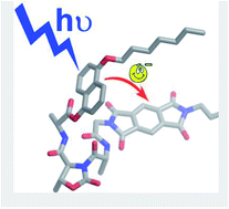
We have designed and prepared three pseudopeptide foldamers, called dyads 1, 2 and 3, equipped with a donor and an acceptor unit to promote intramolecular electron transfer after light excitation. All the three dyads contain the same donor and acceptor, which are a derivative of 1,5-dihydroxynaphthalene and a derivative of pyromellitic diimide, respectively. The donor and acceptor units are separated by hybrid foldamers of different length in order to vary both their distance and relative orientation. Specifically, one, two or three L-Ala-D-Oxd (Ala = alanine, Oxd = 4-carboxy-5-methyl-oxazolidin-2-one) units are contained in dyads 1, 2, and 3, respectively. Dyad 1 folds in a bent conformation in which the donor and acceptor units lie one close to the other, while dyads 2 and 3 preferentially assume an extended conformation. In all the three dyads both the donor and acceptor emissions are efficiently quenched via intramolecular electron transfer, as suggested by photophysical and electrochemical investigations. Because of its bent conformation dyad 1 exhibits a charge-transfer (CT) band at 410 nm in CH2Cl2 solution and a photoinduced electron transfer that occurs more efficiently than in dyads 2 and 3. Upon dissolving dyad 1 in DMSO, a competitive solvent for hydrogen bonds that establish in the pseudopeptide linker, the CT band disappears and the efficiency of electron transfer slightly decreases, in agreement with an unfolded conformation in which donor and acceptor units are no longer in close contact.

Introduction

Long-range electron transfer (ET) through proteins is a fundamental reaction in living organisms, playing a role in energy-conversion processes like photosynthesis and respiration1 as well as in enzymatic reactions.2 Intramolecular electron-transfer reactions between donor and acceptor sites separated by a synthetic peptide or a protein fragment have provided deep insight into the role played by the bridge in mediating electron transfer.3 To carry out these studies the supramolecular chemistry4 and photochemistry5 approach can be very useful because it enables to assemble prefabricated molecular components that carry the desired photophysical and redox properties. Indeed, in a suitably designed supramolecular species A–L–D (where the electron acceptor A and electron donor D units are covalently bound with a linker L) light excitation can cause an intramolecular electron transfer from the excited state of D to A. This approach offers the advantage that the properties of the supramolecular system can be predicted by knowing the properties of the isolated components or of suitable model molecules.

Over the last 50 years several donor and acceptor chromophores have been examined in the context of their capability to form charge-transfer (CT) complexes.6 In the frame of these investigations, it has also been evidenced that the linker plays a key role to put in communication the two chromophores. In principle, a lot of different linkers could be used for this purpose, providing pathways through peptide bonds, aromatic side chains, weak noncovalent hydrophobic interactions, hydrogen-bonding networks associated with helices and sheets and/or other secondary structural features that change the electronic structure and induce low-energy pathways across polypeptides.7 Among the peptide bridging groups studied, the oligoproline building blocks were used as a key model for systematic study of the distance dependence of the electron transfer process.8 The advantage of the oligoprolines over other naturally occurring amino acids and peptides is the predictability of their secondary structure, which imparts significant rigidity upon the spatial separation between the donor and acceptor.9

In the last few years, the design and synthesis of oligomers based on proline units, both in the presence and absence of stabilizing hydrogen bonds, have been extensively pursued. Interesting new molecules capable of folding into defined secondary structures may be prepared by replacing the proline moieties with pseudoproline (ΨPro) units.10

Simplified artificial systems based on backbones designed to fold in secondary structures are called “foldamers”, and they have recently received considerable attention because they hold promises for addressing chemical, physico-chemical and biological problems and represent a new frontier in research.11 We have extensively studied the conformational behavior of foldamers containing a pseudoproline scaffold as 4-carboxy-5-methyl-oxazolidin-2-one (Oxd)12 On acylation of this pseudoproline unit, imides are obtained: the two carbonyls lie apart from one another and form the peptide bond in a trans conformation.13 We have also reported hybrid foldamers, where the Oxd moiety is alternated with an α- or a β-amino acid.14 The relative configuration of the Oxd and the alternated amino acid is very important, as the L-Ala-D-Oxd series tends to form β-bend ribbon spirals, while the L-Ala-L-Oxd series does not.

In this paper, we report the synthesis, the conformational characterization and the photophysical investigation of a series of hybrid oligomers, called dyads 1, 2 and 3 (Fig. 1). They contain a derivative of pyromellitic diimide as electron acceptor group and a derivative of 1,5-dihydroxynaphthalene as the electron donor one. These two moieties are linked by one, two, or three L-Ala-D-Oxd units in dyads 1, 2 and 3.


image file: c4ra13978j-f1.tif
Fig. 1 Formulas of the investigated dyads 1, 2 and 3 and the acceptor and compounds 4 and 5 taken as models of the acceptor and donor units, respectively.

The aim of this work is to check if the photophysical properties, and in particular the interaction between the donor and acceptor units, are affected by the different bridge and by the resulting conformation of the foldamers.

Results and discussion

Synthesis

Dyads 1, 2 and 3 have been prepared in solution by a convergent synthesis, where the donor group, the acceptor group and the spacer are assembled.

The acceptor group, a pyromellitic diimide, has been prepared in one step as white powder by reaction of pyromellitic dianhydride with dodecylamine and glycine in refluxing DMF in 28% yield.15 Besides 6, that was used for the dyads preparation, the corresponding methyl ester 4 (Fig. 1) was synthesized as acceptor model for the photophysical analysis, by reaction of 6 with SOCl2 in CH3OH (Scheme 1).


image file: c4ra13978j-s1.tif
Scheme 1 Reagents and conditions: (i) Gly (1.0 equiv.), dodecylamine (1.0 equiv.), DMF, 120 °C, 6 h; (ii) SOCl2 (excess), MeOH, r.t., 24 h.

The donor 8 was prepared in two steps starting from the commercially available 1,5-dihydroxynaphthalene (Scheme 2). After reaction with potassium carbonate, potassium iodide and n-heptyl bromide in acetone, a mixture of the desired 5-(heptyloxy)naphthalen-1-ol 7 and 1,5-bis(heptyloxy)naphthalene 5 was prepared. After purification, 7 and 5 were obtained in 37% and 30% yield respectively. While 7 was used for the dyad preparation, 5 was used as donor model for the photophysical analysis. Finally 7 was coupled with Boc-L-Ala in the presence of DCC and DMAP to give the final product 8 in 55% yield.


image file: c4ra13978j-s2.tif
Scheme 2 Reagents and conditions: (i) 1-bromoheptane (1.0 equiv.), K2CO3 (2.1 equiv.), KI (1.0 equiv.), acetone, reflux, 10 h; (ii) DCC (1.1 equiv.), DMAP (0.1 equiv.), DCM, r.t., 24 h.

As mentioned before, the donor and the acceptor units were linked together by the spacers, that are oligomers of the Boc-(L-Ala-D-Oxd)n-OH series (n = 1, 2, 3), whose synthesis was previously reported.16 The preparation of dyads 1, 2 and 3 was obtained by two steps, by standard peptide coupling reactions with the acceptor group and the donor group (Scheme 3).


image file: c4ra13978j-s3.tif
Scheme 3 Reagents and conditions: (i) TFA (18 equiv.), dry CH2Cl2, r.t., 4 h; (ii) HATU (1.1 equiv.), DIEA (3.0 equiv.), dry CH3CN, r.t., 1 h; (iii) HBTU (1.1 equiv.), DIEA (3.0 equiv.), dry DMF, r.t., 2 h.

Conformational analysis

Information on the preferred conformations of dyads 1, 2 and 3 in solution was obtained by the analysis of FT-IR and 1H NMR spectra.

IR spectroscopy

The analysis of the N–H stretching regions enables to detect if intramolecular N–H⋯O[double bond, length as m-dash]C hydrogen bonds are formed, because non-hydrogen-bonded amide NH groups exhibit a stretching signal above 3400 cm−1, while hydrogen-bonded amide NH ones17 produce a stretching band below 3400 cm−1. The FT-IR spectra were recorded in CH2Cl2 at a compound concentration of 3 mM; such a concentration was chosen to avoid compound self-aggregation.

NH stretching bands of dyads 1, 2 and 3 are shown in Fig. 2: the spectrum of 1 clearly shows the presence of two bands at 3420 and 3340 cm−1, that account for an hydrogen-bonded and a non-hydrogen-bonded amide NH group. The stretching band at 3340 cm−1 becomes weaker for 2 and totally disappears in the case of 3. This finding suggests that a folded conformation is strongly favoured in the case of the shortest dyad 1, whereas it occurs only partially in dyad 2 and it does not take place at all in dyad 3.


image file: c4ra13978j-f2.tif
Fig. 2 N–H stretching regions of the FT-IR absorption spectra in pure CH2Cl2 at room temperature for 3 mM concentration of dyads 1 (black line), 2 (red line) and 3 (green line).

1H NMR spectroscopy

The 1H NMR analysis of the NH chemical shifts supports the interpretation of the IR spectroscopy results, as the NH of dyad 1 resonates at 7.45 ppm, while the NH's of dyads 2 and 3 resonate between 7.18 and 7.32 ppm, thus showing that NH is more deshielded for 1. In contrast compounds 9a–c show the opposite behaviour as the NH's chemical shifts resonate at 7.35 for 9a, increasing to 7.35–7.57 for 9b and 7.50–7.60 for 9c (for details see ESI).

A further confirmation of the preferred bent conformation assumed by 1 was obtained by analyzing the chemical shifts of the pyromellitic diimide and 1,5-dihydronaphthalene hydrogens. These chemical shifts for dyads 1, 2 and 3 together with those found for the model compound 4 and compound 8 are reported in Table 1.

Table 1 Proton chemical shifts (ppm) of aromatic hydrogens for compounds 1, 2, 3, 4 and 8 in different solvents

image file: c4ra13978j-u1.tif

  Solvent δH1 (ppm) δH2 (ppm) δH3 (ppm) δH4 (ppm) δH5 (ppm) δH6 (ppm) δH7 (ppm)
1 CDCl3 8.02 7.26 7.13 6.74 7.22 7.02 7.85
2 CDCl3 8.18 7.36 7.20 6.79 7.36 7.32 8.25
3 CDCl3 8.17 7.39 7.18 6.79 7.36 7.31 8.27
4 CDCl3 8.33
8 CDCl3 8.21 7.44 7.23 6.83 7.38 7.42
1 DMSO, d6 8.02 7.46 7.20 6.99 7.38 7.32 8.15
4 DMSO, d6 8.03 7.47 7.22 6.97 7.38 7.44
8 DMSO, d6 8.22


We can notice that H7 resonates as a singlet for both hydrogens at 8.33 ppm in the spectrum of 4 (Fig. 3). In dyads 1, 2 and 3 there is no skeleton modification that could be responsible for the variation of this chemical shift. In fact, both 2 and 3 spectra show a singlet at 8.25 and 8.27 ppm respectively. In contrast, in the dyad 1 spectrum, the chemical shift of the pyromellitic diimide hydrogens resonates at 7.85 ppm, suggesting that these hydrogens are shielded by an aromatic ring (Fig. 3). The same effect was observed for the chemical shifts of the 1,5-dihydronaphtalene hydrogens (compound 8), that are all shielded only in the case of dyad 1. This outcome suggests that all the aromatic hydrogens of dyad 1 suffer from the shielding effect of a nearby aromatic ring.


image file: c4ra13978j-f3.tif
Fig. 3 Superimposition of the 1H NMR spectra in CDCl3 of 1, 4 and 8 in the aromatic region. All hydrogens of dyad 1 are shielded compared with 4 and 8, as the two aromatic moieties suffer mutually from a shielding effect.

Interestingly, when the 1H NMR spectra of compounds 1, 4 and 8 are registered in DMSO, d6, the chemical shifts variations are negligible as Δδ ranges from 0 to 0.12 ppm, while in CDCl3 it ranges between 0.09 and 0.47 ppm (Table 1 and ESI for details). Thus the bent conformation of 1 is favoured by CHCl3, that is a structure supporting solvent and is disfavoured by DMSO, that is a competitive solvent for N–H⋯O[double bond, length as m-dash]C bonds (see below).18

ROESY experiments performed on dyads 1 and 2 (as a model of the unfolded dyads), in CDCl3 using a mixing time of 0.400 s, proved this hypothesis.

Besides the trivial couplings, in the ROESY spectrum of 1 in CDCl3, several cross peaks accounting for the interactions between the pyromellitic diimide and 1,5-dihydronaphthalene hydrogens and NH are visible and are highlighted (see ESI for more details). In contrast, in the ROESY spectrum of 2 these signals are totally absent (see ESI for more details). This finding is in agreement with a preferred bent conformation of 1, that favours the proximity between the donor and the acceptor groups in a range of about 4 Å. The preferred conformation of dyad 1, together with all the cross peaks registered in the ROESY spectrum, is summarized in Fig. 4.


image file: c4ra13978j-f4.tif
Fig. 4 Preferential conformation of dyad 1, that accounts for the formation of a NH hydrogen bond and for the NOE enhancements as gathered from the analysis of its ROESY spectrum (10 mM solution in CDCl3, mixing time 0.400 s).

Photophysical characterization

The photophysical properties of the three donor–acceptor dyads 1, 2 and 3 are compared to those of the compounds 4 and 5 (Fig. 1) taken as models for the electron acceptor and donor units respectively (Table 2).
Table 2 Photophysical data for dyads 1, 2 and 3 and model compounds 4 and 5 in air equilibrated CH2Cl2 solution at room temperature
  Absorption Emissionb ηqd
λ (nm) ε (M−1 cm−1) λ (nm) ϕ τ (ns)c
a Shoulder of the absorption band.b For the three dyads data are reported only for the residual emission of the donor subunit (λex = 280 nm). No sensitized or residual emission of the acceptor subunit is observed.c The fitting equation is I = A1[thin space (1/6-em)]exp(−t/τ1) + A2[thin space (1/6-em)]exp(−t/τ2) + B. The values in brackets are the pre-exponential factors.d Donor excited state quenching efficiency.
1 300 10[thin space (1/6-em)]200 330 0.002 0.12 (0.73) 0.99
308a 9300 5.30 (0.02)
410 150  
2 299 10[thin space (1/6-em)]400 330 0.013 0.31 (0.41) 0.95
308a 9600 5.30 (0.03)
3 300 11[thin space (1/6-em)]600 330 0.027 0.52 (0.31) 0.91
308a 10[thin space (1/6-em)]400 5.30 (0.03)
4 309 1950 447 0.003 9.45
319 2000
5 298 9950 330 0.25 6.30
313 8200
327 6100


Absorption properties

The absorption spectra of 1, 2 and 3 (Fig. 5 and Table 2) in CH2Cl2 solution are similar but not exactly superimposable to that obtained by the sum of the acceptor and donor model compounds 4 and 5 (grey lines in Fig. 5 and Table 2). These spectra show the characteristic band of the naphthalene derivative19 donor compound (280–320 nm). However, the vibronic structure is less pronounced, likely because of the different substituents of the naphthalene chromophore in the dyads compared to compound 5, which bring about a decrease of the symmetry.
image file: c4ra13978j-f5.tif
Fig. 5 Absorption spectra of dyad 1 (black full line), dyad 2 (red full line) and dyad 3 (green full line) and of donor (5, grey dashed line) and acceptor (4, grey full line) model compounds in CH2Cl2 solution. In the inset an enlargement of the absorption spectra in the low energy part of the three dyads in CH2Cl2 solution and of the dyad 1 in DMSO solution (black dotted line) are shown. For more details, see text.

In addition, the spectrum of 1 is characterized by an unstructured, large and weak band centered at 410 nm (inset of Fig. 5) that is not present in the spectra of dyads 2 and 3. This band can be assigned to a CT transition between donor and acceptor units (see below), as previously reported for similar donor–acceptor components.20 The presence of this CT band only in 1 is in line with the previously reported results showing that only for the shortest dyad the donor and acceptor units undergo a noticeable interaction because of their close proximity. This band is not sensitive to concentration (the molar absorption coefficient does not change in the range 3.7 × 10⁻⁷ to 3.7 × 10⁻⁴), thus excluding intermolecular interactions, but it is sensitive to the solvent nature. In fact, the CT band disappears upon dissolution of dyad 1 in DMSO (inset of Fig. 5), a competitive solvent that promote the unfolding of the pseudopeptide (see above),18 a result evidencing that the supramolecular structure of the pseudopeptide foldamers can be easily affected.

Emission properties and evidence for electron transfer quenching mechanism

The emission properties of these systems were investigated upon excitation at 286 or at 320 nm, where the donor unit absorbs 90% and 70% of the light, respectively (Table 2).

From the emission spectra obtained by excitation of isoabsorbing CH2Cl2 solutions of 1, 2, 3 and 5 at 286 nm (Fig. 6, left) we can estimate the quenching efficiency ηq of the luminescent excited state of the donor unit in the dyads, after correction for the amount of light directly absorbed by the acceptor (Table 2, see ESI for more details).


image file: c4ra13978j-f6.tif
Fig. 6 Emission spectra of dyad 1 (black full line), dyad 2 (red full line) and dyad 3 (green full line) and of donor (5, grey dashed line) and acceptor (4, grey full line) model compounds for optically matched CH2Cl2 solutions at the excitation wavelength: 286 nm (left) or 320 nm (right).

The quenching efficiency is higher than 90% in all cases and is decreasing from 1 to 3 in agreement with a longer distance between the donor and the acceptor units. For dyad 1 and 2 in DMSO solution (3 it is not soluble in this solvent) we observe, as expected, a stronger decrease of the quenching efficiency for dyad 1 (95%) than for dyad 2 (93%). These results are in accordance with the absence of the CT band in the absorption spectrum of 1 in DMSO. The lifetimes of the donor residual emission for the three dyads in CH2Cl2 solution are reported in Table 2. They are biexponential and characterized by a major short component (less than a ns) and a minor long component (ca. 5 ns) lifetime. The decay of the shorter lifetime is assigned to the donor molecules linked in the dyads, while the longer one is due to a small amount of free donor impurities present in solution.

The quenching efficiencies obtained comparing the corrected emission intensity are the same (within the experimental error) of those estimated by the analysis of the short component lifetime of the luminescent excited state of the donor in the dyads with respect to the one of 5 (Table 2 and ESI for more details). It is important to underline that, exciting the dyads at 286 nm, we are unable to see any emission of the acceptor unit.

By excitation of isoabsorbing CH2Cl2 solutions of the dyads at 320 nm (where the 30% of the light is absorbed by the acceptor subunit), the emission spectra (Fig. 6, right) are characterized only by a very weak tail due to the residual emission of the directly excited donor unit. These emissions are completely different from the one obtained exciting at 320 nm an isoabsorbing solution of the acceptor model 4 (Fig. 6, black dashed-dotted line), demonstrating that in these systems also the luminescent excited state of the acceptor subunit is highly quenched.

On the basis of the redox potentials of the donor and the acceptor model compounds (see ESI),§ the energy of the charge transfer (CT) state, in which the donor is oxidized (Ep.a. = +1.23 V vs. SCE) and the acceptor is reduced (E1/2 = −0.83 V vs. SCE), is lower (ca. 1.5 eV) than the energy of the excited state both of the donor and acceptor units. A schematic energy level diagram is reported in Fig. 7. The presence of this low CT state suggests that the quenching mechanism of both the donor and the acceptor excited state is via electron transfer.


image file: c4ra13978j-f7.tif
Fig. 7 Schematic representation of the energy levels of model compounds 4 and 5 and for the charge transfer (CT) state of the dyads. Absorptions are indicated by full lines, non radiative transition are indicated by dashed lines.

In order to get a deeper insight into the electron transfer mechanism, ultrafast spectroscopic experiments in CH2Cl2 solution were performed by using 266 nm as excitation wavelength (see ESI). No transient spectral changes with distinctive features of the formation of the charge separated product were observed in the 1–1000 ps time scale. We attribute this result to kinetic reasons: the charge recombination process to the ground state is faster than the photoinduced charge separation and, as a consequence, the charge separated product does not accumulate.

Conclusions

We have designed, prepared, and characterized three pseudopeptide foldamers, called dyads 1, 2 and 3. They contain a derivative of pyromellitic diimide as electron acceptor group and a derivative of 1,5-dihydroxynaphthalene as the electron donor one, linked together by one, two or three L-Ala-D-Oxd units, respectively.

IR and NMR analyses clearly evidence that in CH2Cl2 and in CDCl3 solution dyad 1 folds in a bent conformation in which the donor and acceptor units lie in close proximity, while dyads 2 and 3 lie in a extended conformation.

The photophysical properties of the dyads in CH2Cl2 solution strongly support this conformational characterization. Indeed, the absorption spectrum of dyad 1 shows a weak and large band at 410 nm assigned to a charge transfer transition that is not present in the spectra of dyads 2 and 3 in agreement with the fact that the donor and the acceptor are in close proximity only in the shortest dyad. Furthermore, in all the three dyads both the donor and acceptor emissions are efficiently quenched most likely via intramolecular electron transfer that, because of its bent conformation, occurs more efficiently in dyad 1 than in dyads 2 and 3. The degree of interaction between the donor and acceptor units in dyad 1 can also be modulated by changing the folding of the oligopeptide bridge. Dissolving 1 in a competitive solvent like DMSO the bridge is unfolded (as pointed out by NMR experiments and by the disappearing of the CT band) bringing the donor and acceptor units at a longer distance.

To better investigate this remarkable outcome, further work is under progress in our laboratory to design and prepare new compounds with the same spacer of dyad 1, but carrying a wide plethora of acceptor and donor units.

Experimental section

Synthesis

The melting points of the compounds were determined in open capillaries and are uncorrected. High quality infrared spectra (64 scans) were obtained at 2 cm−1 resolution using a 1 mm NaCl solution cell and a ATR-FT-IR Bruker Alpha System spectrometer. All spectra were obtained in 3 mM solutions in dry CH2Cl2 or in nujol at 297 K. All compounds were dried in vacuo and all the sample preparations were performed in a nitrogen atmosphere. NMR spectra were recorded with a Varian Inova 400 spectrometer at 400 MHz (1H NMR) and at 100 MHz (13C NMR). The measurements were carried out in CD3OD, in CDCl3 or in DMSO, d6. The proton signals were assigned by gCOSY spectra. Chemical shifts are reported in δ values relative to the solvent (CD3OD, CDCl3 or DMSO, d6) peak. Detailed synthetic procedure are reported in ESI.

Photophysics

UV-Vis absorption spectra were recorded with a Perkin Elmer λ650 spectrophotometer. Corrected emission spectra and luminescence lifetime measurements were performed with an Edinburgh FLS920 spectrofluorimeter, equipped with a Hamamatsu H5773-04 phototube and a TCC900 card for data acquisition in time-correlated single-photon counting experiments (22 ps time resolution) with a PicoQuant PLS-8-2-567 pulsed LED laser with emission maximum at 280 nm. Emission quantum yields were measured following the method of Demas and Crosby21 (standard used:22 naphthalene in aerated cyclohexane for 5 and quinine sulfate in H2SO4 0.5 M solution for 4). The estimated experimental errors are: 2 nm on the band maximum, 5% on the molar absorption coefficient and luminescence lifetime, 10% on the emission quantum yield.

Electrochemical measurements

Electrochemical experiments were carried out in argon-purged CH2Cl2 (Romil Hi-Dry™) solutions at room temperature with an EcoChemie Autolab 30 multipurpose instrument interfaced to a personal computer. The working electrode was a glassy carbon electrode (0.08 cm2, Amel); the counter electrode was a Pt spiral and a silver wire was employed as a quasi-reference electrode (QRE). The potentials reported are referred to SCE by measuring the AgQRE potential with respect to decamethyl ferrocene (−0.086 V vs. SCE). The concentration of the compounds examined was of the order of 1 × 10⁻³ M; 0.1 M tetrabutylammonium hexafluorophosphate (TBAPF6) was added as supporting electrolyte. Cyclic voltammograms were obtained with scan rates in the range 0.05–5 V s−1. The experimental error on the potential values was estimated to be ±10 mV.

Acknowledgements

CT and LM thank “Ministero dell'Istruzione, dell'Università e della Ricerca” (MIUR) (program PRIN 2010NRREPL_009) and “Consorzio Spinner Regione Emilia Romagna” for financial support. MV, EM and PC thank “Ministero dell'Istruzione, dell'Università e della Ricerca” (MIUR) (PRIN 2010CX2TLM “InfoChem” and FIRB 2010RBAP11C58Y “Nanosolar”) and MAE (Direzione Generale per la Promozione del Sistema Paese).

Notes and references

  1. M. Cordes and B. Giese, Chem. Soc. Rev., 2009, 38, 892 RSC.
  2. J. Stubbe, D. G. Nocera, C. S. Yee and M. C. Y. Chang, Chem. Rev., 2003, 103, 2167 CrossRef CAS PubMed.
  3. S. S. Isied, M. Y. Ogawa and J. F. Wishart, Chem. Rev., 1992, 92, 381 CrossRef CAS.
  4. J.-M. Lehn, Supramolecular Chemistry: Concepts and Perspectives, Wiley-VCH, Weinheim, 1995 Search PubMed.
  5. V. Balzani, P. Ceroni and A. Juris, Photochemistry and Photophysics: Concepts, Research, Applications, Wiley-VCH, Weinheim, 2014 Search PubMed.
  6. A. Das and S. Ghosh, Angew. Chem., Int. Ed., 2014, 53, 2038 CrossRef CAS PubMed.
  7. (a) A. C. Benniston and A. Harriman, Chem. Soc. Rev., 2006, 35, 169 RSC; (b) S. Bhosale, A. L. Sisson, P. Talukdar, A. Fürstenberg, N. Banerji, E. Vauthey, G. Bollot, J. Mareda, C. Röger, F. Würthner, N. Sakai and S. Matile, Science, 2006, 313, 84–86 CrossRef CAS PubMed; (c) N. Sakai, A. L. Sisson, S. Bhosale, A. Furstenberg, N. Banerji, E. Vautheyb and S. Matile, Org. Biomol. Chem., 2007, 5, 2560–2563 RSC; (d) S. Bhosale, A. L. Sisson, N. Sakai and S. Matile, Org. Biomol. Chem., 2006, 4, 3031–3039 RSC.
  8. A. Vassilian, J. F. Wishart, B. van Hemelryck, H. Schwarz and S. S. Isied, J. Am. Chem. Soc., 1990, 112, 7278 CrossRef CAS.
  9. M. Kuemin, S. Schweizer, C. Ochsenfeld and H. Wennemers, J. Am. Chem. Soc., 2009, 131, 15474 CrossRef CAS PubMed.
  10. (a) A. Wittelsberger, M. Keller, L. Scarpellino, L. Patiny, H. Acha-Orbea and M. Mutter, Angew. Chem., 2000, 112, 1153 (Angew. Chem., Int. Ed., 2000, 39, 1111) CrossRef; (b) M. Mutter and T. Haack, Tetrahedron Lett., 1992, 33, 1589 CrossRef; (c) J. P. Tam and Z. Miao, J. Am. Chem. Soc., 1999, 121, 9013 CrossRef CAS; (d) M. Kümin, L.-S. Sonntag and H. Wennemers, J. Am. Chem. Soc., 2007, 129, 466 CrossRef PubMed; (e) M. Keller, C. Sager, P. Dumy, M. Schutkowski, G. S. Fischer and M. Mutter, J. Am. Chem. Soc., 1998, 120, 2714 CrossRef CAS; (f) P. Dumy, M. Keller, D. E. Ryan, B. Rohwedder, T. Wo and M. Mutter, J. Am. Chem. Soc., 1997, 119, 918 CrossRef CAS; (g) M. Mutter, A. Chandravarkar, C. Boyat, J. Lopez, S. Dos Santos, B. Mandal, R. Mimna, K. Murat, L. Patiny, L. Saucède and G. Tuchscherer, Angew. Chem., 2004, 116, 4264 (Angew. Chem., Int. Ed., 2004, 43, 4172) CrossRef; (h) G. Tuchscherer and M. Mutter, J. Pept. Sci., 2005, 11, 278 CrossRef CAS PubMed; (i) Y. K. Kang, H. S. Park and B. J. Byun, Biopolymers, 2009, 91, 444 CrossRef CAS PubMed.
  11. (a) D. Seebach and J. L. Matthews, J. Chem. Soc., Chem. Commun., 1997, 2015 RSC; (b) S. H. Gellman, Acc. Chem. Res., 1998, 31, 173 CrossRef CAS; (c) R. P. Cheng, S. H. Gellman and W. F. DeGrado, Chem. Rev., 2001, 101, 3219 CrossRef CAS PubMed; (d) D. J. Hill, M. J. Mio, R. B. Prince, T. S. Hughes and J. S. Moore, Chem. Rev., 2001, 101, 3893 CrossRef CAS PubMed; (e) A. R. Sanford and B. Gong, Curr. Org. Chem., 2003, 7, 1649 CrossRef CAS; (f) I. Huc, Eur. J. Org. Chem., 2004, 17 CrossRef CAS; (g) M. A. Balbo Block, C. Kaiser, A. Khan and S. Hecht, Top. Curr. Chem., 2005, 245, 89 Search PubMed; (h) R. P. Cheng, Curr. Opin. Struct. Biol., 2004, 14, 512 CrossRef CAS PubMed; (i) E. Gatto, A. Porchetta, L. Stella, I. Guryanov, F. Formaggio, C. Toniolo, B. Kaptein, Q. B. Broxterman and M. Venanzi, Chem. Biodiversity, 2008, 5, 1263 CrossRef CAS PubMed; (j) M. Wolffs, N. Delsuc, D. Veldman, N. V. Anh, R. M. Williams, S. C. J. Meskers, R. A. J. Janssen, I. Huc and A. P. H. J. Schenning, J. Am. Chem. Soc., 2009, 131, 4819 CrossRef CAS PubMed.
  12. C. Tomasini, G. Angelici and N. Castellucci, Eur. J. Org. Chem., 2011, 3648 CrossRef CAS.
  13. (a) C. Tomasini and M. Villa, Tetrahedron Lett., 2001, 42, 5211 CrossRef CAS; (b) F. Bernardi, M. Garavelli, M. Scatizzi, C. Tomasini, V. Trigari, M. Crisma, F. Formaggio, C. Peggion and C. Toniolo, Chem.–Eur. J., 2002, 8, 2516 CrossRef CAS.
  14. (a) G. Luppi, C. Soffrè and C. Tomasini, Tetrahedron: Asymmetry, 2004, 15, 1645 CrossRef CAS PubMed; (b) C. Tomasini, G. Luppi and M. Monari, J. Am. Chem. Soc., 2006, 128, 2410 CrossRef CAS PubMed; (c) G. Angelici, G. Luppi, B. Kaptein, Q. B. Broxterman, H.-J. Hofmann and C. Tomasini, Eur. J. Org. Chem., 2007, 2713 CrossRef CAS; (d) G. Longhi, S. Abbate, F. Lebon, N. Castellucci, P. Sabatino and C. Tomasini, J. Org. Chem., 2012, 6033 CrossRef CAS PubMed; (e) N. Castellucci, G. Falini, L. Milli, M. Monari, S. Abbate, G. Longhi, E. Castiglioni, G. Mazzeo and C. Tomasini, ChemPlusChem, 2014, 79, 114 CrossRef CAS.
  15. Q.-Z. Zhou, M.-X. Jia, X.-B. Shao, L.-Z. Wu, X.-K. Jiang, Z.-T. Lia and G.-J. Chen, Tetrahedron, 2005, 61, 7117 CrossRef CAS PubMed.
  16. C. Tomasini, G. Luppi and M. Monari, J. Am. Chem. Soc., 2006, 128, 2410 CrossRef CAS PubMed.
  17. (a) L. Belvisi, C. Gennari, A. Mielgo, D. Potenza and C. Scolastico, Eur. J. Org. Chem., 1999, 389 CrossRef CAS; (b) J. Yang and S. H. Gellman, J. Am. Chem. Soc., 1998, 120, 9090 CrossRef CAS; (c) I. G. Jones, W. Jones and M. North, J. Org. Chem., 1998, 63, 1505 CrossRef CAS; (d) C. Toniolo, Crit. Rev. Biochem., 1980, 9, 1 CrossRef CAS; (e) B. Imperiali, R. A. Moats, S. L. Fisher and T. J. Prins, J. Am. Chem. Soc., 1992, 114, 3182 CrossRef CAS.
  18. (a) K. D. Kopple, M. Ohnishi and A. Go, Biochemistry, 1969, 8, 4087 CrossRef CAS; (b) D. Martin and H. G. Hauthal, Dimethyl Sulphoxide, Van Nostrand- Reinhold, Wokingham, London, 1975 Search PubMed.
  19. P. R. Ashton, R. Ballardini, V. Balzani, S. E. Boyd, A. Credi, M. T. Gandolfi, M. Gómez-López, S. Iqbal, D. Philp, J. A. Preece, L. Prodi, H. G. Ricketts, J. F. Stoddart, M. S. Tolley, M. Venturi, A. J. P. White and D. J. Williams, Chem.–Eur. J., 1997, 3, 152 CrossRef CAS.
  20. (a) D. G. Hamilton, J. E. Davies, L. Prodi and J. K. M. Sanders, Chem.–Eur. J., 1998, 4, 608 CrossRef CAS; (b) T. Iijima, S. A. Vignon, H.-R. Tseng, T. Jarrosson, J. K. M. Sanders, F. Marchioni, M. Venturi, E. Apostoli, V. Balzani and J. F. Stoddart, Chem.–Eur. J., 2004, 10, 6375 CrossRef CAS PubMed.
  21. J. N. Demas and G. A. Crosby, J. Phys. Chem., 1971, 75, 991 CrossRef.
  22. M. Montalti, A. Credi, L. Prodi and M. T. Gandolfi, in Handbook of Photochemistry, Taylor & Francis, London, 3rd edn, 2006, ch. 10 Search PubMed.

Footnotes

Electronic supplementary information (ESI) available: Synthetic details, IR, NMR and ROESY spectra, quenching efficiency calculations and electrochemical details. See DOI: 10.1039/c4ra13978j
The light emitted by this component accounts for ca. 60% in the lifetime fittings, but considering its very high emission quantum yield it is possible to estimate that donor free molecule are present in concentration less than 10%. The longer lifetime is slightly different from the one reported for the donor model compound 5 likely because of the different substituents.
§ The electrochemical investigation of the dyads was precluded by the limited amount of sample available.

This journal is © The Royal Society of Chemistry 2015
Click here to see how this site uses Cookies. View our privacy policy here.