
Hierarchical hollow TiO2@CeO2 nanocube heterostructures for photocatalytic detoxification of cyanide†
Yongjun Liu,
Tingting Li,
Weiwei Chen,
Yuanyuan Guo,
Lixiang Liu and
Hong Guo*
School of Chemistry Science and Engineering, Yunnan University, Kunming 650091, Yunnan, China. E-mail: guohongcom@126.com; Fax: +86-871-65036626; Tel: +86-871-65032180
First published on 9th January 2015
Abstract
A facile generic strategy is employed to prepare hierarchical hollow TiO2@CeO2 nanocubes via fast simultaneously coordinating etching and precipitation reactions. The intrinsic hollow cubic nature, as well as the heterojunction effect of the unique nanostructures, contributes greatly to its enhanced performance for photocatalytic detoxification of cyanide.
Numerous efforts have been made to develop highly effective photocatalysts for the photodecomposition of waterborne contaminants such as organic dyes. However, reports on visible light photocatalytic mineralization of cyanide are quite few. In fact, the highly toxic cyanides are extensively present in effluent waters of several industries especially electroplating, blast furnaces, coke producing plants, and precious-metals mining operations.1–3 The alkaline chlorination is the best available proven technique.4–6 Unfortunately, it may result in formation of highly toxic cyanogen chloride gas. Many efforts are used semiconductor photocatalysis to utilize solar energy for photocatalytic oxidation of cyanide with ultraviolet light, such as ZnO and TiO2 materials.7–11 Unfortunately, the wide band gap of these materials limits its light absorption only to the UV light range, while reports on visible light photocatalytic mineralization of cyanide are quite few. The main difficulties are as follows. To avoid volatilization of free CN−1, the reaction must be stable in a highly alkaline basic medium because the value of pKa of HCN is ca. 9.3. The carbon in the CN−1 is more strongly bound to nitrogen than that with atoms in dye molecules, and thus the adsorption of the CN−1 on the photocatalytic surface becomes more difficulty with the increase of negative charge. So, photocatalytic removal of cyanide under visible light is still a great challenge. To solve these problems, designing and regulating coupled semiconductor heterojunction with matching band potentials has been proved as one of the most effective approaches to enhance photocatalytic activity because of the improved charge separation, increased charge carrier lifetime, and enhanced interfacial charge transfer efficiency from catalyst to adsorbed substrate.12–15 For example, Bi2WO6 based hybrid materials and CeO2/TiO2 nanobelt heterostructures show enhanced photocatalytic performance.16,17 Pd–CoFeO2–GE composite nanosheets catalysts exhibit a remarkable catalytic activity towards the reduction of 4-nitrophenol by sodium borohydride at room temperature.18 Sol–gel-prepared CeO2/TiO2 film and CeO2/TiO2 nanoparticles prepared by mechanical grinding, solvothermal, and hydrothermal methods have been shown to degrade dye photocatalytically under visible light.19,20 Furthermore, accurate designing molecular architecture of nano/micro-semiconductors can effectively improve their photocatalytic performance, and thus it is another important approach to enhance the photocatalytic active. As one type of promising architectures, hollow nanostructure with well-defined interior voids, a large surface area, abundant active sites for reaction compared with that of solid counterparts of the same size, have attracted much attention and been investigated for a long time.21–23 For instance, TiO2 based hollow materials with different size and morphologies have enhanced photocatalytic activity.7,10,11 Our previous prepared yolk–shell Ag@TiO2 and Pd@CeO2 materials exhibit enhanced catalytic and photocatalytic.24,25 Though these procedures are effective, each design strategy alone always leads to limited improvement in the photocatalytic properties. And thus, the development of a facile, scalable and controllable fabrication of durable hybrid CeO2 based materials with satisfactory photocatalytic performance is still highly desired, to our best knowledge, reports on the fast synthesis of hollow cubic TiO2@CeO2 heterostructures are quite rare compared with current methods that produced nanostructure, and can be an advantage for chemists to elaborate possible new constructions from all chemical components without any time-restricted conditions.
Herein, we chose coupled TiO2@CeO2 composites to demonstrate our concept and propose a facile fast strategy to prepare hollow nanocubic heterostructures with a high purity, high surface areas, and enhanced photocatalytic activity on detoxification of cyanide as illustrated in Scheme 1. The as-prepared Cu2O nanocube is hired as the template, and then S2O32− is used to self-gather around and coordinating etching of Cu2O. Subsequently, OH− originated from hydrolysis of S2O32− results of the precipitation of Ti and Ce ions, and thus (TiCex)O(OH) precursor formed. Finally, thermal treatment facilitates the products of hollow TiO2/CeO2 nanocubes.
 | ||
| Scheme 1 Representative illustration of the formation of hollow TiO2@CeO2 nanocubes by simultaneous coordinating etching of Cu2O nanocubes. | ||
Compared with conventional template-assisted methods produced CeO2 and TiO2 materials, hollow-shaped TiO2@CeO2 hybrid nanoparticle aggregates prepared as such having relatively lower density, higher surface area and more stable hollow configuration without the destructive effect of template removal on product morphology. The heterojunction effect can lead to enhanced charge separation and interfacial charge transfer efficiency due to the existence of an internal electric field. Moreover, the hollow cubic structure can make a more efficient use of the light source via multiple reflections within the interior cavity. To our best knowledge, the fabrication of hollow TiO2@CeO2 hybrid nanocubes, which make an effective heterojunction between CeO2 and TiO2, has never been reported previously. Hence, a higher degree of metal utilization as enhanced detoxification of cyanide and degradation of dye under visible light can be expected.
The whole fabrication process and experimental section of the hollow TiO2@CeO2 nanocubes are listed in ESI.† The SEM image and XRD pattern of as-prepared Cu2O nanocubes are shown in Fig. 1a and b, showing cuprite structure Cu2O (JCPDS card no. 77-0199) cubes.
 | ||
| Fig. 1 SEM image (a) and XRD pattern (b) of prepared Cu2O nanocubes. | ||
The XRD patterns of the synthesized, ring-shaped TiO2@CeO2 hybrid nanoparticle aggregates and its precursor, shown in Fig. 2a, declare the product after calcinations at 450 °C exhibits higher crystallinity than that of TiCexO(OH) precursor. The synthesized samples display composite materials corresponding to cubic fluorite-type CeO2 (JCPDS card no. 81-0792) and tetragonal anatase TiO2 structure (JCPDS card no. 21-1272). Detailed analysis of the peak broadening of the (1 1 1) reflection of TiO2 using the Scherrer equation indicates an average crystallite size ca. 5 nm, suggesting the particles are composed of nanocrystal subunits. The FTIR spectrum images of the prepared hollow nanocubes and that of the precursor are shown in Fig. 2b. The broad absorption peaks centered at ca. 3381 is associated with the asymmetric and symmetric stretching vibrations of the –OH group of absorbed water molecules, and that at 1572 cm−1 is assigned to the bending vibrations of the water molecules. The broad absorption peaks ranged of 500–1000 cm−1 are assigned to metal–O bond. The peak intensity of metal–O bond for the sample is different from that of precursor, implying the structure of prepared sample has a little discrepancy with its precursor. For the precursor, the weak peaks of 1402 cm−1 originated from the residual carbon species. This peak disappeared in the spectrum of synthesized final samples, indicating these organic groups have decomposed after calcinations. The strongest broad peaks in the range of 450–1100 cm−1 are contributed from the metal-oxide. The peak intensity is different from that of precursor, implying the structure of prepared sample has a little discrepancy with its precursor.
 | ||
| Fig. 2 (a) XRD pattern and FTIR spectra (b) of TiO2@CeO2 hybrid nanocubes and its precursor corresponding to curve (a) and (b), respectively. | ||
SEM images of the hybrid nanocubes of TiO2@CeO2 yielded by calcinations at 450 °C are shown as Fig. 3a and b. It is obvious that the synthesized final samples maintain the morphology of prepared Cu2O except for a little shrinkage in size ca. 400 nm uniformly according to Fig. 3a. The obtained product is not a solid ball but a hollow cubic microstructure characteristic. Observation on part particles with partially broken shell, as shown in Fig. 3b indicates that the thickness of shell is estimated to ca. 30–50 nm and the surface of the synthesized powder is made up from nano-sized small particles. The cleft of these nanoboxes might be caused by rapid mass-transport across the shells during fast dissolution of the Cu2O. The unique hollow crossed morphology of TiO2@CeO2 nanocube is also characterized by TEM and HR-TEM, as illustrated in Fig. 3c–f. The low-magnification TEM image in Fig. 3c–d shows a hollow crossed nanocube, which is a visible hollow interior structure obviously. Especially, a typical nanobox with well-defined interior and very thin shell can be detected as Fig. 3d, which is in good agreement with SEM analysis. The thin thickness of shell of nanocubes is ca. 40 nm. This structure is almost the same as the expected model shown in Scheme 1. The selected area electron diffraction (SAED) pattern is displayed in Fig. 2e, which clearly reveals the presence of cubic fluorite-type CeO2 and anatase TiO2 structure. The (2 2 0) and (3 1 1) planes of cubic CeO2, and (1 0 1) plane of anatase TiO2 structure are seen through their interplanner spacing. Moreover, the detected lattice spacing of 0.353 nm and 0.192 nm agree with TiO2 (1 0 1) and CeO2 (2 2 0) plane spacing, respectively. These results are in total agreement with the observed XRD analysis. The unique cubic TiO2@CeO2 heterojunction is expected to be favorable for enhanced catalytic activity for detoxification of cyanide.
 | ||
| Fig. 3 SEM (a and b), TEM (c and d) images, HR-TEM micrographs (e) and (f) are the energy-dispersive X-ray spectroscopy (EDX) image and the selected area electron diffraction (SAED) of as-synthesized hollow nanocube 1 wt% TiO2/CeO2 sample at different magnifications. | ||
The BET surface area of the sample is 136.52 m2 g−1 (seeing Fig. S1a†). The room temperature UV-vis absorption spectrum of the ample exhibits a wide visible-light absorption in the range of 400–550 nm (seeing Fig. S2a†). We adopt the synthesis of CeO2 to analyze the formation mechanism of hollow nanocubes, which can be described as schematically illustrated in Scheme 1 by the following formula:
| Cu2O + XS2O32− + H2O → [Cu2(S2O3)x]2−2x + 2OH | (1) |
| S2O32− + H2O ↔ HS2O3− + OH− | (2) |
| 2xCe3+ + 2OH− + O2 → 2CexO(OH) | (3) |
| CexO(OH) → CeO2 + H2O | (4) |
Firstly, coordinating etching reaction of S2O32− to Cu2O nanocubes was taken place under ambient conditions from its out to core, and offered OH− as eqn (1). At the same time, those OH− originated from hydrolysis of excess S2O32− (eqn (2)) can make the precipitation of Ce ions (eqn (3)). And thus the Ce3+ around with etching interface of Cu2O are self-gather and precipitate in aqueous solution with high concentration of OH−, which process results in the formation of the shell of amorphous CexO(OH) precursor. The shell of CexO(OH) potentially thickens according to the concentration of Ce ions with the proceeding of reaction as eqn (3). It is interesting that the dissolution process of Cu2O also undergoes even the CexO(OH) shell fully forms. The process is tracked to investigate the formation mechanism according to Fig. 4a–f, which is SEM image of simultaneous coordinating etching Cu2O nanocubes at room temperature from 0 to 180 min. It is found clearly that nanocube structures can be detected initially (Fig. 4a). While the reaction is conducted for 30 min (Fig. 4b), the surface of sample becomes a little coarse and was transformed into hollow ones gradually. With the increase of reaction time (Fig. 4c, 90 min), the hollow structures become more significant. Further prolonging the reaction time, the interior of the nanocubes change empty as shown in Fig. 4d (120 min) and Fig. 4e (150 min). Finally, the hollow nanocubes are broken gradually, such as Fig. 4f (180 min). This dynamic process is different from the traditional concept of the sacrificial-template. Finally, CeO2 hollow nanoboxes are obtained by thermal-induced dehydration of CexO(OH) (eqn (4)), after annealing in Ar atmosphere. Compared with other reported CeO2 based materials,26–29 our strategy provides a novel route to prepare hollow nanocubes with shorter time, more larger quantity and lower cost. This route can also be used to prepare other cage-bell advanced materials, such as TiO2 (Fig. S2†) and NiO (Fig. S3†), for details see in ESI.†
 | ||
| Fig. 4 SEM images of CeO2 precursors by simultaneous coordinating etching of Cu2O nanocubes at room temperature for 0 min (a), 30 min (b), 90 min (c), 120 min (d), 150 min (e) and 180 min (f). | ||
Cyanide ion is not degraded under illumination in the absence of photocatalyst, nor in the dark in the presence of the photocatalyst. The time profiles of cyanide oxidation catalyzed TiO2@CeO2 hybrid materials under visible light are shown as Fig. 5, implying a steady and continuous degradation of the cyanide ion. The concentration of cyanide ion decreased sharply from the initial 4.70 mM to 0.18 mM corresponding to the removal rate of 96.17% with the exposure time in 90 min. However, the commercial CeO2 and P25 (TiO2) have not significant effect. The hollow cubic structures make multiple reflections of light within the chamber, allowing more efficient use of the light source. Moreover, the heterojunction effect can lead to enhanced charge separation and interfacial charge transfer efficiency. All the factors contribute greatly to the improved visible light catalytic activity. Compared with the other reports,7–11,22,30,31 our strategy provides a novel route to prepare cube-shaped hybrid materials with higher efficiency for photocatalytic detoxification of cyanide.
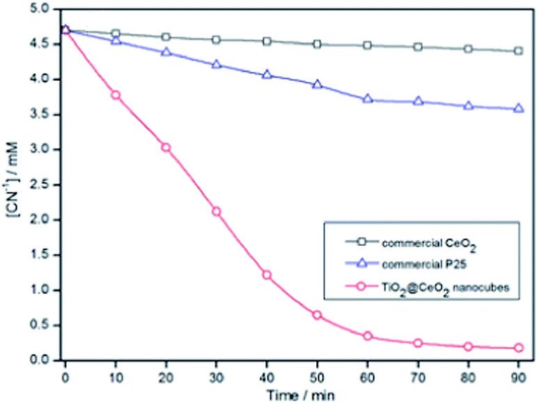 | ||
| Fig. 5 Photodecomposition results: comparison of detoxification of cyanide in the presence of commercial P25 and CeO2. pH = 12.5, catalyst loading = 0.02 g, airflow rate = 8.5 mL s−1, [O2]dissolved = 28.8 ppm. | ||
In summary, hollow TiO2@CeO2 hybrid nanocubes are successfully synthesized by a facile and fast benign procedure by a simple simultaneous coordinated etching reaction and subsequent calcinations. It exhibits a remarkable activity for photocatalytic detoxification of cyanide. This strategy is simple, cheap and mass-productive, which may shed light on a new avenue for large-scale synthesis of hollow cube-shaped structural nano/micro functional hybrid materials for catalyst, energy and other applications.
Acknowledgements
The authors would like to acknowledge financial support provided by the National Natural Science Foundation of China (no. 51474191 and no. 21467030) and the Natural Science Foundation of Yunnan Province (no. 2014FB103).Notes and references
- R. R. Dash, C. Balomajumder and A. Kumar, J. Chem. Eng., 2009, 146, 408 CrossRef CAS PubMed.
- J. Ma and P. K. Dasgupta, Anal. Chim. Acta, 2010, 673, 117 CrossRef CAS PubMed.
- J. N. Smith, A. Keil, J. Likens and R. G. NollCooks, Analyst, 2010, 135, 994 RSC.
- R. R. Dash, A. Gaur and C. Balomajumder, J. Hazard. Mater., 2009, 163, 1 CrossRef CAS PubMed.
- R. C. Rocha-e-Silva, L. A. V. Cordeiro and B. Soto-Blanco, Comp. Biochem. Physiol., Part C: Toxicol. Pharmacol., 2010, 151, 294 CrossRef PubMed.
- M. Banea, G. Nahimana, C. Mandombi, J. H. Bradbury, I. C. Denton and N. Kuwa, Food Chem. Toxicol., 2012, 50, 1517 CrossRef PubMed.
- J. Marugan, R. V. Grieken, A. E. Cassano and O. M. Alfano, Catal. Today, 2009, 144, 87 CrossRef CAS PubMed.
- J. Marugan, R. V. Grieken, A. E. Cassano and O. M. Alfano, Appl. Catal., B, 2008, 85, 48 CrossRef CAS PubMed.
- H. Guo, D. Tian, L. Liu, Y. Wang, Y. Guo and X. Yang, J. Solid State Chem., 2013, 201, 137 CrossRef CAS PubMed.
- A. Bozzi, I. Guasaquillo and J. Kiwi, Appl. Catal., B, 2004, 51, 203 CrossRef CAS PubMed.
- F. Mou, L. Xu, H. Ma, J. Guan, D. Chen and S. Wang, Nanoscale, 2012, 4, 4650 RSC.
- L. S. Zhang, K. H. Wong, Z. G. Chen, J. C. Yu, J. C. Zhao, C. Hu, C. Y. Chan and P. K. Wong, Appl. Catal., A, 2009, 363, 221 CrossRef CAS PubMed.
- M. C. Long, W. M. Cai, J. Cai, B. X. Zhou, X. Y. Chai and Y. H. Wu, J. Phys. Chem. B, 2006, 110, 820211 Search PubMed.
- S. Y. Chai, Y. J. Kim, M. H. Jung, A. K. Chakraborty, D. Jung and W. I. Lee, J. Catal., 2009, 262, 144 CrossRef CAS PubMed.
- Y. Bessekhouad, D. Robert and J.-V. Weber, J. Photochem. Photobiol., A, 2004, 163, 569 CrossRef CAS PubMed.
- J. Tian, Y. Sang, G. Yu, H. Jiang, X. Mu and H. Liu, Adv. Mater., 2013, 25, 5075 CrossRef CAS PubMed.
- J. Tian, Y. Sang, Z. Zhao, W. Zhou, D. Wang, X. Kang, H. Liu, J. Wang, S. Chen, H. Cai and H. Huang, Small, 2013, 9, 3864 CrossRef CAS PubMed.
- X. Lu, L. Yang, X. Bian, D. Chao and C. Wang, Part. Part. Syst. Charact., 2014, 31, 245 CrossRef CAS.
- J. Xie, D. Jiang, M. Chen, D. Li, J. Zhu, X. Lu and C. Yan, Colloids Surf., A, 2010, 372, 107 CrossRef CAS PubMed.
- Y. Liu, P. Fang, Y. Cheng, Y. Gao, F. Chen, Z. Liu and Y. Dai, Chem. Eng. J., 2013, 219, 478 CrossRef CAS PubMed.
- Z. F. Bian, J. Zhu, J. G. Wang, S. X. Xiao, C. Nuckolls and H. X. Li, J. Am. Chem. Soc., 2012, 134, 2325 CrossRef CAS PubMed.
- C. Karunakaran and P. Gomathisankar, ACS Sustainable Chem. Eng., 2013, 1, 1555 CrossRef CAS.
- H. Gnayem and Y. Sasson, ACS Catal., 2013, 3, 186 CrossRef CAS.
- H. Guo, W. Wang, L. Liu, Y. He, C. Li and Y. Wang, Green Chem., 2013, 15, 2810 RSC.
- H. Guo, Y. He, Y. Wang, L. Liu, X. Yang, S. Wang, Z. Huang and Q. Wei, J. Mater. Chem. A, 2013, 1, 7494 CAS.
- R. V. Gulyaev, A. I. Stadnichenko, E. M. Slavinskaya, A. S. Ivanova, S. V. Koscheev and A. I. Boronin, Appl. Catal., A, 2012, 439, 41 CrossRef PubMed.
- L. Q. Liu, F. Zhou, L. G. Wang, X. J. Qi, F. Shi and Y. Q. Deng, J. Catal., 2010, 274, 1 CrossRef CAS PubMed.
- Y. S. Bi, L. Chen and G. X. Lu, J. Mol. Catal. A: Chem., 2007, 266, 173 CrossRef CAS PubMed.
- L. L. Cai, G. Z. Lu, W. C. Zhan, Y. Guo, Y. L. Guo, Q. S. Yang and Z. G. Zhang, J. Mater. Sci., 2011, 46, 5639 CrossRef CAS.
- F. Mou, C. Chen, J. Guan, D. Chen and H. Jing, Nanoscale, 2013, 5, 2055 RSC.
- J. R. Parga, V. Vázquez, H. M. Casillas and J. L. Valenzuela, Chem. Eng. Technol., 2009, 32, 1901 CrossRef CAS.
Footnote |
| † Electronic supplementary information (ESI) available. See DOI: 10.1039/c4ra13898h |
| This journal is © The Royal Society of Chemistry 2015 |