The influence of pH and temperature on the stability of flutamide. An HPLC investigation and identification of the degradation product by EI+-MS

R. N. El-Shaheny*ab and K. Yamadaa
aDepartment of Pharmaceutical Sciences, Graduate School of Biomedical Science, Nagasaki University, 1-14 Bunkyo-machi, Nagasaki 852-8521, Japan. E-mail: rania_n2010@yahoo.com; Tel: +81-80-91096880
bAnalytical Chemistry Department, Faculty of Pharmacy, Mansoura University, Mansoura 35516, Egypt

Received 1st November 2014 , Accepted 2nd December 2014

First published on 2nd December 2014


Abstract

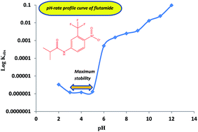
The chemical stability of flutamide (FLT) was investigated using a new validated stability-indicating HPLC method. Separation of FLT from its degradation product was achieved on a C18 column using a mobile phase of methanol–phosphate buffer (0.04 M, pH 4.0) (75[thin space (1/6-em)]:[thin space (1/6-em)]25, v/v) with UV-detection at 240 nm. The method exhibited excellent linearity for FLT over the concentration range of 0.2–25.0 μg mL−1. FLT was found to be labile to degradation in buffered, acidic and alkaline solutions. The degradation kinetics of FLT in aqueous solutions was evaluated as a function of pH and temperature. Degradation of FLT followed first-order kinetics and Arrhenius behavior over the temperature ranges of 70–100 and 60–90 °C under acidic and alkaline conditions, respectively. The pH-rate profile was studied over the pH range of 2.0–12.0 with a maximum stability at pH 3.0–5.0. The activation energies for hydrolysis of FLT were calculated as 79.4 and 52.0 kJ mol−1 at pH 0.5 (0.3 M HCl) and 12.5 (0.03 M NaOH), respectively. 4-Nitro-3-trifluoromethyl aniline was identified by mass spectrometry to be the degradation product resulting from the hydrolysis of FLT. The proposed HPLC method was validated according to ICH guidelines and applied for the quality control of FLT in commercial tablets with a mean percentage recovery of 100.09 ± 0.20%.


Introduction

Flutamide (FLT), propanamide, 2-methyl-N-[4-nitro-3-trifluoromethyl-phenyl],1 is a non-steroidal drug with anti-androgenic properties which appears to act by inhibiting the uptake and/or binding of androgens in target tissues. It is used, usually with gonadorelin analogues, in the palliative treatment of prostatic carcinoma. It is also used in combination with testolactone for the treatment of congenital adrenal hyperplasia.2

FLT is an official drug in the United States Pharmacopoeia (USP)1 that recommends an HPLC method for its determination in pure state and capsules. Also, it is authorized in the British Pharmacopoeia (BP)3 that describes a direct spectrophotometric assay at 295 nm for its determination in pure from. Reviewing the literature revealed some analytical methods for the determination of FLT such as spectrophotometry,4–7 HPLC,8–10 voltammetry,11–13 and GC-electron capture detection.14 Few analytical methods studied the stability of FLT.15,16 Nevertheless, such methods did not study the kinetics of the degradation process or elucidate the chemical structure of FLT degradant. So far, no study elucidated the pH-rate profile of FLT despite it provides useful information for the optimal formulation and storage conditions of pharmaceutical products.17 This encouraged us to develop a simple, rapid and accurate stability-indicating HPLC method for FLT with main targets of (i) studying its degradation kinetics, (ii) identification of possibly formed degradation products under different International Conference on Harmonisation (ICH)-outlined stress conditions,18,19 and (iii) elucidation of its pH-rate profile curve.

Experimental

Instruments

Chromatographic separation was achieved with a Hitachi HPLC system (Tokyo, Japan) equipped with 655A-11 liquid chromatograph, a high sensitivity series L-4000H UV-detector, D-2500 chromato-integrator, LC-organizer and a Rheodyne injector valve with a 50 μL sample loop. Positive electron ionization mass spectra (EI+-MS) were recorded using JMS DX-303 mass spectrometer (Joel Ltd., Japan). SK-620 pH meter (Sato Keiryoki MFG Co. Ltd, China) was used for pH adjustment. A BT-15 Yamato thermostatically controlled water bath (Tokyo, Japan) was used in the stability studies. Asone ultrasonic bath (Osaka, Japan) was used for sonication of solutions and mobile phase.

Chemicals and reagents

A gift sample of flutamide with a certified purity of 98.82% (batch # 9007) was kindly provided by Schering-Plough Co. (Cairo, Egypt). Cytomid® tablets labeled to contain 250 mg FLT per tablet (product of Cipla Ltd., Mumbai, India) were purchased from a local Egyptian pharmacy. Methanol (HPLC grade) was obtained from Kanto Chemical Co., INC. (Tokyo, Japan). Orthophosphoric acid (85% w/v) and potassium dihydrogen phosphate were obtained from Wako Pure Chemical Industries, Ltd. (Osaka, Japan). Hydrochloric acid (35–37%) and sodium hydroxide were obtained from Chameleon Reagent (Osaka, Japan). Hydrogen peroxide (30% w/v) was obtained from Nacalai Tesque, Inc. (Kyoto, Japan). Purified water was obtained using Millipore direct-Q 3UV water purification system (Molsheim, France). Aqueous solutions of 0.3 M HCl and 0.03 M NaOH were prepared. Phosphate buffer of pH 4.0 was prepared by adjusting the pH of 0.04 M potassium dihydrogen phosphate with few drops of orthophosphoric acid (85% w/v). Britton–Robinson buffer (BRB) containing 0.04 M boric acid, 0.04 M orthophosphoric acid, and 0.04 M acetic acid was prepared and the pH was adjusted to 2.0–12.0 with 0.2 M NaOH.20

Standard solutions

A stock solution of FLT containing 2000.0 μg mL−1 was prepared in methanol and a standard solution (200.0 μg mL−1) was prepared by dilution with the same solvent. Working solutions of appropriate concentrations were prepared by dilution with methanol. The solutions were stable for one week when stored in a refrigerator at 4 °C.

Chromatographic conditions

Separation was performed on a Cosmosil 5C18-MS column (150 mm × 4.6 mm id, 5 μm particle size) from Nacalai Tesque, Inc. A mobile phase consisted of methanol–phosphate buffer (0.04 M; pH 4.0) (75[thin space (1/6-em)]:[thin space (1/6-em)]25, v/v) was pumped at a flow rate of 1 mL min−1 and UV-detection was set at 240 nm.

Calculation of degradation kinetics parameters

Kinetics parameters for the degradation of FLT were calculated.21 The observed first-order degradation rate constants (k) were calculated from the slopes of semi-logarithmic plots of image file: c4ra13617a-t1.tif versus time (t) in accordance with eqn (1):
 
image file: c4ra13617a-t2.tif(1)
Where (a) is the initial drug concentration and (ax) is the remaining drug concentration.

Half-life times (t1/2), for the first order degradation reactions, were calculated according to eqn (2):

 
image file: c4ra13617a-t3.tif(2)

Activation energy (Ea) was calculated from the slope of Arrhenius plot of log[thin space (1/6-em)]k versus 1/T in accordance to eqn (3):

 
image file: c4ra13617a-t4.tif(3)
Where A is the frequency factor, R is the gas constant = 8.314 J mol−1 K−1 and T is the absolute temperature (°K = 273 + °C).

General recommended procedures

Calibration graph. Accurately measured volumes of FLT standard solution were diluted with the mobile phase to obtain final concentrations over the range of 0.2–25.0 μg mL−1. Twenty μL aliquots were injected (triplicate) and eluted with the mobile phase under the optimum chromatographic conditions. The average peak areas of FLT were plotted versus the corresponding drug concentrations (μg mL−1) and the regression equation was derived.
Assay of tablets. Ten tablets were accurately weighed, finely pulverized and thoroughly mixed. An accurately weighed amount of the powder equivalent to 10.0 mg FLT were transferred into 100 mL volumetric flask and diluted to the mark with methanol. The solution was sonicated for 30 min then filtered. Accurately measured volumes of the filtrate were diluted with the mobile phase and chromatographed under the optimum conditions. Nominal contents of FLT were calculated using the regression equation.
Degradation protocol. 0.5 mL aliquots of FLT standard solution (200.0 μg mL−1) were transferred into a series of 10 mL glass vials followed by 2 mL of 0.3 M HCl or 0.03 M NaOH. Solutions were then incubated in a thermostatically controlled water bath at different temperature settings (70–100 and 60–90 °C for acidic and alkaline degradation, respectively). Samples were taken at appropriate times (10–40 min), neutralized and made up to 5.0 mL with the mobile phase and mixed well. Twenty μL aliquots were injected and eluted under the optimum chromatographic conditions and the remaining drug concentrations were calculated using the regression equation. A plot of image file: c4ra13617a-t5.tif versus time (t) was constructed. The observed first-order degradation rate constants (k) and half-lives (t1/2), were calculated using eqn (1) and (2), respectively. Arrhenius plots for acidic and alkaline degradation were constructed by plotting log[thin space (1/6-em)]k versus 1/T (°K) and the activation energies (Ea) for FLT degradation were calculated from the slopes according to eqn (3).

Oxidative stress as well as neutral hydrolysis studies were also carried out as follow: 2 mL of H2O2 (30 w/v) or distilled water were added to 0.5 mL of FLT standard solution (200.0 μg mL−1) and heated for 2 h at 80 °C. For the photo-degradation study; 0.5 mL of FLT standard solution (200.0 μg mL−1) was transferred into a series of glass vials and diluted with 2 mL of methanol, water, or methanol–water mixture (1[thin space (1/6-em)]:[thin space (1/6-em)]1, v/v), then exposed to UV-lamp emitting radiation at a wavelength of 254 nm for 24 h. Then, the solutions were made up to 5 mL with the mobile phase, mixed well and eluted under the optimum chromatographic conditions.

pH-rate profile for FLT. 1 mL of FLT stock solution (2000.0 μg mL−1) was transferred into a series of 10 mL glass vials, 9 mL of BRB (pH = 2.0–12.0) were added and mixed well. Solutions were heated under reflux in a boiling water bath. 1 mL aliquots were taken at appropriate times (10–40 min), neutralized and diluted to 10 mL with the mobile phase then analyzed. The remaining drug concentrations were calculated from the regression equation. A plot of image file: c4ra13617a-t6.tif versus time (t) was constructed and the observed reaction rate constants (k) were calculated using eqn (1). The pH-rate profile curve was constructed by plotting log[thin space (1/6-em)]k versus pH of BRB.

Results and discussion

HPLC degradation studies of FLT revealed its stability under neutral, oxidative and photolytic conditions. Meanwhile, it was found susceptible to degradation under acidic and alkaline conditions yielding the same degradation product (DP). Hence, a kinetic study was conducted to explore the degradation process and pH-rate profile curve was also constructed.

Method development

The pH, composition, and flow rate of the mobile phase as well as detection wavelength were optimized to achieve good separation of FLT from its stress induced degradation product (DP) with high sensitivity. For optimization of HPLC conditions, stressed samples of FLT under acidic (0.3 M HCl) and alkaline (0.03 M NaOH) conditions at 80 °C for 30 min were chromatographed.

The pH of phosphate buffer was tested over the range of 3.0–7.0. It was found that, changing the pH exhibits a negligible effect on the retention of the drug and its degradation product. Hence, this study was conducted using phosphate buffer of pH 4.0 to confirm maximum stability and durability of the column. The percentage of methanol in the mobile phase was also studied over the range of 60–80%, v/v. A mobile phase containing 75%, v/v methanol was selected as the optimal yielding good resolution between FLT and DP within a short analysis time (less than 5 min). At lower concentrations of methanol, the retention times of both FLT and DP increased and peak broadening was observed, with a distinct effect on FLT (with a mobile phase containing 60% methanol, retention times of FLT and DP were 18 and 5 min, respectively). Meanwhile, on increasing the percentage of methanol to 80%, v/v, the retention of both components decreased (especially of FLT) and the resolution between FLT and DP was adversely affected. The influence of ionic strength of phosphate buffer was studied over the range of 0.01–0.05 M. 0.04 M phosphate buffer was used in the present study yielding highest number of theoretical plates (NTP) with good resolution (Rs). Flow rate of the mobile phase was also studied (0.8–1.2 mL min−1) and a flow rate of 1.0 mL min−1 was selected as optimal giving good resolution within short analysis time and highest NTP. Detector wavelength was investigated by monitoring the eluents responses at different wavelength settings (215, 240, 254 and 295 nm). Best sensitivities for both FLT and degradation product were attained at 240 nm. A summary of the influence of different chromatographic parameters on system suitability is illustrated in Table 1. Under the optimum chromatographic conditions, good resolution was achieved between the drug (tR = 4.0 min) and its degradation product (tR = 2.44 min) within a short analysis time.

Table 1 Effect of different chromatographic conditions on system suitability parameters of the proposed methoda
Parameter No of theoretical plates (NTP) Capacity factor (k′) Resolution (Rs) Relative retention (α)
FLT DP FLT DP
a System suitability parameters were calculated as per USP.1b Conditions selected as optimum.
Ratio of methanol (%, v/v)
60 11[thin space (1/6-em)]507 14[thin space (1/6-em)]578 10.47 2.23 21.83 4.70
65 9132 6180 4.11 1.10 13.66 3.74
70 4282 4343 2.50 0.76 11.04 3.29
75b 8864 3972 1.52 0.52 6.50 2.92
80 5744 2633 1.03 0.37 4.24 2.78
[thin space (1/6-em)]
pH of phosphate buffer
3.0 8132 3271 1.54 0.52 6.04 2.96
4.0b 8864 3972 1.52 0.52 6.50 2.92
5.0 8383 3164 1.27 0.40 6.00 3.18
6.0 8732 3191 1.34 0.41 5.71 3.27
7.0 8680 3462 1.49 0.49 6.72 3.04
[thin space (1/6-em)]
Ionic strength of phosphate buffer (M)
0.01 7659 3380 1.55 0.54 4.63 2.87
0.02 9540 3463 1.58 0.56 6.04 2.82
0.04b 10[thin space (1/6-em)]007 3631 1.70 0.60 6.26 2.83
0.05 8864 3972 1.52 0.52 6.50 2.92
[thin space (1/6-em)]
Flow rate (mL min−1)
0.8 5697 5053 1.61 0.64 5.11 2.52
1.0b 10[thin space (1/6-em)]010 3642 1.72 0.62 6.30 2.77
1.2 5816 2305 1.59 0.63 4.53 2.52


A comparison between the analytical performance of the proposed HPLC method and methods reported in the literature for the determination of FLT is presented in Table 2.

Table 2 Comparison between the proposed and reported methods for FLTa
Method Linearity range (μg mL−1) LOD (μg mL−1) Remarks Reference
a N/A: data are not available.
Spectrophotometry 0.25–7.0 N/A Harsh conditions, multiple derivatization steps, time consuming, not stability indicating assays 4
0.25–6.0
Spectrophotometry 0.8–6.0 0.132 Harsh conditions, multiple derivatization steps, time consuming, not stability indicating assays 5
0.8–14.0 0.288
Spectrophotometry N/A N/A Harsh conditions, multiple derivatization steps, time consuming, not stability indicating assay 6
Spectrophotometry 0.5–12 0.1197 Harsh conditions, multiple derivatization steps, time consuming, not stability indicating assays 7
0.5–10 0.1047
0.3–10 0.2587
HPLC-UV 2.9–11.6 N/A Poor sensitivity, narrow linearity range, not stability indicating assay 8
HPLC-FL 0.1–0.6 8.697 × 10⁻³ Very narrow linearity range, harsh and multiple steps derivatization conditions, not stability indicating assay 9
HPLC-UV 2.0–12.0 0.02 Narrow linearity range, not stability indicating assay 10
Voltammetry 20.0–160.0 0.05 Needs special polymer film modified carbon paste electrode, not stability indicating assay 11
Voltammetry N/A 0.052 Not stability indicating assays 12
0.024
0.003
Voltammetry 0.06–0.28 N/A Not stability indicating assay 13
GC-electron capture (63Ni) detection 0.002–0.3 N/A Not stability indicating assay, require special instrumentation and expertise, high cost 14
0.003–0.5
HPLC-UV 0.5–20.0 N/A Longer retention time, alkaline, oxidative, photolytic degradation, and pH-rate profile were not studied, DP wasn't identified 15
HPLC-diode array detection spectrophotometry voltammetry 0.138–138.0 N/A Time consuming, degradation kinetics and pH-rate profile were not studied, DP was not identified 16
0.276–276.0
0.524–276.0
HPLC-UV 0.2–25.0 0.05 Short retention time, wide linearity range, high sensitivity, stability indicating assay, degradation under different ICH-conditions was conducted, degradation kinetics was studied, pH-rate profile was constructed Present method


Method validation

A validation procedure was conducted according to ICH guidelines22 to evaluate linearity, range, limit of detection, limit of quantification, accuracy, precision, selectivity, robustness, system suitability, and stability of standard solution and mobile phase.
Linearity and range. Eight concentration levels were considered to study the linearity. Regression analysis of the data23 proved the excellent linearity of the proposed method over a wide dynamic range (0.2–25.0 μg mL−1) as revealed by high value of correlation coefficient (r = 0.9999), small values of the standard deviations of the residuals (Sy/x), slope (Sb), and intercept (Sa), and the % relative error. Table 3 illustrates the linear regression analysis data for FLT.
Table 3 Linear regression analysis data for the determination of FLT by the proposed method
Parameter Result
Concentration range (μg mL−1) 0.2–25.0
Limit of detection (LOD) (μg mL−1) 0.05
Limit of quantification (LOQ) (μg mL−1) 0.16
Correlation coefficient (r) 0.9999
Slope 9.83 × 10³
Intercept 1.22 × 10²
Standard deviation of the residuals (Sy/x) 6.65 × 10²
Standard deviation of the intercept (Sa) 1.61 × 10²
Standard deviation of the slope (Sb) 26.01
% RSD 1.39
% Error (% RSD/image file: c4ra13617a-t7.tif) 0.5


Limit of detection (LOD) and limit of quantification (LOQ). Both LOD and LOQ were calculated as per ICH guidelines22 according to the following equations:
 
LOQ = 10Sa/b (4)
 
LOD = 3.3Sa/b (5)
where Sa = the standard deviation of the intercept of regression line and b = the slope of regression line. The results are also presented in Table 3.
Accuracy. Accuracy of the proposed method was determined by recovery studies. The method was demonstrated to be accurate giving average recovery of 100.63 ± 1.40%. Statistical comparison of the obtained results with those of the reference HPLC method1 using the Student's t-test and the variance ratio F-test was done.23 Both the t- and the F-values obtained at the 95% confidence level, did not exceed the theoretical tabulated values.23 Therefore, there is no significant difference between the proposed and reference methods1 regarding the accuracy and precision (Table 4).
Table 4 Application of the proposed and reference methods for the determination of FLT in pure form and tablets
Matrix Proposed method Reference method1
Conc. taken (μg mL−1) Conc. found (μg mL−1) % Founda % Founda
a Each result is the average of three separate determinations.b Values between parentheses are the tabulated t- and F-values, respectively.23
Pure form 0.2 0.202 101.00 99.05
  0.4 0.407 101.75 100.16
1.0 1.027 102.70 100.25
4.0 4.033 100.83  
8.0 7.892 98.65  
15.0 14.882 99.21  
20.0 20.296 101.48  
25.0 24.863 99.45  
Mean ± SD     100.63 ± 1.40 99.82 ± 0.67
t     0.942(2.262)b
F     4.40(19.35)b
Cytomid® tablets (250 mg FLT per tablet) 1.0 1.003 100.30 99.68
15.0 14.987 99.91 98.25
25.0 25.365 101.46 100.25
Mean ± SD     100.09 ± 0.20 99.39 ± 1.03
t     1.54(2.776)b
F     1.63(19.00)b


Precision. The repeatability (intra-day precision) studies were performed by analysis of FLT at three concentration levels at three successive times on the same day, whereas, intermediate precision (inter-day precision) was checked by repeating the analysis of the three drug concentrations on three consecutive days. The precision was expressed as % RSD. Results of precision studies summarized in Table 5 confirm the precision of the method.
Table 5 Precision data of the proposed method for determination of FLT in pure form
Conc. (μg mL−1) % Found ± SD % RSD % Error
Intra-day precision
0.4 101.20 ± 0.34 0.34 0.20
10.0 100.54 ± 1.34 1.33 0.77
25.0 99.87 ± 1.50 1.50 0.90
[thin space (1/6-em)]
Inter-day precision
0.4 99.54 ± 0.92 0.93 0.53
10.0 100.20 ± 1.35 1.35 0.62
25.0 100.54 ± 0.53 0.53 0.30


Selectivity. Results of the forced degradation studies revealed the ability of the proposed method to entirely separate the drug from its degradation product with a good resolution factor (Rs). In addition no interference from common pharmaceutical excipients in the tablets with the peak of the drug was observed. These results indicated the selectivity of the proposed method.
Robustness. To determine the robustness of the proposed method with deliberated minor changes in chromatographic conditions, four parameters were varied: pH of the mobile phase (4.0 ± 0.2), ratio of methanol (75 ± 2%, v/v), molarity of phosphate buffer (0.04 ± 0.001 M) and detection wavelength (240 ± 2 nm). Introducing these small but deliberate variations didn't significantly affect the resolution or peak area of the analyte. These results demonstrated the robustness of the developed method.
System suitability. The system suitability parameters including Rs, NTP, capacity factor (), and selectivity factor (α) were calculated according to USP guidelines.1 The results are illustrated in Table 6.
Table 6 Final system suitability test parameters for FLT and DP by the proposed methoda
Parameter No of theoretical plates (NTP) Capacity factor (k′) Resolution (Rs) Relative retention (α)
FLT DP FLT DP
a System suitability parameters were calculated as per USP.1
Result 10[thin space (1/6-em)]010 3642 1.65 0.5 6.30 2.75


Stability of standard solution and mobile phase. Comparing the analytical response of freshly-prepared standard solution of FLT with that of an aged solution (24 h at room temperature) revealed the stability of the standard solution over this period. Similarly, the mobile phase was found to be stable up to 48 h.

Application

Degradation behavior of FLT under ICH conditions. FLT exhibited a high stability under photolytic, neutral hydrolysis and oxidative stress conditions, while it underwent rapid degradation under acidic and alkaline conditions yielding the same degradation product (DP). Fig. 1 illustrates representative chromatograms of FLT after acidic and alkaline treatments. Degradation of FLT in acidic and alkaline conditions followed first-order kinetics (Fig. 2), and the degradation process obeyed Arrhenius behavior over the temperature ranges of 70–100 and 60–90 °C under acidic and alkaline conditions, respectively. Linear plots for Arrhenius equations for the acidic and alkaline degradation were obtained as illustrated in Fig. 3. Table 7 represents a summary of the calculated k, t1/2, Ea and Arrhenius equations for FLT degradation in 0.3 M HCl and 0.03 M NaOH. The activation energies Ea for the acidic and alkaline degradation of FLT were found to be 79.4 and 52 KJ mol−1. These values are within the range of 50–96 KJ mol−1 most commonly reported and represents an intermediate sensitivity of the degradation process to temperature.21
image file: c4ra13617a-f1.tif
Fig. 1 Representative chromatograms for FLT (20.0 μg mL−1) after exposure to (A) alkaline degradation (0.03 M NaOH at 90 °C for 30 min) and (B) acidic degradation (0.3 M HCl at 90 °C for 30 min), where: (a) is the solvent front, NTMA is 4-nitro-3-trifuoromethyl aniline (DP).

image file: c4ra13617a-f2.tif
Fig. 2 Semi-logarithmic plots for the first-order degradation of FLT under (A) acidic (0.3 M HCl) and (B) alkaline (0.03 M NaOH) conditions at different temperature settings.

image file: c4ra13617a-f3.tif
Fig. 3 Arrhenius plots for (A) acidic (0.3 M HCl) and (B) alkaline (0.03 M NaOH) degradation of FLT.
Table 7 Kinetic parameters for the degradation of FLT under acidic and alkaline conditions
Condition Temperature (°C) k (min−1) t1/2 (min) Ea (kJ mol−1) Arrhenius equation
HCl (0.3 M) 70 2.78 × 10⁻³ 249 79.4 Log[thin space (1/6-em)]k = 9.60 − (4.15 × 10³/T)
80 9.06 × 10⁻³ 77
90 1.37 × 10⁻² 51
100 2.89 × 10⁻² 24
NaOH (0.03 M) 60 1.03 × 10⁻² 68 52.0 Log[thin space (1/6-em)]k = 6.14 − (2.72 × 10³/T)
70 1.63 × 10⁻² 43
80 3.25 × 10⁻² 22
90 4.46 × 10⁻² 15


The liability of the drug to hydrolysis under alkaline condition is very high compared with its behavior under acidic condition, as revealed from k and Ea values (Table 7), noting that the concentration of HCl is 10 times that of NaOH. FLT is an anilide derivative, in which the amide nitrogen is linked to an aromatic ring which can “pull” electron density from this atom, weakening the amide C–N bond. This ring can also stabilize the leaving group by delocalization of electron pair on nitrogen. As a result, the amide nitrogen could be attacked easier by the hydroxide ion (ŌH) than hydronium ion (H3O+), which rational the liability of the drug to hydrolysis under basic conditions more than under acidic conditions.

pH-rate profile of FLT. The degradation of FLT in BRB over pH range of 2.0–12.0 at boiling temperature resulted in the appearance of DP as a degradation product. All the correlation coefficients (r) of the semi-logarithmic plots of image file: c4ra13617a-t8.tif versus time were >0.98 with most of them >0.99 indicating that, the degradation of FLT followed first-order kinetics. The pH-rate profile of FLT over the pH range of 2.0–12.0 is illustrated in Fig. 4, showing maximum stability over pH range of 3.0–5.0. Degradation occurred more rapidly in basic solution with increased rate of degradation upon increasing the pH. At pH 3.0–5.0, the estimated degradation rate constant was in the range of 1.2 × 10⁻⁶–1.3 × 10⁻⁶ min−1 giving an estimated half-life (t1/2) of 13.4–12.3 months at 100 °C. In more acidic solution (pH 2.0) the degradation rate constant increased to 3.3 × 10⁻⁶ min−1.
image file: c4ra13617a-f4.tif
Fig. 4 pH-rate profile curve for FLT in BRB (0.04 M) at 100 °C.

The rapidness of the proposed method (separation was achieved within 5 min) together with its sensitivity, selectivity, and accuracy, permitted the analysis of large number of samples generated from pH-rate profile studies in a short time.

EI+-MS study of degradation product of FLT. HPLC analysis showed that hydrolysis of FLT in BRB and in 0.3 M HCl as well as 0.03 M NaOH solutions produced the same degradation product with a constant retention time (2.44 min). The corresponding EI+-MS spectrum of this compound yielded an m/z ratio of 206 which is consistent with 4-nitro-3-trifuoromethyl aniline (C7H5F3N2O2) formed as a result of hydrolysis of the amide linkage of FLT (Fig. 5). Fig. 6 illustrates the obtained EI+-MS spectra for FLT and its hydrolytic degradation product.
image file: c4ra13617a-f5.tif
Fig. 5 Degradation pathway of FLT under hydrolytic conditions.

image file: c4ra13617a-f6.tif
Fig. 6 EI+-MS spectra of (A) flutamide intact drug and (B) its hydrolytic degradation product.
Quality control of flutamide tablets. The suggested method was successfully applied for the determination of FLT in Cytomid® tablets. The results shown in Table 4 were satisfactory and with good agreement with the labeled amount. The obtained results indicated stability of FLT under storage conditions since no degradation product was detected. The results were statistically compared with those obtained by the official HPLC method1 using Student t- and variance ration F-tests.23 There was no significant difference between the two methods in terms of the mean values and standard deviation at the 95% confidence level (Table 4). The obtained results indicated the suitability of the proposed method for the quality control of FLT without interferences from common tablets excipients.

Conclusion

We developed a sensitive, accurate, precise, and rapid HPLC method able to discriminate between FLT and its degradation product. FLT was found to be very sensitive to alkaline and acidic media. The proposed method was managed to study the degradation kinetics of FLT under alkaline and acidic conditions and to elucidate its Arrhenius plots. In addition, the proposed method was applied to derive the pH-rate profile curve for FLT in BRB which exhibited highest stability over pH 3.0–5.0.

Acknowledgements

R.N.E. appreciated financial support received from the Ministry of Higher Education, Egyptian Government for the short-term scientific mission in Nagasaki University-Japan.

References

  1. The United States Pharmacopeia, The United States Pharmacopeial Convention, NF 29, Rockville, MD, 34th edn, 2011 Search PubMed.
  2. S. C. Sweetman, Martindale: The Complete Drug Reference, The Pharmaceutical Press, London, 36th edn, 2009, p. 725 Search PubMed.
  3. The British Pharmacopoeia, British Pharmacopoeial Commission, Her Majesty Stationery Office, London, 2013 Search PubMed.
  4. P. Nagaraja, K. R. Sunitha and M. F. Silwadi, J. Pharm. Biomed. Anal., 2000, 23, 617 CrossRef CAS.
  5. P. Nagaraja, H. R. Arun Kumar, R. A. Vasantha and H. S. Yathirajan, Int. J. Pharm., 2002, 235, 113 CrossRef CAS.
  6. P. Nagaraja, H. S. Yathirajan, H. R. A. Kumar and R. A. Vasantha, Indian J. Pharm. Sci., 2002, 64, 272 CAS.
  7. H. N. Deepakumari and H. D. Revanasiddappa, ISRN Spectrosc., 2012, 728594,  DOI:10.5402/2012/728594.
  8. H. R. N. Salgado, M. de Menezes and M. P. B. Storti, Acta Farm. Bonaerense, 2005, 24, 246 CAS.
  9. A. A. Smith, R. Manavalan, K. Kannan and N. Rajendiran, Int. J. PharmTech Res., 2009, 1, 360 Search PubMed.
  10. N. Bujji Babu and R. Ramesh Raju, J. Pharma Res., 2011, 4, 4556 Search PubMed.
  11. P. K. Brahman, R. A. Dar, S. Tiwari and K. S. Pitre, Colloids Surf., A, 2012, 396, 8 CrossRef CAS PubMed.
  12. E. Hammam, H. S. El-Desoky, K. Y. El-Baradie and A. M. Beltagi, Can. J. Chem., 2004, 82, 1386 CrossRef CAS.
  13. K. Pecková, M. Průchová, J. C. Moreira, J. Barek, J. Fischer and V. Vyskočil, Collect. Czech. Chem. Commun., 2011, 76, 1811 CrossRef.
  14. M. Schulz, A. Schmoldt, F. Donn and H. Becker, Eur. J. Clin. Pharmacol., 1988, 34, 633 CrossRef CAS.
  15. A. Miranda, I. Caraballo and M. Millán, Drug Dev. Ind. Pharm., 2002, 28, 413 CrossRef CAS PubMed.
  16. A. Álvarez-Lueje, C. Peña, L. J. Núñez-Vergara and J. A. Squella, Electroanalysis, 1998, 10, 1043 CrossRef.
  17. O. A. G. J. van der Houwen, M. R. de Loos, J. H. Beijnen, A. Bult and W. J. M. Underberg, Int. J. Pharm., 1997, 155, 137 CrossRef CAS.
  18. ICH Harmonised Tripartite Guideline, Stability Testing of New Drug Substances and Products, Q1A (R2), Geneva, 2003 Search PubMed.
  19. ICH Harmonised Tripartite Guideline, Stability Testing: Photostability Testing of New Drug Substances and Products, Q1B, Geneva, 1996 Search PubMed.
  20. H. T. K. Britton and R. A. Robinson, J. Chem. Soc., 1931, 1456–1462 RSC.
  21. A. T. Florence and D. Attwood, Physicochemical Principles of Pharmacy, The Pharmaceutical Press, London, 4th edn, 2006, pp. 93–138 Search PubMed.
  22. ICH Harmonised Tripartite Guidline, Validation of analytical procedures: text and methodology, Q2(R1), Geneva, 2005 Search PubMed.
  23. J. N. Miller and J. C. Miller, Statistics and Chemometrics for Analytical Chemistry, Pearson Education Limited, Harlow, England, 5th edn, 2005, pp. 39–73, 107–149, 256 Search PubMed.

This journal is © The Royal Society of Chemistry 2015
Click here to see how this site uses Cookies. View our privacy policy here.