Exploring the substrate-assisted acetylation mechanism by UDP-linked sugar N-acetyltransferase from QM/MM calculations: the role of residue Asn84 and the effects of starting geometries

Guangcai Maa, Na Chenga, Hao Sua and Yongjun Liu*ab
aSchool of Chemistry and Chemical Engineering, Shandong University, Jinan, Shandong 250100, China. E-mail: yongjunliu_1@sdu.edu.cn; Fax: +86 531 885 644 64; Tel: +86 531 883 655 76
bKey Laboratory of Tibetan Medicine Research, Northwest Institute of Plateau Biology, Chinese Academy of Sciences, Xining, Qinghai 810001, China

Received 28th October 2014 , Accepted 22nd December 2014

First published on 22nd December 2014


Abstract

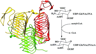
WlbB, one of the enzymes required for the biosynthesis of UDP-2,3-diacetamido-2,3-dideoxy-D-mannuronic acid (UDP-ManNAc3NAcA), is an N-acetyltransferase that catalyzes the N-acetylation of UDP-2-acetamido-3-amino-2,3-dideoxy-D-glucuronic acid (UDP-GlcNAc3NA) to form UDP-2,3-diacetamido-2,3-dideoxy-D-glucuronic acid (UDP-GlcNAc3NAcA). In this paper, based on the crystal structure, the detailed reaction mechanism of WlbB has been studied by using a combined QM/MM method. In particular, six snapshots taken from MD trajectories were used as the computational models to investigate how the starting geometries influence the calculation results. Our calculations suggest that the WlbB-catalyzed process involves two sequential steps. The nucleophilic attack of the C3-amino group of the substrate on the carbonyl carbon of acetyl-CoA occurs in concert with the departure of CoA from acetyl-CoA, generating a negatively charged CoA and a positively charged intermediate, which is inconsistent with the previous proposals that the catalytic reaction undergoes an oxyanion tetrahedral intermediate. Subsequently, the sulfur anion of CoA accepts the proton of the positively charged intermediate to yield the final product. Although Asn84 is not essential, it is important for promoting the catalysis by forming a hydrogen bond with the C3-amino group to position the lone pair of the electrons of the C3-amino group in an ideal orientation for nucleophilic attack and stabilize the transition states and intermediate. The cautious selection of initial geometries was found to be important for exploring the enzymatic mechanism and getting reliable energy barriers of the reaction pathways.


1. Introduction

N-Acetyltransferases (NATs), one of the largest enzyme superfamilies, catalyze the transfer of the acetyl group from acetyl coenzyme A (acetyl-CoA) to the amine group of various acceptor substrates ranging from simple small molecules to large proteins.1–3 NATs have been shown to be indispensable to numerous physiological processes, such as the activation and detoxification of carcinogens in humans,4 the inactivation of amino glycoside antibiotics in bacteria,5 the post-translational modification of histone proteins6 and the circadian rhythm in vertebrates.7

Up to now, enormous quantities of NATs have been identified, which can be sorted into two major classes: the Gcn5-related N-acetyltransferase (GNAT) and the left-handed parallel β-helix (LβH) superfamilies. The former usually function as dimers, and each monomer exhibits a structurally conserved fold composed of a mixed β-sheet flanked by irregular α-helices.7,8 Members of LβH superfamily typically function as trimers and adopt a conserved LβH structure with rare left-handed crossover connections and repeated isoleucine-rich hexapeptide motif, which was first observed in UDP-N-acetylglucosamine (UDP-GlcNAc) acyltransferase.9,10 In recent years, a number of members of the LβH superfamily have been structurally and mechanistically characterized, which often operate on dTDP-, UDP- or GDP-linked sugar substrates.11–17

ManNAc3NAcA, a reasonably rare di-N-acetylated deoxysugar, is found in the outer membranes of some Gram-negative pathogenic bacteria, such as Bordetella petrii, Pseudomonas aeruginosa and Bordetella pertussis.14,18,19 In these bacteria, the precursor of ManNAc3NAcA is UDP-ManNAc3NAcA, which is synthesized by the actions of five distinct enzymes, starting from UDP-GlcNAc. The fourth enzyme, also referred to as WlbB in Bordetella petrii, is an N-acetyltransferase that catalyzes the biotransformation of UDP-2-acetamido-3-amino-2,3-dideoxy-D-glucuronic acid (UDP-GlcNAc3NA) into UDP-2,3-diacetamido-2,3-dideoxy-D-glucuronic acid (UDP-GlcNAc3NAcA) by transferring an amino group from acetyl-CoA to UDP-GlcNAc3NA, as shown Scheme 1.


image file: c4ra13278e-s1.tif
Scheme 1 Schematic showing of the acylation reaction catalyzed by WlbB from Bordetella petrii.

The X-ray crystal structure of WlbB in complex with either UDP and acetyl-CoA or substrate and CoA has been determined to a high resolution of 1.43 Å.14 On the basis of amino acid sequence analysis, WlbB has been identified as a member of LβH superfamily of NATs. Similar to other LβH members, WlbB functions as a trimer, as displayed in Fig. 1a, and each active site is constructed at the interface of the two subunits. Structural analysis of WlbB reveals that only the side chain of Asn84 and the backbone amide group of Arg94 lie within hydrogen bonding distances to the substrate sugar ring.14


image file: c4ra13278e-f1.tif
Fig. 1 (a) Crystal structure of WlbB in the presence of CoA and substrate UDP-GlcNAc3NA. (b) Selected model for the QM/MM calculations in the present study.

Although all members of LβH superfamily have similar left-handed parallel β-helix structures, their active sites are remarkably different.11–16 So far, two possible N-acetylation mechanisms have been proposed. In some members, such as PglD and PerB, it has been suggested that an active site histidine residue acts as a general base to abstract the proton of the amino group of substrate to catalyze the formation of an oxyanion tetrahedral intermediate, and site-directed mutagenesis has confirmed its importance for catalysis.12,16 However, inspection of the active site structures of QdtC and WlbB demonstrates that there were no potential proton acceptors.13,14 In the active site of AntD, although Asp94 is located at hydrogen bonding distance to the sugar amino group, site-directed mutagenesis suggested that it may only play role in binding substrate, but does not serve as a catalytic base.15 In view of the experimental results, Holden and co-workers proposed that the QdtC-, WlbB- and AntD-catalyzed reactions proceed through a substrate-assisted mechanism, that is, the sulfur of CoA acts as the catalytic base to deprotonate the amino group of substrate.15 Note that in QdtC, WlbB and AntD, the substrates have been assumed in their deprotonated states to bind the active sites.13–15

Several theoretical studies have been done to explore the reaction mechanisms of human arylamine N-acetyltransferases and histone acetyltransferases of NATs.20–23 Although some experimental advances have been made in understanding the structures and mechanisms of the LβH superfamily, to the best of our knowledge, no theoretical study was reported yet. Furthermore, some key questions regarding the reaction mechanism of catalysis still remain unresolved: (1) what is the detailedreaction pathway? Whether the proposed oxyanion tetrahedral intermediate exists and whether the sulfur of CoA serves as the proton acceptor? (2) what are the energetics of the whole catalytic cycle? (3) how do the protein environments, especially some key residues in the active site influence the catalytic reaction? Answering the above questions can help us to understand the reaction mechanism of the LβH superfamily. Therefore, further theoretical study at the atomistic level is highly deserved.

In the present work, the detailed acetylation mechanism of WlbB from Bordetella petrii has been studied by using combined quantum mechanical/molecular mechanical (QM/MM) method. In recent years, QM/MM methodology has been successfully employed to investigate the chemical reactions, especially in the biomolecular systems.24–27 Previous theoretical studies of some enzymatic reactions have revealed the importance of selection of starting structure used for the QM/MM calculations.28,29 From the MD simulation, a large number of snapshots were usually observed; however, many of them might be irresponsible for the “reactive” conformations. Lonsdale et al. found that the unreasonable enzyme–substrate complex brings about unrealistically high energy barriers that are not representative of the true enzymatic reactivity.30 Actually, the QM/MM energy barriers are quite sensitive to small difference of the starting geometries. Thus, extensive analysis of the MD trajectories and careful screening of the snapshots are crucial to uncover the enzymatic reactions. Based on our calculations, the previously proposed mechanism was revised, and the effect of starting geometries on the reaction pathways was explored by extracting a series of snapshots. In addition, the energy barriers of the overall reaction, all the stable states and transition states along the reaction pathway as well as the specific role of Asn84 have been illuminated.

2. Computational details

System preparations

The enzyme model was constructed on the basis of the X-ray crystal structure of WlbB in complex with substrate UDP-GlcNAc3NA and CoA (PDB entry 3MQH).14 As mentioned above, WlbB is known as a trimer and exhibits three identical active sites wedged between each two subunits. Since the three reactive centers are remote from each other, therefore, only two of the three subunits with one substrate and one CoA were employed for our QM/MM calculations. The selected model is shown in Fig. 1b. The preparation of the system follows the following steps: (1) the CoA was decorated with an acetyl group to generate an acetyl-CoA; (2) the protonation states of all titratable residues were determined on the basis of the calculated pKa values using PROPKA program31 and verified by VMD program;32 (3) the force field parameters and atomic charges for substrate and acetyl-CoA were derived by analogy to the parameterized compounds presenting in the CHARMM22 force field;33 (4) the missing hydrogen atoms were added by using HBUILD facility implemented in the CHARMM package,34 and the water molecules presented in crystal structure were frozen at their original positions; (5) the system was hydrated with 36 Å sphere of pre-equilibrated TIP3 water molecules, and finally (6) seven Na+ were added at random positions to ensure an overall charge neutrality of the system. The whole solvated system was composed of 21[thin space (1/6-em)]120 atoms, including 5086 TIP3 water molecules, as shown in Fig. S1.

MD simulation

The solvated system was first submitted to a series of energy minimizations. Then MD simulation was carried out using the CHARMM22 all-atom force field33 included in CHARMM program.34 The system was first heated to 300 K for 200 ps, and then followed by 200 ps of equilibration at 300 K. Subsequently, a total of 20 ns MD simulation was performed with stochastic boundary condition. During the simulation, all the water molecules were allowed to move and the system was kept at 300 K and 1 bar. The integration time step is 1 fs. The SHAKE algorithm was used to constrain the covalent bonds involving the hydrogen atoms.35 The system was divided into two regions: the inner reaction (r < 33 Å) and outer buffer zone (33 Å < r < 36 Å). The inner region was treated with full Newtonian dynamics, whereas the buffer region was treated with Langevin dynamics. The root-mean-squared deviation (RMSD) for the backbone atoms of protein during the MD simulation was derived, as shown in Fig. S2. After 8 ns, the solvated system was basically equilibrated.

As mentioned above, the choice of initial structure can largely influence the accuracy of the QM/MM calculation results.28–30 To test how the initial structure influences the calculation results, six snapshots were randomly chosen as the initial models for the following QM/MM calculations from the MD trajectories at an interval of 3 ns from 5 ns to 20 ns, which were labeled as NAT-1 to NAT-6, respectively. These snapshots were used to explore the detailed reaction mechanism and the effects of starting structures on the reaction pathways, and to model the most reactive structure.

QM/MM calculations

All QM/MM calculations were performed on the basis of the six selected snapshots using ChemShell package36 integrating TURBOMOLE program37 for QM calculations and the DL-POLY program38 for MM calculations. The electronic embedding scheme39 was used to polarize the QM part by the MM point charges of the force field. No electrostatic cutoff was introduced for the MM/MM and QM/MM interactions. Hydrogen link atoms were employed to saturate the valences of the QM boundary atoms with the charge shift scheme.40 The QM calculations were carried out using B3LYP functional in combination with 6-31G(d,p) basis set for geometry optimizations. Although more benchmark studies have suggested that B3LYP is not one of the best functionals, it still remains a valid and particularly efficient alternative for the investigation of most chemical properties.41–43 The MM region was described at CHARMM22 force field as included in DL-POLY program.

Unless noted otherwise, the QM region contains the glycosyl moiety of substrate UDP-GlcNAc3NA, a part of acetyl-CoA and the side chain of Asn84 (56 atoms), as shown in Fig. 2. The remaining atoms of the solvated model were assigned to the MM region. In some additional calculations, to investigate how the selection of QM region influences the calculation results, the Asn84 was left out (47 atoms) or the backbone amino group of Arg94, Gln59 and three solvent water molecules (85 atoms) were added to the QM region. During the QM/MM calculations, we defined an active region including the QM region and the MM atoms within 15 Å of the N3 atom of substrate. The active region was fully optimized, whereas all the remaining atoms were fixed. Geometry optimizations were performed with the hybrid delocalized internal coordinates (HDLC) optimizer,44 in which the quasi-Newton limited memory Broyden–Fletcher–Goldfarb–Shanno (L-BFGS) method45,46 was used to locate minima and the partitioned rational function optimization (P-RFO) algorithm47 was used for transition state searches.


image file: c4ra13278e-f2.tif
Fig. 2 The QM region (in ball and stick model) that contains the sugar ring of UDP-GlcNAc3NA, a part of acetyl-CoA and the side chain of Asn84.

Considering six MD snapshots were used to investigate the detailed reaction pathways, we calculated the average values of the energy barriers of the pathways to analyze the results of the QM/MM optimizations. The averages were calculated according to the Boltzmann-weighted average equation:

 
image file: c4ra13278e-t1.tif(1)
where ΔEave is the average barrier, R is the gas constant, T is the temperature (300 K), n is the number of energy profiles, and ΔEi refers to the energy barrier of pathway i. Note that this approach can effectively lower the contributions of unreasonably high barriers. Assuming that all initial geometries are thermally accessible, it is expected that the smaller barrier corresponds to the more likely reaction pathway.

3. Results and discussion

3.1. Catalytic mechanism

Using six MD snapshots as initial models, we investigated the detailed reaction mechanism of WlbB. On the basis of our QM/MM calculations, the previously proposed acetylation mechanism has been revised, as outlined in Scheme 2. The whole WlbB-catalyzed process contains two sequential reaction steps: nucleophilic attack and proton transfer. The C3-amino group of substrate UDP-GlcNAc3NA (ES) first attacks the carbonyl carbon of acetyl-CoA and simultaneously the CoA departs from acetyl-CoA to form a negatively charged CoA and a positively charged intermediate (EI), which is inconsistent with previous proposal that reaction undergoes an oxyanion tetrahedral intermediate.13–15 The sulfur anion of CoA subsequently acts as the general base to deprotonate the amino group of the intermediate to yield the final product UDP-GlcNAc3NAcA (EP).
image file: c4ra13278e-s2.tif
Scheme 2 Proposed substrate-assisted acetylation mechanism of WlbB based on the QM/MM calculations.

Energetic data for the reaction pathways of the six models are listed in Table 1. The energy barriers range from 16.9 to 22.1 kcal mol−1 for nucleophilic attack process and from 6.6 to 12.0 kcal mol−1 for proton transfer process. Ignoring the possible errors in the QM/MM calculations, the wide fluctuation of the energy barriers for these models is mainly due to the conformational variability of snapshots. The Boltzmann-averaged energy barrier is 17.9 kcal mol−1 for nucleophilic attack process and 10.4 kcal mol−1 for proton transfer process. Based on the Boltzmann-weighted average equation, the lowest barrier contributes most to the average. Note that a simple sample of six snapshots should not be expected to provide an accurate average. The effects of starting geometries on the reaction pathways will be discussed in Section 3.3. The calculated barriers show that the nucleophilic attack step is rate-limiting.

Table 1 Summary of the calculated QM/MM relative energies (kcal mol−1) of WlbB-catalyzed processes for the six models from NAT-1 to NAT-6
Model ES TS1 EI TS2 EP
NAT-1 0.0 22.1 10.9 14.1 −11.2
NAT-2 0.0 19.8 12.0 13.7 −10.3
NAT-3 0.0 20.3 11.4 13.6 −11.1
NAT-4 0.0 18.9 8.6 10.7 −15.1
NAT-5 0.0 16.9 6.6 9.4 −13.6
NAT-6 0.0 19.7 11.0 13.1 −11.0


For NAT-5, the barriers are obviously smaller than those of the other models, suggesting this model should be more preferred to catalyze the acetylation reaction. Here, we utilize model NAT-5 as the example to illustrate the detailed reaction mechanism of WlbB. Optimized structures of reactant complex (ES), transition states (TS1 and TS2), intermediate (EI) and product complex (EP) for NAT-5 are shown in Fig. 3. In ES, the distances of R(N3–C) and R(C–S) are 2.90 and 1.80 Å, respectively. The acyl O atom of Asn84 forms a hydrogen bond with H1 atom of C3-amino group with a distance of 2.07 Å. This hydrogen bond induces the amino nitrogen to position its lone pair of electrons in an ideal orientation for nucleophilic attack. The detailed discussion on the specific role of Asn84 is described in Section 3.2. Two-dimensional potential energy surface (PES) for the nucleophilic attack process has been mapped out using the distances of R(N3–C) and R(C–S) as reaction coordinates (see Fig. 4). On this contour diagram, the horizontal axis characterizes the formation of N3–C bond, and the vertical axis characterizes the cleavage of C–S bond. On the basis of the optimized ES, the R(N3–C) was scanned with a decrement of 0.05 Å and the R(C–S) was scanned with an increment of 0.05 Å. In TS1, the distance of R(N3–C) is 1.73 Å, while the distance of R(C–S) is 1.97 Å. This suggests that the nucleophilic attack process proceeds through a concerted mechanism, in the transition state the N3–C bond was partially formed and simultaneously the C–S bond was partially broken. The energy barrier of this step is calculated to be 16.9 kcal mol−1, which is smaller than those of the other models. The calculated barrier is slightly overestimated compared with the experimental estimations of 15–16 kcal mol−1, which is deduced from the kcat values of 8 and 72 s−1 for the QdtC- and AntD-catalyzed N-acetylation reactions, respectively.13,15 Another local energy minimum located in the PES corresponds to the intermediate EI. In EI, the C–S bond has been completely broken, and the negatively charged sulfur of CoA adjusts its position to form a hydrogen bond with H2 atom of the protonated amino group with a distance of 2.13 Å. Compared with ES, this intermediate is quite unstable. The calculation results confirm that the sulfur of CoA functions as a general base to remove the H2 proton via a low-energy transition state (TS2). In TS2, the distance of H2–S is shortened to 1.73 Å and the distance of N3–H2 is changed to 1.21 Å. The calculated barrier of proton transfer is 2.8 kcal mol−1 relative to EI, suggesting this step is easy to occur. The final product (EP), UDP-GlcNAc3NAcA, has been demonstrated to be the substrate of 2-epimerase (WlbD), which is the last enzyme required for the biosynthesis of UDP-ManNAc3NAcA.14


image file: c4ra13278e-f3.tif
Fig. 3 Optimized structures of reactant (ES), transition states (TS1 and TS2), intermediate (EI) and product (EP) for model NAT-5.

image file: c4ra13278e-f4.tif
Fig. 4 PES calculated at the B3LYP/6-31G** level for the nucleophilic attack process using distances R(N3–C) and R(C–S) as reaction coordinates.

3.2. Role of residue Asn84

The WlbB sequence shows 36% similarity with QdtC, and structural analysis demonstrates that both of them have no potential proton acceptor, except for an asparagine residue (Asn159 in QdtC and Asn 84 in WlbB) which is located within hydrogen-bonding distance to the amino group of substrate in the active pocket.13,14 In QdtC, mutation of Asn159 to Ala159 only slightly lowers the enzymatic activity, indicating this residue was not essential for catalysis.14 Taking these into consideration, further theoretical calculations were performed to explore the specific role of Asn84 for the WlbB-catalyzed reaction. On the basis of the optimized reactant complex of NAT-5, Asn84 was mutated into Ala84 (model A). The optimized structures of stably local minima and transition states for this model are shown in Fig. S3. The absence of Asn84 makes the amino group of substrate away from the carbonyl carbon of acetyl-CoA and causes the lone pair of electrons of amino nitrogen in an improper position for the subsequent nucleophilic attack reaction. The energy barrier for the nucleophilic attack process of Asn84Ala mutant is calculated to be 24.9 kcal mol−1, which is considerably higher than that of NAT-5, as shown in Table 2. Mutation of Asn84 into Ala84 significantly reduces but does not completely abolish the enzymatic activity, demonstrating Asn84 being important for promoting catalysis. Meanwhile, switching Asn84 from the QM region to MM region (model B) slightly raises the barrier heights of the nucleophilic attack and proton transfer processes compared with NAT-5. Besides, we further constructed a larger model (model C) in which the backbone NH group of Arg94, the side chain of Gln59, and three solvent water molecules were added to the QM part. These groups form hydrogen bonds to the glycosyl moiety of substrate. Compared with NAT-5, the energy barrier heights of model C are only slightly decreased, suggesting these groups have no crucial impact on the barrier heights of the reaction pathway, which in turn demonstrates that the original selection of QM region is reasonable. The optimized structures of stable minima and transition states for model B and C are shown in Fig. S4 and S5, respectively.
Table 2 Summary of QM/MM relative energies (kcal mol−1) calculated for the different models based on the optimized reactant complex of model NAT-5
Model ES TS1 EI TS2 EP
a Asn84 was removed from the QM part and mutated into Ala84.b Asn84 was left out to the QM region.c backbone NH group of Arg94, Gln59 and three solvent water molecules were added to the QM part.
Aa 0.0 24.9 14.2 16.0 −12.2
Bb 0.0 17.7 9.9 12.6 −11.2
Cc 0.0 16.2 6.1 8.0 −15.0


Based on the experimental observations and our QM/MM calculation results, we come to a conclusion that although Asn84 does not directly take part in the WlbB-catalyzed reaction, it is important for promoting the activity of the enzyme by forming hydrogen bond with the C3-amino group of the substrate. On the one hand, the hydrogen bond stabilizes the transition states and intermediate along the reaction pathway. On the other hand, the presence of Asn84 positions the lone pair of electrons of C3-amino group in a right orientation for nucleophilic attack. As mentioned above, mutation of Asn84 into Ala84 remarkably reduces the catalytic efficiency.

3.3. Effects of starting geometries

It is well known that a reasonable conformation of enzyme–substrate complex is crucial for depicting the reaction pathway. To explore how the starting geometries influence the QM/MM calculation results, we extracted six snapshots from the MD trajectories as the initial models (NAT-1 to NAT-6). Table 3 shows the relative QM/MM (ΔEQM/MM), QM with and without MM point charges (ΔEQM+ptch and ΔEQM) and MM energies (ΔEMM) for the six optimized reactant complex. A substantial energy spread is observed in the ΔEQM/MM, which is mainly attributed to the variation in ΔEMM. It is predictable, since the relative orientations of protein side chains constantly change during the MD simulation, as shown in Fig. S6. The impact of the conformational variability on the energy differences is significant. However, the variations in the ΔEQM+ptch and ΔEQM are minor, indicating the atoms of QM parts are well superposed (Fig. S6). We also note that although the total energy of the reactant of NAT-5 is much lower than that of other models, the EQM+ptch and EQM are not the smallest ones.
Table 3 Relative QM/MM energies (ΔEQM/MM), QM energies of the QM/MM-optimized structure with and without MM point charges (ΔEQM+ptch and ΔEQM) and MM energies (ΔEMM) (kcal mol−1) for the six optimized reactant complexes from NAT-1 to NAT-6
  ΔEQM/MM ΔEQM+ptch ΔEQM ΔEMM
NAT-1 368.1 −17.2 0.6 385.3
NAT-2 402.3 −14.3 2.2 416.6
NAT-3 215.7 9.3 0.5 206.4
NAT-4 445.1 2.2 −0.5 443.0
NAT-5 0.0 0.0 0.0 0.0
NAT-6 135.9 8.4 −1.0 127.5


As aforementioned, the calculated barriers for nucleophilic attack processes range from 16.9 to 22.1 kcal mol−1 and from 6.6 to 12.0 kcal mol−1 for proton transfer processes (Table 1). This illustrates that the initial geometries remarkably influence the individual energy barriers of the WlbB-catalyzed processes. Key distances in the optimized structures of all species during the reactions for the six models are given in Table S1. Comparison of the enzyme–substrate complexes (ESs) reveals that in NAT-1 the distances of R(N3–C) and R(O–H1) are 2.96 and 2.18 Å, respectively, which are longer than those of the other models. This result in the NAT-1 desires more nucleophilic activation energy than the others. In contrast, NAT-5 exhibits more suitable N3–C distance and stronger hydrogen-bonding interaction between Asn84 and the amino group, thereby inducing the nucleophilic attack reaction to occur more readily. Whereas in NAT-2 the distance of R(N3–C) is only 0.01 Å shorter than that of NAT-5, the hydrogen-bonding distance of R(O–H1) is much longer, which increases the energy barrier for nucleophilic attack process and in turn affirms the importance of Asn84 for catalysis. Previous QM/MM calculations have revealed that not only the active pocket residues but also the residues in considerable distance to active center have important influence on the reaction barriers.48 Although no residue is involved in the acetylation reaction, the residues and water molecules that locate within hydrogen bonding distances to the substrate and cofactor are important for substrate and cofactor binding. Undoubtedly, unreasonable conformational changes of these residues may more or less influence the enzymatic reaction. In these six models, NAT-5 shows the relatively lower energy barriers for both nucleophilic attack and proton transfer processes (16.9 and 9.4 kcal mol−1) than those of the others. Furthermore, the total energy of NAT-5 is significantly lower than that of the other models. All these results suggest that the reaction path described by NAT-5 should be the most preferred one. Given the conformational complexity of the enzyme–substrate complex and the unexpected errors in the QM/MM calculations, the careful selection of MD snapshots is vital for exploring the reaction mechanism and getting accurate results.

4. Conclusions

In this article, an extensive QM/MM study of the acetylation mechanism of WlbB was reported. Based on our QM/MM calculation results, the previously proposed substrate-assisted acetylation mechanism has been revised. The WlbB-catalyzed process involves two sequential steps: nucleophilic attack and proton transfer processes. The nucleophilic attack of the C3-amino group of substrate on the carbonyl carbon of acetyl-CoA and the departure of CoA from acetyl-CoA occur simultaneously to form a negatively charged CoA and a positively charged intermediate, which is incompatible with the previous proposals that the reaction undergoes an oxyanion tetrahedral intermediate. The sulfur anion of CoA subsequently accepts the extra proton of the intermediate to yield the final product. Intriguingly, although Asn84 is not essential for catalysis, it is not a residue to be trifled with. Our calculations confirmed that Asn84 plays important roles in positioning the substrate and stabilizing the intermediate and transitions states by forming a hydrogen bond to the C3-amino group of substrate.

Six snapshots taken from MD trajectories were used as the initial models to explore the effects of starting geometries. The calculation results insist on the fact that the selection of initial geometries significantly affects the individual energy barriers of the enzymatic reaction, in other words, a reasonable starting geometry is crucial for exploring the enzymatic activity.

Despite the fact that WlbB demonstrates a lack of a catalytic base to deprotonate the amino group of substrate, some other members of LBH superfamily, such as AntD from Bacillus cereus, PerB from Caulobactercrescentus and PglD from Campylobacter jejuni, have an active site residue probably functions as the potential proton acceptor. However, whether the residue is directly involved in enzymatic reaction is still open to debate, which deserves further theoretical studies.

Acknowledgements

This work was supported by the Natural Science Foundation of China (21373125, 21173129).

References

  1. D. A. P. Evans, Pharmacol. Ther., 1989, 42, 157–234 CrossRef CAS.
  2. F. Dyda, D. C. Klein and A. B. Hickman, Annu. Rev. Biophys. Biomol. Struct., 2000, 29, 81–103 CrossRef CAS PubMed.
  3. E. Sim, K. Walters and S. Boukouvala, Drug Metab. Rev., 2008, 40, 479–510 CrossRef CAS PubMed.
  4. N. Brockton, J. Little, L. Sharp and S. C. Cotton, Am. J. Epidemiol., 2000, 151, 846–861 CrossRef CAS.
  5. J. Davies and G. D. Wright, Trends Microbiol., 1997, 5, 234–240 CrossRef CAS.
  6. S. Timmermann, H. Lehrmann, A. Polesskaya and A. Harel-Bellan, Cell. Mol. Life Sci., 2001, 58, 728–736 CrossRef CAS.
  7. D. C. Klein and J. L. Weller, Science, 1970, 169, 1093–1095 CAS.
  8. M. W. Vetting, L. P. S. de Carvalho, M. Yu, S. S. Hegde, S. Magnet, S. L. Roderick and J. S. Blanchard, Arch. Biochem. Biophys., 2005, 433, 212–226 CrossRef CAS PubMed.
  9. C. R. Raetz and S. L. Roderick, Science, 1995, 270, 997–1000 CAS.
  10. G. Parisi and J. Echave, BMC Evol. Biol., 2004, 4, 41 CrossRef PubMed.
  11. E. S. Rangarajan, K. M. Ruane, T. Sulea, D. C. Watson, A. Proteau, S. Leclerc, M. Cygler, A. Matte and N. M. Young, Biochemistry, 2008, 47, 1827–1836 CrossRef CAS PubMed.
  12. N. B. Olivier and B. Imperiali, J. Biol. Chem., 2008, 283, 27937–27946 CrossRef CAS PubMed.
  13. J. B. Thoden, P. D. Cook, C. Schaffer, P. Messner and H. M. Holden, Biochemistry, 2009, 48, 2699–2709 CrossRef CAS PubMed.
  14. J. B. Thoden and H. M. Holden, Biochemistry, 2010, 49, 4644–4653 CrossRef CAS PubMed.
  15. R. L. Kubiak and H. M. Holden, Biochemistry, 2012, 51, 867–878 CrossRef CAS PubMed.
  16. J. B. Thoden, L. A. Reinhardt, P. D. Cook, P. Menden, W. W. Cleland and H. M. Holden, Biochemistry, 2012, 51, 3433–3444 CrossRef CAS PubMed.
  17. D. P. Chantigian, J. B. Thoden and H. M. Holden, Biochemistry, 2013, 52, 8374–8385 CrossRef CAS PubMed.
  18. E. L. Westman, A. Preston, R. A. Field and J. S. Lam, J. Bacteriol., 2008, 190, 6060–6069 CrossRef CAS PubMed.
  19. E. L. Westman, D. J. McNally, A. Charchoglyan, D. Brewer, R. A. Field and J. S. Lam, J. Biol. Chem., 2009, 284, 11854–11862 CrossRef CAS PubMed.
  20. Q. A. Qiao, C. L. Yang, R. J. Qu, Y. Q. Jin, M. S. Wang, Z. H. Zhang, Q. Xu and Z. X. Yu, Biophys. Chem., 2006, 122, 215–220 CrossRef CAS PubMed.
  21. E. Y. Lau, J. S. Felton and F. C. Lightstone, Chem. Res. Toxicol., 2006, 19, 1182–1190 CrossRef CAS PubMed.
  22. J. F. Jiang, J. Y. Lu, D. Lu, Z. J. Liang, L. C. Li, S. S. Ouyang, X. Q. Kong, H. L. Jiang, B. R. Shen and C. Luo, PLoS One, 2012, 7, e36660 CAS.
  23. X. L. Zhang, S. S. Ouyang, X. Q. Kong, Z. J. Liang, J. Y. Lu, K. K. Zhu, D. Zhao, M. Y. Zheng, H. L. Jiang, X. Liu, R. Marmorstein and C. Luo, J. Phys. Chem. B, 2014, 118, 2009–2019 CAS.
  24. C. S. Li and S. Shaik, RSC Adv., 2013, 3, 2995–3005 RSC.
  25. K. Gkionis, S. T. Mutter and J. A. Platts, RSC Adv., 2013, 3, 4066–4073 RSC.
  26. M. Zheng and D. G. Xu, J. Phys. Chem. B, 2013, 117, 10161–10172 CrossRef CAS PubMed.
  27. X. L. Pan, F. C. Cui, W. Liu and J. Y. Liu, J. Phys. Chem. B, 2012, 116, 5689–5693 CrossRef CAS PubMed.
  28. F. Claeyssens, K. E. Ranaghan, F. R. Manby, J. N. Harvey and A. J. Mulholland, Chem. Commun., 2005, 5068–5070 RSC.
  29. A. Lodola, M. Mor, J. Zurek, G. Tarzia, D. Piomelli, J. Harvey and A. Mulholland, Biophys. J., 2007, 92, L20–L22 CrossRef CAS PubMed.
  30. R. Lonsdale, J. N. Harvey and A. J. Mulholland, J. Phys. Chem. B, 2010, 114, 1156–1162 CrossRef CAS PubMed.
  31. M. H. M. Olsson, C. R. Søndergard, M. Rostkowski and J. H. Jensen, J. Chem. Theory Comput., 2011, 7, 525–537 CrossRef CAS.
  32. W. Humphrey, A. Dalke and K. Schulten, J. Mol. Graphics, 1996, 14, 33–38 CrossRef CAS.
  33. A. D. MacKerell Jr, D. Bashford, M. Bellott, R. L. Dunbrack Jr, J. D. Evanseck, M. J. Field, S. Fischer, J. Gao, H. Guo, S. Ha, D. Joseph-McCarthy, L. Kuchnir, K. Kuczera, F. T. K. Lau, C. Mattos, S. Michnick, T. Ngo, D. T. Nguyen, B. Prodhom, W. E. Reiher III, B. Roux, M. Schlenkrich, J. C. Smith, R. Stote, J. Straub, M. Watanabe, J. Wiórkiewicz-Kuczera, D. Yin and M. Karplus, J. Phys. Chem. B, 1998, 102, 3586–3616 CrossRef PubMed.
  34. B. R. Brooks, R. E. Bruccoleri, B. D. Olafson, D. J. States, S. Swaminathan and M. Karplus, J. Comput. Chem., 1983, 4, 187–217 CrossRef CAS.
  35. J. P. Ryckaert, G. Ciccotti and H. J. C. Berendsen, J. Comput. Phys., 1977, 23, 327–341 CrossRef CAS.
  36. P. Sherwood, A. H. de Vries, M. F. Guest, G. Schreckenbach, C. R. A. Catlow, S. A. French, A. A. Sokol, S. T. Bromley, W. Thiel, A. J. Turner, S. Billeter, F. Terstegen, S. Thiel, J. Kendrick, S. C. Rogers, J. Casci, M. Watson, F. King, E. Karlsen, M. Sjøvoll, A. Fahmi, A. Schäfer and C. Lennartz, J. Mol. Struct., 2003, 632, 1–28 CrossRef CAS.
  37. R. Ahlrichs, M. Bar, M. Haser, H. Horn and C. Kolmel, Chem. Phys. Lett., 1989, 162, 165–169 CrossRef CAS.
  38. W. Smith and T. R. Forester, J. Mol. Graphics, 1996, 14, 136–141 CrossRef CAS.
  39. D. Bakowies and W. Thiel, J. Phys. Chem., 1996, 100, 10580–10594 CrossRef CAS.
  40. A. H. de Vries, P. Sherwood, S. J. Collins, A. M. Rigby, M. Rigutto and G. J. Kramer, J. Phys. Chem. B, 1999, 103, 6133–6141 CrossRef CAS.
  41. S. F Sousa, P. A. Fernandes and M. J. Ramos, J. Phys. Chem. A, 2007, 111, 10439–10452 CrossRef PubMed.
  42. R. P. P. Neves, P. A. Fernandes, A. J. Varandas and M. J. Ramos, J. Chem. Theory Comput., 2014, 10, 4842–4856 CrossRef CAS.
  43. N. F. Brás, M. A. Perez, P. A. Fernandes, P. J. Silva and M. J. Ramos, J. Chem. Theory Comput., 2011, 7, 3898–3908 CrossRef.
  44. S. R. Billeter, A. J. Turner and W. Thiel, Phys. Chem. Chem. Phys., 2000, 2, 2177–2186 RSC.
  45. J. Nocedal, Math. Comput., 1980, 35, 773–782 CrossRef.
  46. D. C. Liu and J. Nocedal, Math. Prog., 1989, 45, 503–528 CrossRef.
  47. A. Banerjee, N. Adams, J. Simons and R. Shepard, J. Phys. Chem., 1985, 89, 52–57 CrossRef CAS.
  48. J. B. Rommel and J. Kästner, J. Am. Chem. Soc., 2011, 133, 10195–10203 CrossRef CAS PubMed.

Footnote

Electronic supplementary information (ESI) available. See DOI: 10.1039/c4ra13278e

This journal is © The Royal Society of Chemistry 2015
Click here to see how this site uses Cookies. View our privacy policy here.