Advanced construction of heterostructured InCrO4–TiO2 and its dual properties of greater UV-photocatalytic and antibacterial activity

J. Kamalakkannan, V. L. Chandraboss, S. Prabha and S. Senthilvelan*
Department of Chemistry, Annamalai University, Annamalainagar 608 002, India. E-mail: dr_senthilvel@yahoo.co.in

Received 15th October 2014 , Accepted 15th December 2014

First published on 17th December 2014


Abstract

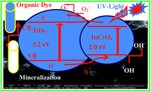
The development of coupled semiconductor oxides has led to significant advances in photocatalytic functional materials. In this article, we report the preparation of a nano-spherical InCrO4-loaded TiO2 photocatalyst by a simple co-precipitation method. The InCrO4–TiO2 (ICT) nanomaterial was characterized using XRD, SEM with EDX, FT-IR, FT-Raman and PL analysis. The photocatalytic activity of InCrO4–TiO2 was much better than that of TiO2 and TiO2–P25 under UV-light irradiation. InCrO4–TiO2 showed better activity than TiO2 and TiO2–P25 in the degradation of methyl green (MEG), malachite green (MAG) and methylene blue (MB) because it has maximum efficiency at neutral pH = 7 under UV-light irradiation. Among all these dyes, the photodegradation of MEG was the fastest. The mechanism for the photocatalytic activity of the InCrO4-loaded TiO2 nanomaterial has been discussed. The production of hydroxyl radicals on the surface of UV-irradiated photocatalysts was determined by fluorescence technique using coumarin as a probe molecule. The photodegradation of dyes is well described by pseudo first order kinetics and high quantum yield. Furthermore, the antibacterial activity of the TiO2 and InCrO4–TiO2 materials was investigated against Gram negative Vibrio cholerae and Gram positive Bacillus subtilis bacterial strains.


1. Introduction

The dye from industrial effluents often poses a major environmental problem. Various dyes have been used in the textile, dyeing, paper, pulp, plastic, leather, cosmetics and food industries.1 The coloured dyestuff discharged by these industries presents certain hazards and environmental problems.2 Photocatalysis is one of the new techniques for the removal of dyes from wastewater.3–6 In this regard, some reported metal oxide nanocomposites have revealed excellent photocatalytic performance.7–9 To date, more than 9000 different types of dye have been incorporated in the colour index.10 MEG is one of the most common dying materials for wood, silk and cotton.11 Titanium dioxide is used in the paper, paint and plastics industries due to its excellent optical properties. The use of TiO2 as a semiconductor photocatalyst is an efficient method for the elimination of environmental pollutants, and the degradation of organic contaminants from water has been reported and researched in various contexts.12 In addition, titania doped with metal molybdates13 was reported to have improved photocatalytic properties. With its enhanced absorption of visible/ultraviolet light, TiO2 presents a relatively high electron–hole recombination rate due to its wide band gap energy (ca. 3.0 eV for rutile and 3.2 eV for anatase), and as a result retards the photoactivity of a multicomponent system, the extent of which also depends on the synthetic procedure used and its composition. In recent years, binary metal oxides, such as TiO2/Fe2O3, TiO2/WO3, TiO2/MoO3, TiO2/SiO2 and TiO2/ZrO2, have been widely studied for their unique chemical, physical and photocatalytic properties.14–18 Jiang Yin et al. reported that MCrO4 (Ba, Sr) has photocatalytic properties,19 but interestingly no-one has studied the photocatalytic properties of SrCrO4/TiO2 mixed oxides. Our results revealed that the photocatalytic activity of InCrO4–TiO2 was much higher than that of TiO2 and TiO2–P25 under UV-light irradiation. InCrO4–TiO2 showed better activity than TiO2 and TiO2–P25 in the degradation of methyl green (MEG), malachite green (MAG) and methylene blue (MB) because it has maximum efficiency at neutral pH = 7. The photodegradation and decolorization of methyl green (MEG) was fast. The photodegradation of MEG was studied using InCrO4–TiO2 calcinated at different temperatures (200, 300, and 450 °C), different concentrations of MEG dye (1 × 10⁻⁴ and 2 × 10⁻⁴ M) and different catalyst loading amounts. Among the catalyst, only InCrO4–TiO2 showed further antibacterial activity. Recently, the World Health Organization reported that at least 75–95% of the populations of developing countries mainly rely on traditional medicines and a major fraction of the traditional therapies involve the use of plant extract products or their active constituents.20 Traditional medicine treatment is a frequent practice in developed and developing countries at the primary healthcare level.21 Due to the amplified and unsystematic use of antibiotics for the treatment of humans and animals, the range of microorganisms with antibiotic resistance and multidrug resistance has increased a great deal in developing countries.22 The requirement for more and more drugs from plant sources is constantly increasing, which necessitates screening medicinal plants with promising biological activity.23 Many metabolites isolated from marine algae have been shown to possess bioactivity,24 and in vitro antiproliferative activity in cancer cell lines.25 In the present investigation, the antibacterial activities of the TiO2 and InCrO4–TiO2 nanomaterials were investigated against Gram positive (Bacillus subtilis) and Gram negative (Vibrio cholerae) bacterial strains.

2. Experimental

2.1. Synthesis of the InCrO4–TiO2 nanomaterial

The mixed oxide InCrO4-loaded TiO2 (ICT) photocatalyst was prepared by the co-precipitation method. The total synthesis was carried out in two steps. A typical synthesis of InCrO4 was conducted in the following way, InCl3 and (NH4)2Cr2O7·2H2O were first dissolved with distilled water respectively, and the as-prepared InCl3 solution was put in a beaker, to which was then slowly added (NH4)2Cr2O7·2H2O until a ratio of 1[thin space (1/6-em)]:[thin space (1/6-em)]1 was reached; continual magnetic stirring for 2 h was required to keep the reactant mixed uniformly, and the pH of the solution was adjusted to about 9 by the addition of ammonia solution. After that, the obtained solution was placed in a sonicator for 20 min and a precipitate was formed. The obtained precipitate was filtered and washed with distilled water three to five times to remove the remaining Cl and NH4. Finally, the sample was dried at 100 °C for 1 h. In the second step, the obtained InCrO4 was added into tetraisopropyl orthotitanate with anhydrous ethanol. The resulting solution was stirred at room temperature for 4 h and ultrasonication was carried out for 20 min, until a precipitate was formed. The obtained precipitate was filtered and washed with distilled water and ethanol until the alkali phases were removed from the precipitate. Then the precipitate was collected and dried in an oven at 100 °C for 12 h. The resulting powder was finally calcined at 200, 300 or 450 °C for 3 h. The heat treatment of the sample at a temperature higher than 300 °C resulted in poor photocatalytic activity, as shown in Scheme 1.
image file: c4ra12453g-s1.tif
Scheme 1 Preparation of the InCrO4–TiO2 nanomaterial.

2.2. Materials

Tetraisopropyl orthotitanate (C12H28O4Ti), ammonium dichromate dihydrate ((NH4)2 Cr2O7·2H2O-analytical reagent), Indium chloride (InCl3), NH3 solution, nitric acid (HNO3-65%), malachite green (C23H25ClN2), methyl green (C26H33N3Cl2) and methylene blue (C18H18N3SCl3H2O) were used as received. A gift sample of TiO2–P25 (80% anatase), ethanol, tBuOH, coumarin (1 mM of 4-hydroxycoumarin) and double distilled water were the guaranteed reagents of Sigma Aldrich and used as such. The chemical structure of methyl green is shown in Fig. 1. The aqueous solutions were prepared by using double distilled water. All glassware was cleaned with chromic acid followed by thoroughly washing with distilled water.
image file: c4ra12453g-f1.tif
Fig. 1 Chemical structure of methyl green.

2.3. Photocatalysis

The reaction was carried out using a multilamp photoreactor with 365 nm UV lamps. The reaction was maintained at an ambient temperature (303 K). In a typical experiment, aqueous suspensions of dye (40 mL, 1 × 10⁻⁴ M) and 0.150 g of the photocatalyst were loaded in reaction tubes. Prior to irradiation, the suspension was magnetically stirred in the dark to ensure the establishment of an adsorption–desorption equilibrium. The suspension was kept under constant air-equilibrated conditions. At given irradiation time intervals, the suspension was measured spectrophotometrically at 630 nm (MEG), 620 nm (MAG) and 665 nm (MB) within the Beer–Lambert law limit.

2.4. Analysis of hydroxyl radicals (˙OH)

The detection of hole and hydroxyl radical species was undertaken. The formation of hydroxyl radicals is similar to their formation in the photocatalytic experiments. A PL technique with coumarin as a probe molecule was used to investigate the formation of holes and ˙OH molecules on the surface of the InCrO4-TiO2 illuminated by UV-irradiation for 60 min.

2.5. Determination of the antibacterial activity by the disc diffusion method

Nutrient agar plates were prepared under sterile conditions and incubated overnight to identify contamination. About 0.2 mL of working stock culture was shifted into separate nutrient agar plates and spread thoroughly using a glass spreader. Whatmann no. 1 discs (6 mm in diameter) were impregnated in the testing compounds dissolved in DMSO (200 mg mL−1) for about half an hour. A commercially available drug disc, ciprofloxacin (10 mg per disc), was used as a positive reference standard. All the compounds were tested at dose levels of 1000 μg and DMSO was used as a control. The solutions of each test compound, control and reference standard were added to the wells separately, and the plates were left undisturbed for at least 2 hours in a refrigerator to allow the diffusion of the solution properly into the nutrient agar medium. Petri dishes were subsequently incubated at 37 ± 1 °C for 24 hours. After incubation, the diameter of the zone of inhibition surrounding each of the wells was measured with the help of an antibiotic zone reader.26

2.6. Characterization

X-ray diffraction (XRD) spectra were recorded on an X’PERT PRO model X-ray diffractometer from Pan Analytical instruments operated at a voltage of 40 kV and a current of 30 mA with Cu Kα radiation.

Scanning electron microscopy (SEM) with elementary dispersive X-ray (EDX) analysis was carried out on a FEI Quanta FEG 200 instrument with an EDX analyzer facility at 25 °C. The sample was prepared by placing a small quantity of the prepared material on a carbon coated copper grid and allowing the solvent to evaporate.

Fourier transform-infrared (FT-IR) spectroscopy was performed using a SHIMADZU FT-IR spectrometer with a KBr pellet.

FT-Raman spectra were recorded with an integral microscope Raman system RFS27 spectrometer equipped with a 1024 – 256 pixels liquefied nitrogen-cooled germanium detector. A 1064 nm Nd:YAG laser (red laser) was used for excitation. To avoid intensive heating of the sample, the laser power at the sample was no more than 15 mW. Each spectrum was recorded with an acquisition time of 18 s.

Photoluminescence (PL) spectra were recorded at room temperature using a Perkin-Elmer LS 55 fluorescence spectrometer. Nanoparticles were dispersed in carbon tetrachloride and excited using light of wavelength 300 nm. Ultraviolet and visible (UV-vis) absorbance spectra were measured over a range of 800–200 nm with a Shimadzu UV-1650PC recording spectrometer using a quartz cell with a 10 mm optical path length.

The fluorescence experiments using coumarin (1 mM of 4-hydroxycoumarin) were carried out using a Hitachi F-7000 fluorescence spectrophotometer.

3. Results and discussion

3.1. XRD analysis

The obtained XRD patterns of the TiO2 and InCrO4–TiO2 nanomaterials are shown in Fig. 2a and b. The TiO2 peaks at 15.62°, 25.22°, 29.02°, 37.54°, 48.12°, 54.27°, and 63.37° are the diffractions of the TiO2 (100), (101), (101), (004), (200), (211) and (204) crystal planes of anatase phase TiO2 (JCPDS no. 21-1272). The InCrO4–TiO2 peaks at 21.02°, 25.27°, 27.37°, 31.07°, 38.05°, 48.12°, 54.82°, and 62.02° are the diffractions of the ICT (100), TiO2 (101), ICT (200), ICT (112), ICT (202), TiO2 (200), TiO2 (211) and ICT (400) crystal planes of InCrO4 (JCPDS no. 01-088-0110). The two samples of TiO2 and InCrO4–TiO2 exhibited the diffraction patterns of FCC crystal structures. The average crystalline size (L) of the TiO2 and InCrO4–TiO2 particles could be calculated from the Debye–Scherrer formula, L = 0.89λ[thin space (1/6-em)]cos[thin space (1/6-em)]θ, where L is the crystalline size (in nm), λ is the wavelength (in nm), β is the full width at half maximum intensity (FWHM, in radians), and θ is the Bragg diffraction angle. The average crystalline sizes of the TiO2 and InCrO4–TiO2 products were calculated to be about 78.9 nm.
image file: c4ra12453g-f2.tif
Fig. 2 XRD patterns of (a) undoped TiO2 and (b) InCrO4–TiO2 nanomaterial.

3.2. SEM with EDX analysis

SEM micrographs of the calcined (450 °C) pure TiO2 and InCrO4–TiO2 nanomaterials are shown in (Fig. 3a and Fig. 4a and b), these images were used to examine the morphology and topography of the prepared materials. In this article, we report the image of the prepared nano spherical-shaped InCrO4-loaded TiO2 photocatalyst, which revealed individual particles of various shapes with an average size of 80 nm. The shape of the InCrO4–TiO2 material revealed by SEM is in agreement with the results obtained using XRD. Since the technique employed for the loaded TiO2 has a strong influence on the morphology of the final material, further EDX analysis confirms that only titanium and oxygen are present in undoped TiO2, whereas indium, chromate, titanium and oxygen are present in InCrO4–TiO2 (Fig. 3b and 4c). An image profile and plot profile of a selected area of the TiO2 and InCrO4–TiO2 nanomaterial are presented in Fig. 3c and d and Fig. 4d and e.
image file: c4ra12453g-f3.tif
Fig. 3 (a) SEM image, (b) EDX elemental analysis, and (c) an image profile and (d) plot profile of the selected area highlighted in (a) of the TiO2 nanomaterial.

image file: c4ra12453g-f4.tif
Fig. 4 (a and b) SEM images, (c) EDX elemental analysis, and (d) an image profile and (e) plot profile of the selected area highlighted in (b) of the InCrO4–TiO2 nanomaterial.

3.3. FT-IR analysis

IR spectra were recorded in order to investigate the surface characteristics of TiO2 and InCrO4–TiO2 before and after use in the treatment of methyl green dye (Fig. 5a). TiO2 shows absorption peaks at 3750, 3370, 1640, 1550, 1400, 1250, 1140, 1180, 967, 852, 787, 683 and 482 cm−1. The band at 3750 cm−1 is due to NH2, while 3370 cm−1 indicates the presence of carboxylic acid OH, 2341 cm−1 is due to a C[triple bond, length as m-dash]C ring, 1640 cm−1 is due to S–H2O, 1550 cm−1 is due to O–H, 1400 cm−1 is due to C–H bonds27 and 1205.55 cm−1 is due to a C–N amine group. 852 cm−1 is due to a C–O28 bond, and the main absorption peaks at 482, 671 and 787 cm−1 were assigned29–32 to Ti–O and Ti–O–Ti bonds. The FTIR spectrum of InCrO4–TiO2 before use in the treatment of methyl green (Fig. 5b) shows peak positions at 3750, 3368, 2922, 2852, 2405, 2328, 2357, 1745, 1629, 1398 and 478 cm−1. The band at 3750–3368 cm−1 is due to NH2, while 2922 cm−1 indicates the presence of an aliphatic C–H band , 2357 cm−1 is due to a C[double bond, length as m-dash]N nitrate group, 1745 cm−1 is due to C[double bond, length as m-dash]O, 1629 cm−1 is due to S–H2O bands,33–35 1398 is due to O–H groups36 and 478 cm−1 is due to Ti–In–Cr–O–Ti bonds. The FTIR spectrum of InCrO4–TiO2 after use in the treatment of methyl green (Fig. 5c) shows peak positions at 3700, 3250, 2920, 1750, 1620, 1400 1300 and 473 cm−1. The band at 3700–3250 cm−1 shows aliphatic and aromatic NH2 groups, 2920 cm−1 indicates the presence of aliphatic C–H groups, 1750 cm−1 is due to C[double bond, length as m-dash]O bands, 1629 cm−1 is due to S–H2O bands, 1300 cm−1 is due to O–H groups, C[double bond, length as m-dash]O, aromatic OR, or C–N amine groups, and 473 cm−1 is due to Ti–In–Cr–O–Ti bonds. The degradation of dye was measured by TOC determination. The degree of mineralization was determined using a total organic carbon (TOC) analyzer. The 40 mL of samples with 0.15 mg L−1 of catalyst were taken at different time intervals. The results show that mineralization is a slow process, requiring a long time for complete mineralization. After 60 minutes, total degradation and decolorization were achieved catalytically, whereas only 92.8% of the TOC was removed by mineralization.
image file: c4ra12453g-f5.tif
Fig. 5 FT-IR spectra of the (a) TiO2 and (b) InCrO4–TiO2 nanomaterial before the photodegradation of MEG and (c) InVO4–TiO2 nanomaterial after the photodegradation of MEG.

3.4. FT-Raman analysis

Anatase TiO2 is body centred tetragonal (space group D4h, I41/amd, with an elongated cell having a = 0.3783 and c = 0.951 nm) and contains two primitive unit cells, each of which contains two formula units of TiO2.37 According to the factor group analysis, six modes of anatase TiO2, A1g + 2B1g + 3 Eg, are Raman active and three modes, A2u + 2Eu, are infrared active, while one vibration, B2u, will be inactive in both infrared and Raman spectra. All of these modes account for the 15 normal modes of vibration. Thus, group theory predicts six Raman active modes for the tetragonal anatase phase: three Eg modes centred at 145, 197, and 639 cm−1; two B1g modes at 399 and 519 cm−1; and one A1g mode at 513 cm−1.38 Fig. 6 presents the FT-Raman spectrum of InCrO4–TiO2, which shows peaks located at about 1084, 897, 871, 834, 785, 621, 555, 446, 415, 397, 287, 235 and 145 cm−1, with the main peaks at around 145 cm−1 and from 621.68 to 397.97 cm−1. It has been known that a shift in the peak position and change in its width are related to changes in surface oxygen deficiency. The blue shift and decrease in peak width demonstrated that the oxygen deficiency content increased, which might indicate that the InCrO4–TiO2 sample should possess high photocatalytic activity and high antibacterial activity.
image file: c4ra12453g-f6.tif
Fig. 6 FT-Raman spectrum of InCrO4–TiO2 nanomaterial.

3.5. PL analysis

The photoluminescence spectra of TiO2 and InCrO4–TiO2 are shown in Fig. 7a and b, respectively. As photoluminescence occurs due to electron–hole recombination, its intensity is directly proportional to the rate of electron–hole recombination.39 TiO2 gave four emissions at 437, 486, 531 and 595 nm and InCrO4–TiO2 gave four emissions at 437, 486, 539 and 642 nm. Slight shifts in the emissions of InCrO4–TiO2 compared to those of the undoped TiO2 material were observed. The intensity of the InCrO4–TiO2 emission is less when compared to that of the TiO2 nanomaterials. This is because of the suppression of the recombination of electron–hole pairs by InCrO4–TiO2, indicating that the activity difference is not due to the variation of the separation efficiency of the photogenerated electron and hole pairs. As we know, the recombination of electron and hole pairs can release energy in the form of PL emission. A low electron and hole recombination rate implies a lower luminescence emission intensity and higher photocatalytic activity.40–42
image file: c4ra12453g-f7.tif
Fig. 7 PL spectrum of (a) undoped TiO2 and (b) InCrO4–TiO2 nanomaterial.

3.6. Kinetic study of the degradation

A solution of (1 × 10⁻⁴ M) MEG was prepared in doubly distilled water and 0.150 g of InCrO4–TiO2 was added to it. The pH of the reaction mixture was adjusted to 7 and then this solution was exposed to a 200 W tungsten lamp at 60.0 mW cm−2. It was observed that there was a decrease in the absorbance of the methyl green solution with an increasing time of exposure to UV light. A linear plot between 1 + log[thin space (1/6-em)]A vs. time was obtained, which indicates that the photocatalytic degradation of methyl green follows pseudo-first order kinetics. The rate constant for this reaction was measured with the help of the following equation:43,44
 
k = 2.303 × slope (1)

The results are presented in Table 1a.

Table 1 (a) The description of a typical run, showing the effect of time on the absorbance with a particular rate constant value and (b) the effect of pH
(a) A typical run
pH = 7, [Methyl green] = 1 × 10⁻⁴ M InCrO4–TiO2 = 0.150 g
Light intensity = 60.0 mW cm−2
Time (min) Absorbance (A) 1 + log[thin space (1/6-em)]A
0 0.635 0.8027
15 0.355 0.5502
30 0.205 0.3117
45 0.195 0.2900
60 0.130 0.1139
InCrO4–TiO2 – rate constant (k) = 2.0685 × 10⁻⁴ sec−1

(b) Effect of pH
[Methyl green] = 1 × 10⁻⁴ M InCrO4–TiO2 = 0.150 g
Light intensity = 60.0 mW cm−2
pH Rate constant (k) × 10−4 (sec−1)
3 1.13
5 1.15
7 2.06
9 1.68
11 1.13


3.7. Mechanism

On the basis of these observations, a tentative mechanism for the photocatalytic degradation of the methyl green dye is proposed:
 
1MEG0 + → MEG1 (2)
 
1MEG1 + ISC → 3MEG1 (3)
 
InCrO4–TiO2 (SC) + → e (CB) + h+ (VB) (4)
 
OH + h+ →˙OH (5)
 
˙OH + 3MEG1 → leuco MG (6)
 
Leuco MEG → products (7)

Methyl green absorbs radiation of desired wavelengths and it is excited, giving its first excited singlet state. Then, it undergoes intersystem crossing (ISC) to give its more stable triplet state. Along with this, the semiconducting indiumchromate–TiO2 (SC) also utilizes this energy to excite its electron from the valence band to the conduction band. An electron can be removed from the hydroxyl ion by a hole (h+) present in the valence band of the semiconductor, generating an ˙OH radical. This hydroxyl radical will oxidize methyl green to its leuco form, which may ultimately degrade further. It was confirmed that the ˙OH radical participates as an active oxidizing species in the degradation of methyl green, as the rate of degradation was appreciably reduced in the presence of a hydroxyl radical scavenger (2-propanol),43 as shown in Scheme 2.


image file: c4ra12453g-s2.tif
Scheme 2 Schematic representation for the photogeneration of holes and electrons in the InCrO4–TiO2 nanomaterial with UV light exposure in the mineralization of MEG.

3.8. Photodegradation and decolorization of MEG

3.8.1. Primary analysis: photodegradation and decolorization of MEG with artificial UV-light. The photoirradiation of MEG in aqueous medium in the presence of the catalyst and in atmospheric air was studied using a multilamp photoreactor with mercury UV lamps of wavelength 365 nm. The reference wavelength of the MEG reaction solution is 630 nm. The initial dye concentration was 1 × 10⁻⁴ M and the pH of the dye is neutral (pH = 7). It was shown to be dark green in colour. ICT was found to be most efficient for photodegradation and decolorization. The reaction time afforded the photodegradation and decolorization of MEG. Thus, InCrO4–TiO2 exhibited much higher photocatalytic activity than undoped TiO2 and TiO2–P25, and better performance was obtained than in the experiments carried out in the dark or without a catalyst, as shown in Fig. 8a. MEG underwent 0, 40, 63, 72 and 88% degradation and 0, 43, 65, 74.5 and 92% decolorization in the presence of TiO2 under UV-light over 60 min irradiation, as shown in Fig. 8b. The dye is resistant to photolysis and, for the same experiment with InCrO4–TiO2 in 1 × 10⁻⁴ M dye, MEG underwent 0, 45.7, 67.7, 84.2 and 92.8% degradation and 0, 47, 70, 87.2 and 98.5% decolorization under UV-light over 60 min irradiation, as shown in Fig. 8c. The reaction time afforded the photodegradation and decolorization of MEG on InCrO4–TiO2, which exhibited much higher photocatalytic activity than the TiO2 nanomaterial.
image file: c4ra12453g-f8.tif
Fig. 8 (a) Primary analysis: UV-light, dark, no catalyst, TiO2–P25, and % of photodecolorization and degradation of MEG by (b) TiO2 and (c) InCrO4–TiO2 nanomaterial.
3.8.2. Effect of pH on the photocatalytic efficiency of InCrO4–TiO2 during degradation under UV-light. The pH of the solution may also affect the degradation of methyl green, malachite green and methylene blue. The effect of pH on the rate of the degradation of methyl green was investigated in the pH range of 3–11, and the results are reported in Table 1b. It was observed that the degradation rate increases with an increase in pH up to 7 and then decreases. After 60 min of irradiation, the MEG degradation values were 51, 70, 92, 80, 76, and 60% at pH 3, 5, 7, 9, and 11, respectively. The optimum pH was found to be 7 for MEG degradation. The low removal efficiency in the acidic pH range may have been due to the dissolution of the TiO2 in InCrO4–TiO2. InCrO4–TiO2 is more effective than TiO2 in the degradation of MEG, because it has maximum efficiency at neutral pH = 7. To find out the reason for the effect of pH on the degradation efficiency, the effects of the prepared photocatalysts under UV-light irradiation on different colored dye (MEG-pH = 7), (MG-pH = 7) and (MB-pH = 7) solutions were investigated, as shown in Fig. 9a. The time required to obtain a clear, colourless solution (92.8% degradation of MEG) by InCrO4–TiO2 was found to be 60 min. It was observed that the rate of the photocatalytic degradation of methyl green by ICT increased as the pH was increased and the optimum pH value was 7. On further increasing pH, the rate of the reaction was decreased. This behavior may be explained on the basis that, as the pH was increased, there was a greater probability of hydroxyl radicals being formed. The hydroxyl radicals are produced from the reaction between an OH ion and hole (h+) in the valence band of the semiconductor; with the formation of more ˙OH radicals, the rate of the photocatalytic degradation of the dye increases. Above pH 7, a decrease in the rate of the photocatalytic degradation of MEG was observed, which may be due to the fact that the cationic form of methyl green is converted to its neutral form, which experiences no attraction towards the semiconductor surface, which is negatively charged due to the adsorption of OH ions.
image file: c4ra12453g-f9.tif
Fig. 9 (a) The effect of ICT on different dyes at pH = 7 (MEG-pH = 7, MAG-pH = 7, MB-pH = 7). (b) The effect of the calcination temperature (200, 300 and 450 °C) on the degradation of the MEG dye. (c) The effects of using different concentrations of MEG dye and (d) the effects of using different catalyst loading amounts on the degradation of MEG dye under UV-light.
3.8.3. Effect of heat-treatment temperatures. The effect of different calcination temperatures on the photocatalytic activity of the mixed oxides was also investigated. Fig. 9b shows the photodegradation of MEG under UV light irradiation using InCrO4–TiO2 mixed oxides calcined at 200 °C, 300 °C or 450 °C. The samples calcined at 200 °C showed the highest photocatalytic activity. It seems that the increase in the calcination temperature decreased the number of defect states on the surface.45 Above a temperature of 200 °C, the activity of the catalyst decreased. It was also observed that the adsorption of the MEG dye decreased with an increase in the calcination temperature of the catalyst. The heat treatment of the sample at a temperature higher than 300 °C resulted in poor photocatalytic activity.
3.8.4. Effect of different concentrations of MEG. The effect of dye concentration was observed by using different concentrations of methyl green. The results are summarized in Table 2a. It is important from an application point of view to study the dependence of degradation and adsorption on the concentration of dyes. Fig. 9c shows that the increase in the dye concentration from 1 to 2 × 10⁻⁴ M decreases the rate constant from 0.034 to 0.02139 min−1. The rate of degradation relates to ˙OH (hydroxyl radical) formation on the catalyst surface and the probability of ˙OH reacting with a dye molecule. As the initial concentration of the dye increases, the path length of the photons entering the solution decreases. Thus, the photocatalytic degradation efficiency decreases,46,47 while at low concentrations the opposite effect is observed due to increased photon absorption by the catalyst. The large amount of adsorbed dye may also have a competing effect on the adsorption of oxygen and OH onto the surface of the catalyst. Different concentrations of MEG were prepared and used for the photodegradation and decolorization process using UV light. The photodegradation and decolorization of MEG was higher in lower concentrations than in higher concentrations.
Table 2 (a) The effect of methyl green concentration and (b) effect of catalyst loading in the presence of a rate constant value
(a) Effect of methyl green concentration
pH = 7 Light intensity = 60.0 mW cm−2
[Methyl green] × 10−4 (M) Rate constant (k) × 10−4 (sec−1)
1.00 2.068
2.00 1.2834

(b) Effect of catalyst loading
pH = 7 [Methyl green] = 1 × 10⁻⁴ M
Light intensity = 60.0 mW cm−2
InCrO4–TiO2 (g) Rate constant (k) × 10−4 (sec−1)
0.015 g 2.068
0.200 g 1.1958


3.8.5. Effect of catalyst loading. The catalyst loading may also affect the degradation of dye, and hence different amounts of ICT were used. The results are reported in Table 2b. The catalyst loading in photocatalytic processes is an important factor that can strongly influence dye degradation. Variation of the amount of InCrO4–TiO2 suspended in the reaction medium leads to increase of the degradation rate. The pseudo-first-order rate constants are 0.034 and 0.0593 min−1 for InCrO4–TiO2 at catalyst loading amounts of 0.150 g, 0.200 g and 0.250 g, and the total volume of solution used in each case was 40 mL (Fig. 9d). This observation can be explained in terms of the availability of active sites on the catalyst surface and penetration of UV-light into the suspension. The total active surface area increases with increasing catalyst dosage.48 However, with excess catalyst dosage there is a decrease in UV-light penetration as a result of increased light scattering by catalyst particles.49 As a result, the photoactivated volume of the suspension decreases. Additionally, it is important to keep the treatment expenses low for industrial use. Thus, we used 0.150 g as the optimal catalyst amount in our work.
3.8.6. Data analysis. We tested the efficiency of this catalyst for MEG dye degradation. The degradation was highly effective with InCrO4–TiO2, and the influence of operational parameters had been carried out to find out the optimum conditions. The photocatalytic degradation of MEG dye using InCrO4–TiO2 obeys pseudo-first-order kinetics. At a low initial dye concentration, the rate expression is given by
 
d[C]/dt = k′[C] (8)
where k′ is the pseudo-first-order rate constant. The dye was adsorbed onto the InCrO4–TiO2 surface, and the adsorption–desorption equilibrium was reached in 60 min. After adsorption, the equilibrium concentration of the dye solution was determined and taken as the initial dye concentration for kinetic analysis. Integration of eqn (8) (with the limit of C = C0 at t = 0, with C0 being the equilibrium concentration of the bulk solution) gives eqn (9)
 
ln(C0/C) = kt (9)
where C0 is the equilibrium concentration of dye and C is the concentration at time t. The photonic efficiency under optimum conditions for MEG dye degradation by InCrO4–TiO2 was calculated using the reported method.50 The quantum yield of a photocatalytic reaction is defined as the number of MEG dye molecules that are decomposed (degraded) per photon absorbed (eqn (10)).
 
Φ = number of molecules decomposed/number of photons of light absorbed (10)

The photodegradation rate constant (k′) of the MEG dye under the monochromatic light source can also be used for the calculation of its reaction quantum yield50–52 using eqn (11).

 
image file: c4ra12453g-t1.tif(11)
where Φ is the quantum yield of the reaction (dimensionless), I0 is the light intensity of the incident light in the 200–800 nm range (1.381 × 10⁻⁶), ε is the molar absorptivity of MEG at 630 nm (1.0 × 105 cm−1 M−1), and l is the path length (1 cm) of the reaction and is for 50 mL of irradiated solution. The degradation quantum yields for undoped TiO2 and InCrO4–TiO2 are 1.1 × 10⁻² and 6.5 × 10⁻² moles per Einstein,32 respectively. These results indicate that the quantum yield of the process using InCrO4–TiO2 is high when compared to other processes (Table 3).

Table 3 Kinetic data of the photocatalytic degradation of MEG in aqueous solution after 1 hour UV irradiation on TiO2 and InCrO4–TiO2
Catalyst Degradation (%) k (min−1) t1/2 (min) R2 Quantum yield (moles per Einstein)
TiO2 88 0.065 12.158 0.040 1.1 × 10⁻²
InCrO4–TiO2 92.8 0.034 12.501 0.014 6.5 × 10⁻²


3.8.7. Stability and reusability. The stability and reusability of the InCrO4–TiO2 (ICT) photocatalyst was investigated by repeating the MEG degradation experiments five times. After each cycle, the InCrO4–TiO2 photocatalysts were washed thoroughly with water, and a fresh solution of MEG was made before each photocatalytic run under UV-light in the photoreactor. The results are shown in Fig. 10. Complete degradation occurred in the 1st cycle, with the 2nd, 3rd, 4th and 5th cycles yielding 99, 98, 96.5 and 94.4% degradation, respectively. The results indicate that the prepared catalysts are stable and reusable. They also indicate that the photocatalytic efficiency of InCrO4–TiO2 was decreased slowly with reuse. In the fifth cycle, a 5.6% loss in catalytic activity was observed. Hence the catalyst can be reused for continuous treatment of dye containing waste water. This suggests that ICT photocatalysts have excellent stability and reliability for the photodegradation of pollutants. After the completion of the degradation process, the solution was tested for In3+ leaching with sodium sulfide. There was no precipitation of indium sulfide (black color). As there was no further leaching of In3+, this catalyst is nontoxic for wastewater treatment. This suggests that InCrO4–TiO2 photocatalysts have excellent stability and reusability for the photodegradation of pollutants.
image file: c4ra12453g-f10.tif
Fig. 10 Reusability of the InCrO4–TiO2 catalyst for MEG dye (1 × 10⁻⁴ M) degradation under UV-light irradiation over 60 min.
3.8.8. Hydroxyl radical analysis. The photocatalytic activity of the as-prepared samples was further confirmed by the detection of ˙OH, as shown by the changes in the fluorescence spectra of a coumarin solution under UV-light irradiation as a function of irradiation time (ICT – 0, 30 and 60 min). This result suggests that the fluorescence arises53 from the chemical reactions between the coumarin solution and ˙OH formed on the illuminated InCrO4–TiO2. Further observation reveals that the fluorescence intensity at 365 nm further confirms the existence of hydroxyl radicals. The hydroxyl radicals formed on the surface of the InCrO4–TiO2 samples illuminated using UV light were detected using the fluorescence technique. The emission spectra of coumarin solutions with InCrO4–TiO2 samples excited at 320 nm were recorded. Fig. 11 shows fluorescence signals at 365 nm for different times after the addition of InCrO4–TiO2 samples. The maximum fluorescence intensity was found for InCrO4–TiO2 at 60 min. This suggests that the fluorescence is caused by the chemical reaction of coumarin with hydroxyl radicals formed in photocatalytic reactions.54 Hence, the hydroxyl radical is the reactive oxidation species in the InCrO4–TiO2 samples and finally induces the degradation of MEG. Moreover, at 60 min the InCrO4–TiO2 sample with maximal photocatalytic activity produced many more reactive hydroxyl radicals than other samples,55 which is also consistent with the results of the photocatalytic decomposition. Using photocatalytic reactions, the hydroxyl radical analysis further confirms that the hydroxyl radicals are the active species. The formation rate of ˙OH is directly related to the photocatalytic activity of the InCrO4–TiO2 nanomaterial.
image file: c4ra12453g-f11.tif
Fig. 11 Fluorescence spectra measured at λmax = 320 nm with InCrO4–TiO2 samples, obtained using various times (0, 30 and 60 min) in coumarin solution (with the sample illuminated for 60 min under UV light).

3.9. Determination of antibacterial activity using the disc diffusion method

3.9.1. Antibacterial activity of the InCrO4–TiO2 nanomaterial. The antibacterial activity of the InCrO4–TiO2 nanomaterial was assessed against two pathogenic bacteria using the disc diffusion method. The antibacterial activity of InCrO4–TiO2 against the bacterial strains was assessed by the zone of inhibition around the material.

Fig. 12 shows the activity of TiO2 and InCrO4–TiO2 against the tested bacterial strains. The antibacterial activity of the materials was assessed based on their zones of inhibition. The TiO2 material showed very poor activity against Bacillus subtilis (Gram positive) and Vibrio cholerae (Gram negative) and the InCrO4–TiO2 material showed significantly higher activity against Bacillus subtilis (Gram positive) and Vibrio cholerae (Gram negative), as shown in Fig. 12. The TiO2 material exhibited moderate and poor activity against all of the tested bacterial strains. InCrO4–TiO2 was found to have better antibacterial activity and higher photocatalytic activity, and the comparison of its antibacterial activity with that of the TiO2 material is shown in Table 4.


image file: c4ra12453g-f12.tif
Fig. 12 Antibacterial activity, assessed using the disc diffusion method, of (1) TiO2 and (2) InCrO4–TiO2 against (a) Bacillus subtilis (Gram positive) and (b) Vibrio cholerae (Gram negative). S denotes the standard antibiotic disc (ciprofloxacin) used as a positive control.
Table 4 Antibacterial activity, assessed using the disc diffusion method, against Bacillus subtilis (Gram positive) and Vibrio cholerae (Gram negative) bacterial strains
S. no. Bacteria Standard antibiotic disca Zone of inhibition (mm)
TiO2 InCrO4–TiO2 Control (DMSO)
a Ciprofloxacin.
1 Bacillus subtilis (positive) 28 14 16
2 Vibrio cholerae (negative) 30 13 16


4. Conclusions

A heterostructured InCrO4-loaded TiO2-coupled semiconductor photocatalyst was synthesized by a co-precipitation technique and characterized using XRD, SEM with EDX, FT-IR, FT-Raman and PL analysis. These techniques confirmed the formation of InCrO4–TiO2 material, XRD and SEM showed the average particle sizes to be 78.9 nm and 80 nm, and EDX analysis identified In, Cr, Ti and O in the material. It showed increased absorption in the UV region. SEM analysis showed that InCrO4–TiO2 has a nanosphere-like structure with high porosity. The photocatalytic activity of the catalyst was evaluated by the degradation of MEG, MAG and MB in aqueous solution under UV-light. InCrO4–TiO2 was more efficient in the dye degradation than the TiO2 and TiO2–P25. The rate is linearly related to the amount of catalyst and the optimum pH for the efficient removal of dye was determined. The photodegradation of MEG on InCrO4–TiO2 in aqueous medium has been studied as a function of the heat-treatment temperature, concentration of dye and amount of catalyst loading, and improved activity was found under these conditions than when the TiO2 material was used. The mechanism for the photocatalytic degradation of MEG by InCrO4–TiO2 was found to be due to the formation of hydroxyl radicals, which are active species in the photocatalytic reactions. The formation rate of ˙OH radicals is directly related to the photocatalytic activity of the InCrO4–TiO2 nanomaterial, which was confirmed by fluorescence analysis. The photocatalysis process in the presence of InCrO4–TiO2 offered the highest quantum yield. The material also showed better antibacterial activity again the strains Bacillus subtilis (Gram positive) and Vibrio cholerae (Gram negative). The prepared catalyst was found to be reusable. Furthermore it had superior photocatalytic activity. The reported work would be more useful for industrial effluent treatment due to its dual advantages and its simplicity, improved stability, reusability and antibacterial activity for advanced performance.

Conflicts of interest

The authors declare no competing financial interest.

References

  1. O. Gulnaz, A. Kaya, F. Matyar and B. Arikan, J. Hazard. Mater., 2004, 108, 183–188 CrossRef CAS PubMed.
  2. J. Liquiang, S. Xiaojun, S. Jing, C. Weimin and X. Zili, Sol. Energy Mater. Sol. Cells, 2003, 70, 133–151 CrossRef.
  3. E. Forgacs, T. Csarhati and G. Oros, Environ. Int., 2004, 30, 953–971 CrossRef CAS PubMed.
  4. R. Tayade, K. P. Surolia, R. G. Kulkarni and V. Raksh, Sci. Technol. Adv. Mater., 2007, 8, 455–462 CrossRef CAS PubMed.
  5. S. Senthilvelan, V. L. Chandraboss, B. Karthikeyan, M. Murugavelu and B. Loganathan, AIP Conf. Proc., 2012, 1461, 395,  DOI:10.1063/1.4736928.
  6. V. L. Chandraboss, S. Senthilvelan, L. Natanapatham, M. Murugavelu, B. Loganathan and B. Karthikeyan, J. Non-Cryst. Solids, 2013, 368, 23–28 CrossRef CAS PubMed.
  7. V. L. Chandraboss, L. Natanapatham, B. Karthikeyan, J. Kamalakkannan, S. Prabha and S. Senthilvelan, Mater. Res. Bull., 2013, 48, 3707–3712 CrossRef CAS PubMed.
  8. S. Senthilvelan, V. L. Chandraboss, B. Karthikeyan, L. Natanapatham and M. Murugavelu, Mater. Sci. Semicond. Process., 2013, 16, 185–192 CrossRef CAS PubMed.
  9. V. L. Chandraboss, B. Karthikeyan, J. Kamalakkannan, S. Prabha and S. Senthilvelan, J. Nanopart., 2013, 2013, 507161 Search PubMed.
  10. J. D. Joshi, J. Vora, S. Sharma, C. Chirag, A. Patel and B. Patel, Asian J. Chem., 2004, 2, 1069–1075 Search PubMed.
  11. E. Haque, J. W. Jun and S. H. Jhung, J. Hazard. Mater., 2011, 185, 507–511 CrossRef CAS PubMed.
  12. H. K. Shon, S. Phuntsho and S. Vigneswaran, Desalin. Water Treat., 2008, 225, 235–248 CrossRef CAS PubMed.
  13. T. K. Ghorai, D. Dhak, S. K. Biswas, S. Dalai and P. Pramanik, J. Mol. Catal. A: Chem., 2007, 273, 224–229 CrossRef CAS PubMed.
  14. T. K. Ghorai, M. Chakraborty and P. Pramanik, J. Alloys Compd., 2011, 509, 8158–8164 CrossRef CAS PubMed.
  15. Y. R. Do, W. Lee, K. Dwight and A. Wold, J. Solid State Chem., 1994, 108, 198 CrossRef CAS.
  16. J. Papp, S. Soled, K. Dwight and A. Wold, Chem. Mater., 1994, 6, 496–500 CrossRef CAS.
  17. L. Xu, E. M. P. Steinmiller and S. E. Skrabalak, J. Phys. Chem. C, 2012, 116, 871–877 CAS.
  18. X. Fu, L. A. Clark, Q. Yang and M. A. Anderson, Environ. Sci. Technol., 1996, 30, 647–653 CrossRef CAS.
  19. J. Yin, Z. Zou and J. Ye, Chem. Phys. Lett., 2003, 378, 24–28 CrossRef CAS.
  20. Robinson M. M. Classifications, Terminology and Standards, WHO, Geneva: Xiaorui Zhang Traditional Medicines, WHO. Traditional medicines: global situation, issues and challenges, 3rd edn, 2011.
  21. T. Essawi and M. Srour, J. Ethnopharmacol., 2000, 70, 343–349 CrossRef.
  22. G. Mijovic, B. Andric, D. Terzic, M. Lopicic and B. Dupanovic, Journal of IMAB-Annual Proceeding (Scientific Papers), 2012, 18, 216–219 CrossRef.
  23. P. Sumathi and A. Parvathi, J. Med. Plants Res., 2010, 4, 316–321 Search PubMed.
  24. K. B. Oh, J. H. Lee, S. C. Chung, J. Shin, H. J. Shin and H. K. Kim, et al., Bioorg. Med. Chem. Lett., 2008, 18, 104–108 CrossRef CAS PubMed.
  25. K. Vallinayagam, R. Arumugam and G. Ragupathy Raja kannan, Global J. Pharmacol., 2009, 3, 50–52 Search PubMed.
  26. T. M. Lopez Goeme, M. A. Alvarez Lemus, V. Angeles Morales, E. Gomez Lopez and P. Castillo Ocampo, J. Nanomed. Nanotechnol., 2012, 1, 7 Search PubMed.
  27. L. M. Matuana, D. P. Kamdem and J. Zhang, J. Appl. Polym. Sci., 2001, 80, 1943–1950 CrossRef CAS PubMed.
  28. D. L. Pavia, G. M. Lampman, G. S. Kriz and J. A. Vyvyan, Introduction to Spectroscopy, Brooks/Cole, Belmont, CA, 4th edn, 2009 Search PubMed.
  29. K. M. Joshi and V. S. Shrivastava, Int. J. Nano Dimens., 2012, 2, 241–252 CAS.
  30. W. Jiang, J. A. Joens, D. D. Dionysiou and K. E. O’Shea, J. Photochem. Photobiol., A, 2013, 262, 7–13 CrossRef CAS PubMed.
  31. W. Jiang, 2013, 11, 13, Florida International University, E-mail: wjian001@fiu.edu.
  32. Gusfiyesi, A. Alif, H. Aziz, S. Arief and E. Munaf, Res. J. Pharm., Biol. Chem. Sci., 2014, 5, 918–930 Search PubMed.
  33. B. N. Khare, M. Meyyappan, A. M. Cassell and C. V. Nguyen, J. Nanosci. Lett., 2002, 2, 73–77 CrossRef CAS.
  34. J. Wang, T. Ma, Z. Zhang, X. Zhang, Y. Jiang, W. Sun, R. Li and P. Zhang, Ultrason. Sonochem., 2007, 14, 575–582 CrossRef CAS PubMed.
  35. B. Ding, H. Kim, C. Kim, M. Khil and S. Park, Nanotechnology, 2003, 14, 532–537 CrossRef CAS.
  36. B. P. Singh, S. Nayak, S. Samal, S. Bhattacharjee and L. Besra, Appl. Surf. Sci., 2012, 258, 3524–3531 CrossRef CAS PubMed.
  37. U. Balachandran and N. G. Eror, J. Solid State Chem., 1982, 42, 276–282 CrossRef CAS.
  38. M. Mikami, S. Nakamura, O. Kitao and H. Arakawa, Phys. Rev. B: Condens. Matter Mater. Phys., 2002, 66, 155–213 CrossRef.
  39. B. Subash, B. Krishnakumar, R. Velmurugan, M. Swaminathan and M. Shanthi, Catal. Sci. Technol., 2012, 2, 2319–2326 CAS.
  40. J. Zhong, F. Chen and J. L. Zhang, J. Phys. Chem. C, 2010, 114, 933–939 CAS.
  41. J. Cao, B. D. Luo, H. L. Lin, B. Y. Xu and S. F. Chen, J. Hazard. Mater., 2012, 217, 107–115 CrossRef PubMed.
  42. S. Y. Wu, H. Zheng, Y. W. Lian and Y. Y. Wu, Mater. Res. Bull., 2013, 48, 2901–2907 CrossRef CAS PubMed.
  43. K. Ameta, P. Tak, D. Soni and S. C. Ameta, Sci. Rev. Chem. Commun., 2014, 4, 38–45 CAS.
  44. K. Ameta, A. Porwal, K. L. Ameta and S. C. Ameta, J. Curr. Chem. Pharm. Sci., 2014, 4, 65–72 CAS.
  45. K. T. Ghorai and N. Biswas, J. Mater. Res. Technol., 2013, 2, 10–17 CrossRef PubMed.
  46. B. Subash, B. Krishnakumar, V. Pandiyan, M. Swaminathan and M. Shanthi, Sep. Purif. Technol., 2012, 96, 204–213 CrossRef CAS PubMed.
  47. B. Krishnakumar, K. Selvam, R. Velmurugan and M. Swaminathan, Desalin. Water Treat., 2010, 24, 132–139 CrossRef CAS.
  48. B. Krishnakumar, B. Subash and M. Swaminathan, Sep. Purif. Technol., 2012, 85, 35–44 CrossRef CAS PubMed.
  49. R. Velmurugan, K. Selvam, B. Krishnakumar and M. Swaminathan, Sep. Purif. Technol., 2011, 80, 119–124 CrossRef CAS PubMed.
  50. B. Subash, B. Krishnakumar, M. Swaminathan and M. Shanthi, Langmuir, 2013, 29, 939–949 CrossRef CAS PubMed.
  51. N. Daneshvar, S. Aber, M. S. S. Dorraji, A. R. Khataee and M. H. Rasoulifard, Int. J. Chem. Nucl. Metall. Mater. Eng., 2007, 1, 62–67 Search PubMed.
  52. W. K. Choy and W. Chu, Chemosphere, 2007, 66, 2106–2113 CrossRef CAS PubMed.
  53. Q. Xiang, J. Yu and M. Jaroniec, Phys. Chem. Chem. Phys., 2011, 13, 4853–4861 RSC.
  54. K. I. Ishibashi, A. Fujishima, T. Watanabe and K. Hashimoto, Electrochem. Commun., 2000, 2, 207–210 CrossRef CAS.
  55. L. Wei Cheng, J. Chien Tsai, T. Yun Huang, C. Wei Huang, B. Unnikrishnan and Y. Wei Lin, Mater. Res. Express, 2014, 1, 025023 CrossRef.

This journal is © The Royal Society of Chemistry 2015
Click here to see how this site uses Cookies. View our privacy policy here.