
Preparation of elastic polyurethane microcapsules using CaCO3 microparticles as templates for hydrophobic substances loading†
Xiaofan Liu,
Shupeng She,
Weijun Tong* and
Changyou Gao
MOE Key Laboratory of Macromolecular Synthesis and Functionalization, Department of Polymer Science and Engineering, Zhejiang University, Hangzhou 310027, China. E-mail: tongwj@zju.edu.cn; Fax: +86-571-87951922; Tel: +86-571-87951922
First published on 15th December 2014
Abstract
Elastic polyurethane (PU) microcapsules were successfully fabricated via a simple and well controllable adsorption and crosslinking method on porous CaCO3 templates in organic solvent. The PU capsules possessed some amine groups, enabling well dispersion in water. These PU capsules with hydrophobic interiors could load hydrophobic substances such as drugs and dyes from organic solution spontaneously. And the loaded dyes can be well preserved. Their deformation and recovery behaviours were investigated after being forced to flow through a microchannel. Due to their good elasticity, more than 82% squeezed PU capsules could recover their original spherical shape even at a high deformation ratio of 32%. These elastic PU microcapsules with strong shape recovery ability may find diverse applications as microcontainer or microsensor which could survive through harsh conditions like forced deformation.
Introduction
Microcapsules have opened up fascinating avenues to a broad range of biomedical applications1 such as drug delivery,2,3 biosensors,4 bioreactors,5 catalysis and biomimetic fields6 because of their tailored structures and integrated functionalities. On one hand, most of the capsules or particles have been used to encapsulate hydrophilic substances like proteins. On the other hand, the ability to load poorly water soluble materials (like most drugs) is also very important, because above 40% new pharmacologically active compounds identified through screening of combinatorial libraries are poorly water-soluble.7 More importantly, the diameter of microcapsules (about several micrometers) is relatively big compared with the size of blood capillary (∼3 μm for the smallest) in the body, leading to a barrier to their potential applications in vivo. Although some attempts have been made to solve the problem via the red blood cell-like microparticles,8,9 hydrogels10 and polyelectrolyte multiplayer microcapsules11 with different sizes and shapes possessing good deformation and recovery ability under flow condition, the microcapsules with better elasticity and thereby shape-persistency are much attractive to challenge this barrier, and are deserved extensive investigation.Polyurethane (PU) is widely used in biomaterials and shape memory fields due to their good elasticity and highly flexible chains resulting from their specific micro-phase structure, blood compatibility with strong anticoagulant activity, and its ability to be engineered to have a wide range of mechanical properties through the selection of various soft and hard segments.12–16 PU materials can be used in the forms of micelles, nanoparticles, scaffolds, capsules and so on. Among them, PU microcapsules have been used to encapsulate diverse substances including pesticides, corrosion inhibitor,17 drugs and proteins,18 various dyes19 and especially oils and organic solvents.20 They can be used in painting and coating,21 self-healing materials,22 heat transfer and medical fields. So far the fabrication of PU capsules has been focused on interfacial polymerization,23,24 emulsion and diffusion methods22 and micelles,25 which are hard to precisely control over the size, shape, morphology and inner structure of capsules.
In the various methods26 for fabricating microcapsules, the template synthesis on a sacrificial core27 has attracted considerable attention because of their high surface areas, unique pore structures and tunable particle morphology. It has been widely used to fabricate multifunctional microcapsules and other micro-structured materials. Recently, porous CaCO3 microparticles became the attractive templates due to their simple preparation procedure, biocompatibility, cost-effectiveness and mild removal conditions.28 For example, by layer-by-layer assembly of oppositely charged polyelectrolytes onto porous CaCO3 templates, followed by removal of CaCO3, matrix polyelectrolyte microcapsules have been prepared.29 The inner structure of the porous CaCO3 templates could be tuned by changing synthesis conditions, resulting the cell-like multilayer microcapsules.30 By polymerizing the infiltrated molecules in the porous template, porous microcapsules or microspheres could be fabricated after template removal.31 Yan et al.32 loaded poly-L-lysine (PLL) modified nanoparticles into porous CaCO3 microspheres and followed by crosslinking with glutaraldehyde (GA) or dimethyl pimelimidate dihydrochloride (DMP), and obtained hybrid colloidal spheres integrating the functionalities of bioorganic components and inorganic nano-objects after template removal.
Herein, we integrate the elasticity of PU material and the simple and well controllable adsorption and crosslinking method on porous CaCO3 templates33 for the construction of elastic PU microcapsules, and further study their capability for loading of hydrophobic substances and their deformation and recovery property after squeezed through a self-made microchannel with a height smaller than the capsules' diameter. This approach involves infiltration of PU into the nanopores of the CaCO3 particles in organic solvent, cross-linking of the polymer chains, and removal of the CaCO3 particle templates (Fig. 1). Compared with previous studies, fabrication of capsules in organic solvents greatly expands the range of materials adopted by this technique. More importantly, the new materials such as PU here can endow the capsules with new properties and functions such as the elasticity. The ability of loading of low-molecular-weight hydrophobic dyes into the capsules is also demonstrated. The deformation and recovery behaviors of this kind capsules passing through a microchannel are observed on line by confocal laser scanning microscopy (CLSM). This property can help the microcapsules to pass through the blood capillary vessel when they are used in vivo in the future.
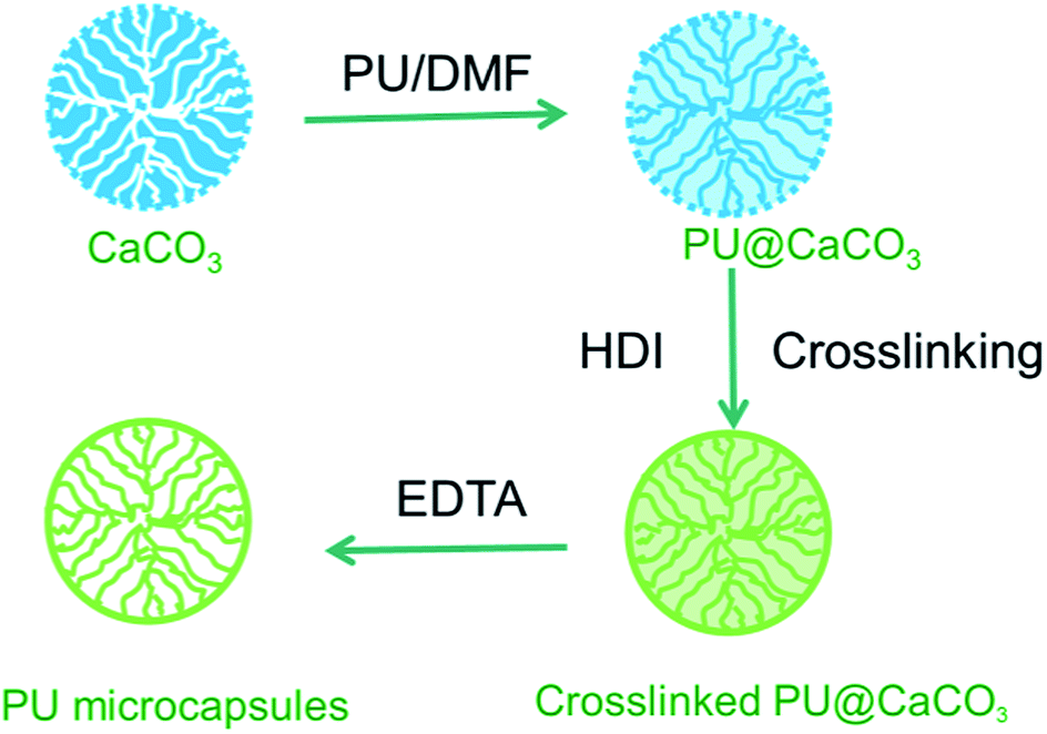 | ||
| Fig. 1 Schematic illustration of the fabrication process of PU microcapsules. | ||
Experimental section
Materials
Polyurethane (PU, Mn ∼ 58 kDa. The shore hardness is 65A, implying elastic) was obtained from Yantai Wanhua Polyurethane Co., Ltd. China. Hexamethylene diisocyanate (HDI) and stannous octoate were obtained from Acros. Coumarin 6 was obtained from Sigma-Aldrich. Dimethyl formamide (DMF), toluene, Ca(NO3)2·4H2O and Na2CO3 were obtained from Sinopharm Chemical Reagent Co., Ltd. Ethylenediaminetetraacetic acid disodium salt (EDTA) was purchased from Guangdong Guanghua Chemical Factory Co., Ltd. All chemicals were used as received. The water used in all experiments was prepared in a Millipore Milli-Q Reference purification system.Preparation of CaCO3 microparticles11
100 mL 0.33 M Na2CO3 solution was added to an equal volume of 0.33 M Ca(NO3)2 solution under mechanical agitation (900 rpm) for 1 min at room temperature. After 20 min, the suspension was centrifuged (2000 rpm, 1 min) and washed with ethanol 3 times, and then the particles were dispersed in ethanol for later use.Preparation of PU microcapsules
PU was first dissolved in DMF with a concentration of 10 wt%. The porous CaCO3 particles (0.5 g) were incubated in 10 mL PU solution for certain time to load the PU into the inner pores of CaCO3 spontaneously. Then the particles were washed by DMF and ethanol 3 times by centrifugation and dried at 70 °C in an oven for 5 h. The obtained PU-loaded CaCO3 particles (1 g) were dispersed in HDI solution (25 mg in 10 mL toluene with 5 mg stannous octoate as catalyst) for 1 h to crosslink the adsorbed PU. After washed by ethanol 3 times and separation by centrifugation (2000 rpm, 1 min), the particles were incubated in 0.2 M EDTA for 2 h to ensure the complete CaCO3 removal. The obtained microcapsules were washed by water 3 times and separation by centrifugation (5500 rpm, 10 min) and dispersed in water.The loading and release of coumarin 6
To load coumarin 6 into the capsules, 0.5 mL microcapsule suspension (∼6 × 10⁶ mL−1 in water) was mixed with 1 mL coumarin 6 solution (1 mg mL−1 in ethyl acetate), and the mixed solution was shaken overnight. After centrifugation (6000 rpm, 10 min) and discard of supernatant, the microcapsules were washed with water 3 times and finally dispersed in water. 1 mL suspension of the dye-loaded capsules was centrifugated, and the supernatant was discarded. The capsules were re-incubated in 1 mL ethyl acetate for 0.5 h under shaking to extract coumarin 6. After centrifugation (5500 rpm, 10 min), the concentration of coumarin 6 in the supernatant was determined with a UV-vis spectrophotometer (UV-2550, Shimadazu, Japan) at 442 nm by referring to a calibration curve. Each value was averaged from three parallel measurements.The release of coumarin 6 from PU microcapsules was studied at 37 °C in PBS (pH = 7.4). In a typical experiment, 1 mL dye-loaded capsules (∼3 × 10⁸ mL−1 in PBS, pH = 7.4) was transferred to a dialysis bag with a cut off Mw of 8–14 kDa, which was immersed in 10 mL of pH 7.4 PBS at 37 °C. Then 1 mL incubation medium was taken out for UV-vis measurement and 1 mL fresh PBS of the same pH was supplemented at desired time intervals. The results were averaged from triplicate experiments.
The microchannel experiment
The microchannel was fabricated via the photolithographic and wet chemical etching methods as described previously.11 Access holes were drilled with a 1.2 mm diameter diamond-tipped drill bit at each terminal of the groove, and followed by permanently covered with a thin cover slip through a thermal bonding procedure and glued to a pipet tip head at each terminal on the back side of the glass slide. Thus, the microchannel with a height of 4.7 μm in Z direction and a function of the sample supply and collection was successfully fabricated. The suspension of microcapsules in water was diluted sufficiently to avoid clogging of capillary and the collision among capsules. 50 μL of the diluted suspension of microcapsules was added to the access hole at the inlet, with the same amount of water in the two sheath fluid inlets. A negative pressure (0.77 atmosphere pressure) was applied at the outlet to force the movement of microcapsules in the microchannel. All the capsules passed through were collected in the small cell at the outlet and used for subsequent characterizations. During the experiment, the capsule could be trapped within the microchannel by releasing the negative pressure for in situ observation.Characterizations
Results and discussion
Preparation of microcapsules with specific size, morphology and structure on CaCO3 templates has received much attention.29,34,35 Via a simple adsorption and crosslinking method, microcapsules can be fabricated after template removal, as shown in this work. The CaCO3 particles had a spherical shape and an average size of 7.2 ± 0.4 μm (Fig. 2a) with porous surface (Fig. 2b) and fiber-like inner structure (Fig. 2c). The average pore size was 70.8 nm, which is comparable with previous report.29 After PU adsorption and crosslinking, the pores of the template were blocked and the particle surface became smoother (Fig. 2d–e). The cross section of PU-adsorbed particle (Fig. 2f) shows that a new layer was likely formed on the particle surface by comparison with Fig. 2c. The average pore size decreased to 21.3 nm, indicating the successful loading of PU into the pores of templates.32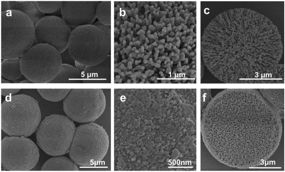 | ||
| Fig. 2 SEM images of (a) CaCO3 microparticles, and (b) their surface and (c) cross section morphology, and (d) crosslinked PU@CaCO3 microparticles, and (e) their surface and (f) cross section morphology. | ||
After template removal, the PU microcapsules were obtained. SEM images (Fig. 3a) reveal that the capsules collapsed with a non-flat morphology and considerably high folds in the middle parts of the capsules, which is substantially different from that of capsules templated on colloids of smoother surface texture.9 This special morphology is most possibly induced by matrix inside the capsules or the thicker shells,29,36 which may prevent the full collapse of the shells. The template of CaCO3 is porous and the PU can be easily adsorbed into the pores, leading to an matrix microcapsule.29 TEM image (Fig. 3b) indicates that there are some substances inside of the microcapsules, forming a network structure from the center to the periphery.37 This is consistent with the adsorption of PU inside the porous CaCO3 particles, which is then fixed in situ by HDI. Such a structure is the inverse replica of the CaCO3 template. Energy dispersive X-ray spectroscopy (EDX) results showed that there was only trace amount of Ca (0.01% by atom) in the microcapsules, meaning the template was removed (Fig. S1†). Some other methods such as interfacial polymerization can also prepare PU capsules, whose size is always in the order of dozens of micrometers21,23 or even bigger.24 Smaller capsules even in the order of nanometers can be prepared by a mini-emulsion method.18 However, the common problems of these methods are the large distribution of the capsule size, and hard control over the surface morphology, not even to say the inner structure. The present method can easily control the PU capsule size by the template, and even adjust the inner and surface morphology as shown by the above results.
 | ||
| Fig. 3 (a) SEM image, (b) TEM image of a PU microcapsule fabricated by incubation in 10% PU solution for 24 h followed by 1 h HDI crosslinking. | ||
The resulted microcapsules could be well dispersed in water with a diameter of about 7.2 μm, almost same to that of the template. The surface zeta potential of the microcapsules was +17.1 mV. The well dispersity and the positive surface charge can be attributed to the amino groups on the microcapsules due to the hydrolysis of unreacted isocyanate groups in HDI,38 which is revealed by FTIR characterization. Compared with that of the PU raw material, the absorbance peaks (3342, 1635 and 1565 cm−1) of amine or amide39 in the PU microcapsules (PU cap) were enhanced significantly (Fig. 4). By contrast, the ester vibration at 1733 cm−1 was weakened apparently.
 | ||
| Fig. 4 FTIR spectra of PU raw material (PU) and PU microcapsules (PU cap, fabricated by incubation in 10% PU solution for 24 h followed by 1 h HDI crosslinking). | ||
Although the PU capsules possess some amine groups which facilitate the dispersion of the microcapsules, the major matrix of PU materials is intrinsically hydrophobic and water-insoluble. This feature enables the loading of hydrophobic substances such as coumarin 6 and Nile red (Fig. 5). The fluorescence spectra of Nile red can be used to monitor the domain environment of the microcapsules, as this dye is solvatochromic, and the position of the peak implies the hydrophobicity of the domain environment where Nile red exists.40 The peak in microcapsules of 620 nm was blue-shifted compared with that in water at 660 nm (Fig. 6), indicating the hydrophobic domain environment in the microcapsules.40 Such a hydrophobic environment is very important for the encapsulation of hydrophobic substances. Different from other types of microcapsules, the as-prepared PU microcapsules can be well dispersed in water, but possess the ability to suck hydrophobic substances from organic solution.
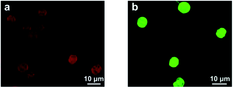 | ||
| Fig. 5 CLSM images of PU microcapsules loaded with hydrophobic dyes of (a) Nile red and (b) coumarin 6. | ||
 | ||
| Fig. 6 Fluorescence emission spectra of PU microcapsules loaded with Nile red, and Nile red in water. | ||
The influences of the experimental parameters such as the concentrations of PU(1%, 2%, 6% and 10%), the adsorption time of PU (20 min, 2 h and 24 h), and crosslinking time (20 min, 1 h, 3 h) on the obtained PU microcapsules have been studied. The lower concentration of PU, shorter adsorption time of PU and cross-linking time resulted thinner capsules with fewer folds, indicating less PU was adsorbed and crosslinked, and the microcapsules became less intact (data not shown). When the crosslinking time was longer than 20 min and the concentration of PU was above 2%, they would not obviously influence the structure of the obtained capsules. However, at the fixed PU concentration of 10% and crosslinking time of 1 h, the adsorption time had significant impact on the resulted microcapsules. When the adsorption time increased form 20 min to 24 h, the surface area of PU@CaCO3 decreased from 10.1 m2 g−1 to 7.1 m2 g−1, suggesting more PU was deposited into the template. After template removal, the shell became thicker and the number of typical folds and creases increased (Fig. S2†), meaning a typical sign of mass increase in the capsules. The larger mass in the capsules enhanced the loading amount of hydrophobic substances as well. For example, the loading amount of coumarin 6 in one capsule fabricated via 20 min PU adsorption was 3.4 ± 0.4 pg. When the adsorption time increased to 24 h, this value also increased greatly to 15.0 ± 0.5 pg (Fig. S3†). The release of coumarin 6 was negligible in PBS at 37 °C for even 48 h, and no peak value was detected by the UV-vis. This is due to the negligible solubility of coumarin 6 in water and it prefers to stay in the relatively hydrophobic interior of capsules (Fig. 6). This result reveals that the capsules can suck hydrophobic substances from organic solution and the hydrophobic dye molecules are well preserved inside the capsules.
The PU microcapsules are assumed to have a good elasticity because of the elastic PU materials. To verify this assumption, the PU microcapsules with a diameter of 7.2 ± 0.4 μm (fabricated by incubation in 2% PU solution for 20 min followed by 20 min HDI crosslinking) were forced to pass through a microchannel. By laser scanning microscopy, the key geometric features of the microchannel used in this study were characterized with a depth (size in Z direction) of 4.7 μm and a width (size in Y direction) of 35 μm of the constriction region, and the length of the constriction is about 150 μm. At the both sides of the constriction region, the depth and width of the channel are about 25 μm and 75 μm, respectively. By this way, the PU microcapsules are forced to deform in Z-direction (4.7 μm) if they can pass through the microchannel (Fig. 7a). From the experiment, we find the capsules can successfully pass through the channel. If we release the negative pressure at a proper time, the capsules can be stopped within the channel (Fig. S4†). As evidenced by the series of Z-scanning images of the PU microcapsules captured by CLSM, obvious shape deformation in Z direction did occur when they were trapped inside the microchannel (Fig. 7b). After being squeezed from the microchannel, the deformation of the capsules disappeared completely, suggesting the elastic recovery of the microcapsules (Fig. 7c and S5 and S6†). Statistic analysis found a recovery ratio of 82.4%, and the capsule size was identical to the original one. These results demonstrate that the capsules can recover their shape when the deformation ratio is as high as 32% (The deformation ratio is defined as (Dcap − Dcns)/Dcap × 100%, where Dcap and Dcns are the capsule diameter and constriction depth, respectively). Such good elasticity may originate from both the elastic PU materials and the inner network structure of the PU microcapsules, since it is known that the substances inside facilitate the shape recovery after releasing the physical constriction.11 Previous results showed that the recovery percentage of the poly(allylamine hydrochloride) (PAH)/poly(styrene sulfate) (PSS) multiplayer microcapsules was as low as ∼15% at a similar deformation ratio.11 Therefore, The PU capsules have a much stronger ability to maintain their spherical shape subjecting deformation and force release. It is worth mentioning that the PU capsules fabricated by 10% PU, 24 h incubation and 1 h HDI crosslinking could not pass through the microchannel at the same experimental conditions. It is understandable that higher force will be required to deform these stiffer PU capsules with more dense structure and higher crosslinking degree. Therefore, the deformation and recovery property of the PU microcapsules is greatly influenced by the preparation conditions.
 | ||
| Fig. 7 (a) Scheme of the microchannel. Series Z-scanning CLSM images of a microcapsule (b) within the microchannel and (c) out of the microchannel after being squeezed through. The capsules were stained with coumarin 6. | ||
To study the release of dye from the capsules after passing through the microchannel, it is difficult to use the general used protocol for drug release studies because the collected capsules are very few. According to the reference, CLSM was used to measure the fluorescent intensity of capsules before and after they passed through the microchannel, which is correlated to the content of dye inside the capsules.41 After squeezed through the microchannel with a diameter of 4.7 μm, the coumarin 6 content inside the capsules was not changed significantly (Fig. S7†), revealing that the dye molecules are stably loaded regardless of the water flow and capsule deformation. This may be due to the hydrophobic property of coumarin 6. This property is important for future applications such as fluorescent micro-sensors where the dye molecules should be well preserved when the capsules are squeezed through narrow capillaries.
Conclusion
The elastic PU microcapsules were successfully fabricated by a simple and well controllable adsorption and crosslinking method by using porous CaCO3 particles as sacrificial templates. The interior of the capsules had a network structure from the center to the periphery. The PU capsules had some amine groups, enabling well dispersion in water. The matrix of PU capsules could still suck hydrophobic substances such as Nile red and coumarin 6 from organic solution. The hydrophobic dyes can be well maintained inside the capsules. 82% capsules could recover their original shape after they were squeezed through a microchannel with a deformation ratio of 32%, and the low molecular-weight dyes can be stably maintained regardless of squeezing through the microchannel. These elastic PU microcapsules may find promising applications in the adsorption and removal of hydrophobic molecules, microsensors with stable loaded fluorescent dyes and so on.Acknowledgements
The authors thank Prof. Hong Shen from the Department of Chemistry of Zhejiang University for technical help with the preparation of microchannel and Dr Bing Yang from the Department of Information Science & Electronic Engineering of Zhejiang University for the help with the CLSM measurements of the geometry of the microchannel. This study is financially supported by the Natural Science Foundation of China (no. 21174130, 21374101, 51120135001).References
- W. J. Tong, X. X. Song and C. Y. Gao, Chem. Soc. Rev., 2012, 41, 6103–6124 RSC.
- J. W. Cui, Y. Yan, Y. J. Wang and F. Caruso, Adv. Funct. Mater., 2012, 22, 4718–4723 CrossRef CAS.
- R. Palankar, A. G. Skirtach, O. Kreft, M. Bedard, M. Garstka, K. Gould, H. Mohwald, G. B. Sukhorukov, M. Winterhalter and S. Springer, Small, 2009, 5, 2168–2176 CrossRef CAS PubMed.
- P. Rivera-Gil, M. Nazarenus, S. Ashraf and W. J. Parak, Small, 2012, 8, 943–948 CrossRef CAS PubMed.
- W. X. Song, Q. He, Y. Cui, H. Mohwald, S. Diez and J. B. Li, Biochem. Biophys. Res. Commun., 2009, 379, 175–178 CrossRef CAS PubMed.
- Y. Cui, J. B. Fei and J. B. Li, Sci. Sin.: Chim., 2011, 41, 273 CrossRef.
- R. C. Luo, S. S. Venkatraman and B. Neu, Biomacromolecules, 2013, 14, 2262–2271 CrossRef CAS PubMed.
- R. Haghgooie, M. Toner and P. S. Doyle, Macromol. Rapid Commun., 2010, 31, 128–134 CAS.
- S. P. She, Q. Q. Li, B. W. Shan, W. J. Tong and C. Y. Gao, Adv. Mater., 2013, 25, 5814–5818 CrossRef CAS PubMed.
- M. Guvendiren, H. D. Lu and J. A. Burdick, Soft Matter, 2012, 8, 260–272 RSC.
- S. P. She, C. X. Xu, X. F. Yin, W. J. Tong and C. Y. Gao, Langmuir, 2012, 28, 5010–5016 CrossRef CAS PubMed.
- S. A. Guelcher, Tissue Eng., Part B, 2008, 14, 3–17 CrossRef CAS PubMed.
- S. Farzaneh, J. Fitoussi, A. Lucas, M. Bocquet and A. Tcharkhtchi, J. Appl. Polym. Sci., 2013, 128, 3240–3249 CrossRef CAS.
- J. Ling, M. Z. Rong and M. Q. Zhang, Chin. J. Polym. Sci., 2014, 32, 1286–1297 CrossRef CAS.
- Y. Xiang, X. L. Tuo and X. G. Wang, Progr. Chem., 2008, 21, 1546–1552 Search PubMed.
- C.-W. Ou, C.-H. Su, U. S. Jeng and S.-h. Hsu, ACS Appl. Mater. Interfaces, 2014, 6, 5685–5694 CAS.
- E. Koh, S. Lee, J. Shin and Y. W. Kim, Ind. Eng. Chem. Res., 2013, 52, 15541–15548 CrossRef CAS.
- F. Gaudin and N. Sintes-Zydowicz, Colloids Surf., A, 2008, 331, 133–142 CrossRef CAS PubMed.
- H. Souguir, F. Salaun, P. Douillet, I. Vroman and S. Chatterjee, Chem. Eng. J., 2013, 221, 133–145 CrossRef CAS PubMed.
- A. B. W. Brochu, W. J. Chyan and W. M. Reichert, J. Biomed. Mater. Res., Part B, 2012, 100, 1764–1772 CrossRef PubMed.
- E. Koh, N. K. Kim, J. Shin and Y. W. Kim, RSC Adv., 2014, 4, 16214–16223 RSC.
- S. Neuser, E. Manfredi and V. Michaud, Mater. Chem. Phys., 2014, 143, 1018–1025 CrossRef CAS PubMed.
- A. Latnikova, D. O. Grigoriev, H. Mohwald and D. G. Shchukin, J. Phys. Chem. C, 2012, 116, 8181–8187 CAS.
- J. L. Yang, M. W. Keller, J. S. Moore, S. R. White and N. R. Sottos, Macromolecules, 2008, 41, 9650–9655 CrossRef CAS.
- M. Li and J. M. Xue, Langmuir, 2011, 27, 3229–3232 CrossRef CAS PubMed.
- J. W. Cui, M. P. van Koeverden, M. Mullner, K. Kempe and F. Caruso, Adv. Colloid Interface Sci., 2014, 207, 14–31 CrossRef CAS PubMed.
- Y. J. Wang, V. Bansal, A. N. Zelikin and F. Caruso, Nano Lett., 2008, 8, 1741–1745 CrossRef CAS PubMed.
- D. Volodkin, Adv. Colloid Interface Sci., 2014, 207, 306–324 CrossRef CAS PubMed.
- D. V. Volodkin, A. I. Petrov, M. Prevot and G. B. Sukhorukov, Langmuir, 2004, 20, 3398–3406 CrossRef CAS.
- W. J. Tong, W. F. Dong, C. Y. Gao and H. Mohwald, Macromol. Chem. Phys., 2005, 206, 1784–1790 CrossRef CAS.
- M. Behra, S. Schmidt, J. Hartmann, D. V. Volodkin and L. Hartmann, Macromol. Rapid Commun., 2012, 33, 1049–1054 CrossRef CAS PubMed.
- X. H. Yan, J. B. Li and H. Mohwald, Adv. Mater., 2012, 24, 2663–2667 CrossRef CAS PubMed.
- J. W. Cui, Y. J. Wang, J. C. Hao and F. Caruso, Chem. Mater., 2009, 21, 4310–4315 CrossRef CAS.
- D. V. Volodkin, N. I. Larionova and G. B. Sukhorukov, Biomacromolecules, 2004, 5, 1962–1972 CrossRef CAS PubMed.
- G. B. Sukhorukov, D. V. Volodkin, A. M. Gunther, A. I. Petrov, D. B. Shenoy and H. Mohwald, J. Mater. Chem., 2004, 14, 2073–2081 RSC.
- Z. P. Wang, H. Mohwald and C. Y. Gao, Langmuir, 2011, 27, 1286–1291 CrossRef CAS PubMed.
- W. J. Tong, Y. Zhu, Z. P. Wang, C. Y. Gao and H. Mohwald, Macromol. Rapid Commun., 2010, 31, 1015–1019 CrossRef CAS PubMed.
- A. V. Wisnewski, M. Mhike, J. M. Hettick, J. Liu and P. D. Siegel, Toxicol. In Vitro, 2013, 27, 662–671 CrossRef CAS PubMed.
- Y. Liu, Y. Y. Wei and C. Z. Zong, Colloid Polym. Sci., 2014, 292, 873–884 CAS.
- F. Zhao, G. Shen, C. Chen, R. Xing, Q. Zou, G. Ma and X. Yan, Chem.–Eur. J., 2014, 20, 6880–6887 CrossRef CAS PubMed.
- P. A. L. Fernandes, M. Delcea, A. G. Skirtach, H. Mohwald and A. Fery, Soft Matter, 2010, 6, 1879–1883 RSC.
Footnote |
| † Electronic supplementary information (ESI) available. See DOI: 10.1039/c4ra12193g |
| This journal is © The Royal Society of Chemistry 2015 |