DOI:
10.1039/C4RA11795F
(Paper)
RSC Adv., 2015,
5, 2602-2614
Antiradical–antimicrobial activity and phenolic profile of pomegranate (Punica granatum L.) juices from different cultivars: a comparative study
Received
4th October 2014
, Accepted 24th November 2014
First published on 24th November 2014
Abstract
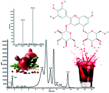
Pomegranate juice (PJ) constituents have shown to exhibit anticarcinogenic, antimicrobial, antioxidant and antiviral activities. In the present study, the concentration of phenolic compounds and the antiradical activity of PJs from the fruits of the two relatively new Greek cultivars “Persephone” and “Porphiroyeneti” were determined in comparison to the “Wonderful” cultivar. Total phenolic content and antiradical activity of the examined juices were found to vary in the same decreasing manner as follows: “Porphiroyeneti” > “Wonderful” > “Persephone”. The antimicrobial activity of PJs was also determined, showing equal or higher effect than commercial antimicrobial agents (streptomycin, ampicillin, bifonazole and ketoconazole). All tested extracts demonstrated noteworthy antibacterial activity with minimal inhibitory concentration ranging from 0.05 to 0.20 mg mL−1 and minimal bactericidal concentration ranging from 0.10 to 0.40 mg mL−1. Moreover, PJ extracts showed satisfactory fungistatic (0.05–0.2 mg mL−1) and fungicidal (0.1–0.3 mg mL−1) activity against all fungi tested. Among the cultivars tested, “Porphiroyeneti” showed slightly better antiradical and antimicrobial activity. In addition, a GC-MS methodology was developed for the determination of the phenolic profile of the extracts of PJ after different types of chemical hydrolysis. Finally, an HPLC-PDA-ESI-MSn analysis was conducted for the identification of the phenolic compounds in the extracts of PJ. In total, more than 30 non-anthocyanidinic and more than 20 anthocyanidinic compounds were identified. Our results confirm the functionality of pomegranate juices and the potential applications of PJ extracts towards novel products as food additives or preservatives.
1. Introduction
Since ancient times, pomegranate fruit (Punica granatum L.) has been an economically important plant, one of the most known medicines in ancient history, and particularly nowadays a ‘hot’ commodity on health product markets.1 Pomegranate has been termed as a ‘superfruit’, along with others, such as blueberry and cranberry mainly because of its remarkable antiradical and antioxidative properties.2 Pomegranate is currently ranked 18th in terms of global annual fruit consumption.3 Its widespread public knowledge of the health attributes has led to a big rise in the demand for this fruit and its by-products during the last years in the Western world.4 Resultantly, commercial pomegranate orchards have increased significantly,4 and pomegranate fruit is cultivated throughout the world in subtropical and tropical areas in many variable climatic conditions.1 Its successful adaptation to the Mediterranean climate has produced a wide diffusion in various countries, thus originating several local genotypes, locally called “cultivars” along the centuries,5 and over 1000 cultivars of P. granatum have been identified globally.3
Among pomegranate derived products, its juice is one of the most popular drinks in the super juice category,6 and is consumed throughout the world because of its pleasant and unique aroma, flavor and color.7 Pomegranate juice provides a simple and convenient way to consume biologically active nutrients.8 The ethnopharmacological relevance of pomegranate is justified by recent findings, indicating its medicinal and nutritional properties against a wide range of human disorders and maladies.9 Clinical research studies suggest that several compounds of the pomegranate juice are characterized by their anticarcinogenic, antimicrobial, antioxidant and antiviral activities. Furthermore, according to biological studies, pomegranate juice is rich in anti-atherosclerotic and anti-atherogenic compounds, which have been shown to reduce blood pressure and low density lipoprotein (LDL) oxidation.10 Interestingly, pomegranate juice has been reported to possess a 3-fold higher antioxidant activity than that of red wine or green tea, and 2-, 6- and 8-fold higher levels than those detected in grape/cranberry, grapefruit, and orange juice, respectively.5 Pomegranate and its extracts are reported to have a positive effect against many cancers, such as skin, colon, prostate and breast cancer. Specifically, phenolic compounds isolated from pomegranate juices, such as punicalagins and ellagic acid, have shown to have antiproliferative activity against cancer cells.11–13 Polyphenols, and particularly flavonoids and ellagitannins in pomegranate juice have antimicrobial activity against Escherichia coli, Salmonella enteritidis, Staphylococcus aureus, Bacillus subtilis, Klebsiella pneumoniae, Pseudomonas aeruginosa, Yersinia enterocolitica, and Listeria monocytogenes.14 Furthermore, it is reported that a mixture of ellagic acid, gallic acid, and punicalins, which had been isolated from pomegranate juice byproducts, revealed antimicrobial activity against pathogenic microorganisms such as Clostridia species, Candida albicans, Cryptococcus neoformans, Staphylococcus aureus, Aspergillus fumigatus and Mycobacterium intra cellulare.15
The health promoting features of the pomegranate juice have been attributed to its phenolic content (e.g. significantly high levels of hydrolyzable and condensed tannins, phenolic acids, anthocyanins, and other flavonoids), which exhibits a high antioxidant activity,16–21 and they play a significant role in its sensory properties (flavour, colour, bitterness, and astringency).17 Furthermore, a group of six anthocyanins, 3-monoglucosides and 3,5-diglucosides of delphinidin, cyanidin, and pelargonidin, originating mostly from the arils, constitute a typical anthocyanin profile of pomegranate juices and could be successfully used in quality-authenticity controls.21,22
The phenolic and polyphenolic content, as well as the overall composition of pomegranate juices, are strongly influenced by a number of factors, including agronomical, environmental and climate conditions such as geographical variables, harvest time, fruit maturity, and postharvest conditions such as storage, processing factors and juice extraction methods.22–24 However, the cultivar per se has been reported to be one of the most influencing factors in the phytochemical content of fruits.25–27
The evaluation of phenolic compounds of the pomegranate juice and its organoleptic characteristics is essential to satisfy current market and processing industry demands for quality fruit and for its potential use as a nutraceutical beverage.28 In this frame, it is highly important to study Punica granatum's cultivar characteristics, particularly its edible parts. This will help to achieve the best germplasm management and cultivar selection, which is important not only for consumers, but also for cultivators, breeders, and food as well as pharmaceutical industries.10,29
In this study, we have performed a series of analyses of P. granatum juice, including its antiradical activity, total phenolic content and antimicrobial properties, and LC-MSn and GC-MS indicated the most prominent phenolic compounds. To our knowledge, this is the first study focused on the novel Greek pomegranate cultivars “Persephone” and “Porphiroyeneti”. A comparison between these two Greek cultivars with the most widespread “Wonderful” cultivar was also performed.
2. Materials and methods
2.1. Chemicals, standards and solvents
All reagents used were of analytical grade and they were purchased from Mallinckrodt Chemical Works (St. Louis, MO, USA), Alfa Aesar GmbH & Co (Germany), Acros Organics (Belgium) and Sigma Chemical Co. (Sigma-Aldrich Company, UK). Folin–Ciocalteu's phenol reagent, DPPH˙ (2,2-diphenyl-1-picrylhydrazyl) free radical, and Trolox (6-hydroxy-2,5,7,8-tetramethylchroman-2-carboxylic acid) were obtained from Sigma Chemical Co. (St. Louis, MO, USA). ABTS [2,2′-azinobis(3-ethylbenzothiazoline-6-sulfonic acid)] was obtained from Tokyo Chemical Industry Co. Ltd (Tokyo, Japan). Standard phenolic compounds, 3,4,5-trihydroxybenzoic acid, trans-4-hydroxycinnamic acid, 3,4-diydroxycinnamic acid, vanillin and quercetin, were purchased from Alfa Aesar (Germany), and L-ascorbic acid, (±)-naringenin and (±)-catechin were supplied from Sigma-Aldrich (Steinheim, Germany). Cinnamic acid and 4-hydroxybenzoic acid were purchased from Merck (Darmstadt, Germany), 2,6-di-tert-butyl-4-methylphenol from Acros Organics (Belgium) and 2-(4-hydroxyphenyl)ethanol from Fluka Analytical (Japan).
All solvents used were GC, HPLC and LC-MS grade and purchased from Sigma Chemical Co (Sigma-Aldrich Company, UK). Lichrosolv hypergrade for LC-MS acetonitrile was supplied by Merck (Darmstadt, Germany), and LC-MS grade water was obtained by Fischer Scientific (UK). Formic acid was purchased from LGC Standards (Germany).
2.2. Sampling and sample preparation
Ripe fresh fruits of pomegranate (Punica granatum L.) of “Persephone”, “Porphiroyeneti” and “Wonderful” cultivars were harvested five times during autumn, from October to December 2012, from different mature trees. All cultivars were obtained from a local producer in Ermioni (Argolis unit, Peloponnese region, Greece) (geographic coordinates 37°23′N 23°15′E) and were grown under the same geographical conditions and by the same applied agronomic practices. The average temperature and the amount of rainfall for the studied crops of 2012 were 17.4–22.3 °C and 16.8–56.4 mm, respectively. Approximately 15 kg of pomegranate fruit samples were randomly picked from each cultivar in each sampling. Fruits were divided into groups according to the sampling and the cultivar. The fruit samples obtained (N = 5 samples per cultivar) were transported to the laboratory after the harvest, where pomegranates with defects (sunburns, cracks, cuts and bruises in peel) were discarded. Fruits of each cultivar were weighted and their length and diameter were measured. Fruit length was taken by measuring the distance between the apex and the end of stem. The maximum width of the fruit was taken by measuring the diameter, which is the direction perpendicular to the polar axis. Fruits were washed in cold tap water and drained, and the top and bottom of the husks were removed with a stainless knife to prevent microbial contamination. Each fruit was hand peeled and only the arils were juiced using a LiquaFresh juice extractor (Mellerware, South Africa). The resulting juices (N = 5 juice samples per cultivar) were filtered through muslin cloth to remove large particles. Physical characteristics of the juices were measured and freeze dried in a Modulyo D Freeze Dryer, equipped with a Thermo Savant ValuPump VLP200, (Thermo Electron Corporation, Thermo Fischer, USA). From each of the freeze dried sample, half the quantity was packaged in vacuum for microbiological analysis and the other half was used for phenolic extraction. All determinations were carried out at least three times per sample.
2.3. Extraction methodology
Ultrasound-Assisted Extraction (UAE) procedure was performed according to the method described by Petrović et al. (2014):30 10.00 g of lyophilized juice sample and 50.00 mL solvent (methanol) were placed in a 250 mL three-neck vessel in ice-bath (maximum temperature 35 °C) and sonicated using Sonics & Material Inc., Vibra-Cell VCX750 (20 kHz, 750 W) ultrasonics processor, equipped with piezoelectric converter and 13 mm diameter probe fabricated from titanium alloy Ti–6Al–4V. The amplitude was 80% and the pulse sonication sequence was 10 s ON and 5 s OFF. After sonication, the extracts were filtered by a Buchner funnel and the filtrates were diluted in 50.00 mL with methanol. The extracts were stored at 4 °C for further analysis.
2.4. Determination of total phenolics
The total phenolic content of each sample was determined applying a micromethod of Folin–Ciocalteu's colorimetric assay.31 Twenty microliters of juice extracts (20–25 mg mL−1), standard solutions or blank (methanol) were added to 1500 μL of water and 100 μL of the Folin–Ciocalteu reagent, mixed thoroughly and allowed to stand for 8 min. Then 300 μL of saturated sodium carbonate solution were added and mixed well. The cuvettes were left at 40 °C for 30 min. The absorbance of the cooled samples at room temperature was measured at 750 nm with a UV-vis spectrophotometer (Novaspek III visible spectrophotometer, Amersham Biosciences, USA). The total phenolic content was expressed as mg gallic acid equivalents (GAE) per 100 mL of juice (and mg GAE per g juice dry matter), using a standard curve with 25–500 mg L−1 gallic acid (y = 0.001x + 0.003, R2 = 0.993).
2.5. Methods for determining antiradical activity
(a) Scavenging activity on 2,2-diphenyl-1-picrylhydrazyl radical (DPPH˙). The antiradical activity of juice extracts was evaluated using the stable 2,2-diphenyl-1-picryl-hydrazyl radical (DPPH˙) according to a modification of the method described by Brand-Williams et al. (1995).32 Fifteen microliters of juice extracts (200 mg mL−1) and 1500 μL of methanolic solution of DPPH˙ (75 μM) were added in a cuvette and the absorbance at 515 nm (till stabilization – plateau) was measured using a UV-vis spectrophotometer (Novaspek III visible spectrophotometer, Amersham Biosciences, USA). The negative and positive errors due to (a) DPPH˙ dilution and (b) the red colour of the extracts, respectively, were taken into consideration using different blank solutions. More specifically, the corrected absorbance is given by the equation Af = Ai + (Aa − Ab) − (Ac − Ad), where Af is the final corrected absorbance for each sample, Ai is the measured absorbance of the sample (1500 μL DPPH˙ with 15 μL extract), Aa is the absorbance of DPPH˙ solution (75 μM), Ab is the absorbance of a solution of 1500 μL DPPH˙ with 15 μL methanol, Ac is the absorbance of a solution of 1500 μL methanol with 15 μL extract and Ad is the absorbance of methanol. The antiradical activity of the pomegranate juices was expressed as mg ascorbic acid equivalents (AAE) per 100 mL of juice (and mg AAE per g juice dry matter), using a standard curve with 40–320 mg L−1 ascorbic acid (y = −0.003x + 1.057, R2 = 0.990).
(b) Scavenging activity on [2,2′-azino-bis-(3-ethylbenzothiazoline-6-sulfonic acid)] radical (ABTS˙+). The antiradical activity of juice extracts was determined according to a minor modification of the method described by Re et al. (1999).33 This assay assesses the capacity of a compound to scavenge the stable ABTS radical (ABTS˙+), in comparison to the antioxidant activity of Trolox, a water soluble form of vitamin E, which is used as a standard. Briefly, the ABTS˙+ stock solution was prepared through the reaction of 7 mM ABTS with 2.45 mM sodium persulfate (Na2S2O8) (final concentrations) in a volume ratio of 1
:
1 and was then incubated in the dark at room temperature for 16 h before use. The concentrated ABTS˙+ solution was diluted with ethanol to a final absorbance of 0.8–0.7 at 734 nm. Fifteen microliters of juice extracts (20–25 mg mL−1) or standard solutions of Trolox were added to 1500 μL of ABTS˙+ diluted solution and the reduction in absorbance was measured after 1 min of stirring in the dark. The stock solution of Trolox (6.0 mM) was prepared in ethanol. Absorbance was measured on a UV-vis spectrophotometer (Novaspek III visible spectrophotometer, Amersham Biosciences, USA). The negative error due to ABTS˙+ dilution was taken into consideration using different blank solutions. More specifically, the corrected absorbance is given by the equation Af = Ai + (Aa − Ab), where Af is the final corrected absorbance for each sample, Ai is the measured absorbance of the sample (1500 μL ABTS˙+ with 15 μL extract), Aa is the absorbance of ABTS˙+ solution and Ab is the absorbance of a solution of 1500 μL ABTS˙+ with 15 μL methanol. The antiradical activity of the pomegranate juices was expressed as mg Trolox Equivalents (TE) per 100 mL of juice (and mg TE per g juice dry matter), using a standard curve with 0.20–1.50 mM Trolox (y = −0.283x + 0.693, R2 = 0.995).
2.6. Chemical hydrolysis of pomegranate juices extracts
In order to identify the phenolic compounds in pomegranate juice by GC-MS analysis, mild alkaline and acidic hydrolysis of the studied extracts were performed using a modified method of Ross et al. (2009).34 During the hydrolysis, the glycosidic bonds of glycosylated phenolic compounds are cleaved and the resulting compounds are analyzed after silylation.
(a) Mild alkaline hydrolysis. In brief, 1.5 mL of pomegranate juice extract was treated with 1.5 mL of a solution consisting of NaOH (4 M), ascorbic acid (2% (w/v)) and EDTA (14 mM). The solution was vortexed for 5 min and remained at room temperature in dark for 16 h. Phenolics were extracted with 1.5 mL of diethyl ether–ethyl acetate solution (DE–EA, 1
:
1, v/v). The mixture was vortexed for 60 s and cooled for 10 min. After phase equilibration, phenolic compounds from alkaline hydrolysis were transferred to the upper DE–EA organic layer.
(b) Post alkaline acidic hydrolysis. The bottom aqueous layer resulting from alkaline hydrolysis was treated with 1.5 mL of a solution consisting of HCl (3 M), ascorbic acid (1% (w/v)) and EDTA (5 mM). The solution was vortexed for 5 min and incubated in a water bath at 85 °C for 60 min. Phenolics were extracted with 2.0 mL of diethyl ether–ethyl acetate solution (DE–EA, 1
:
1, v/v). The mixture was vortexed for 10 min and cooled for 10 min. After phase equilibration, phenolic compounds from acidic hydrolysis were transferred to the upper DE–EA organic layer.
(c) Acidic hydrolysis. In 1.5 mL of pomegranate juice extract, 1.0 mL of a solution was added consisting of HCl (3 M), ascorbic acid (1% (w/v)) and EDTA (5 mM). The experimental procedure followed the protocol described in the previous paragraph.
2.7. Silylation of the phenolic compounds
Silylation procedure was performed according to a modification of the method described by Trigui et al. (2013).35 Specifically, 1000 μL of the DE–EA organic layer after chemical hydrolysis were derivatized after evaporation of the solvent under a nitrogen stream. For the silylation procedure, a mixture of trimethylchlorosilane (80.0 μL) and N,O-bis(trimethylsilyl)trifluoroacetamide (200.0 μL) was added and vortexed in screw-cap glass tubes (previously deactivated with 5% dimethyldichlorosilane in toluene, and rinsed twice with toluene and thrice with methanol), and consecutively placed in a water bath at 80 °C for 45 min.
2.8. Gas chromatography/mass spectrometry analysis of phenolic compounds
Qualitative analysis was performed using a mass spectrometer QP2010 Series (Shimadzu USA MANUFACTURING, Inc., Kyoto, Japan). Electron impact (EI) ionization was produced by accelerating electrons from a filament through a difference of 70 eV. A non-polar column was used (DB-5 MS, 30 m, 0.25 mm i.d. and 0.25 μm film thickness; Agilent, USA). The stationary phase was phenyl arylene polymer virtually equivalent to 5% phenyl-methylpolysiloxane. The carrier gas was helium. The temperature of the injector was 295 °C, of the interface 295 °C and of the ion source 200 °C. The temperature was programmed at 70 °C, raised from 70 °C to 125 °C at a rate of 17 °C min−1, held constant at 125 °C for 1 min, raised from 125 °C to 140 °C at a rate of 9 °C min−1, held constant at 140 °C for 5 min, raised from 140 °C to 200 °C at a rate of 5 °C min−1, held constant at 200 °C for 3 min, raised from 200 °C to 220 °C at a rate of 5 °C min−1, held constant at 220 °C for 3 min. raised from 220 °C to 280 °C at a rate of 15 °C min−1, held constant at 280 °C for 1 min and finally raised from 280 °C to 295 °C at a rate of 15 °C min−1, held constant at 295 °C for 3 min. The duration of the analysis was 40 min.
2.9. Liquid chromatography-mass spectrometry (LC-MS)
(a) Instrumentation. Phenolics separation was carried out using a Thermo Scientific Surveyor Plus HPLC-PDA-ESI-MSn system (San José, CA, USA). The platform comprised of a Thermo Scientific Surveyor HPLC Pump Plus, a Thermo Scientific Surveyor Autosampler Plus Lite, a Thermo Scientific Accela PDA Detector and a LCQ FLEET mass spectrometer with electrospray ionization (ESI). The data were processed using the Xcalibur software program (version 2.1).
(b) Chromatographic conditions and mass spectrometry. The separation of phenolics was carried out using a Finnigan Surveyor system and a Hypersil Gold Column (3 μm, 2.1 × 100 mm, Thermo, Palo Alto, CA) protected with a security guard cartridge (Hypersil Gold, 3 μm, 10 × 2.1 mm i.d.). The gradient mobile phase consisted of solvent A (water – 0.5% (v/v) formic acid) and solvent B (acetonitrile). The flow rate was 0.3 mL min−1 and the injection volume was 5.0 μL. The gradient elution program was initial 5% B, linear 5–9% B at 4 min, linear 9–15% B at 8 min, linear 15–18% B at 11 min, held constant for 1 min, linear 18–50% B at 15 min, held constant for 2 min, purging with 100% B during 6 min and re-equilibration of the column during 10 min. The channels of detection wavelengths were set at 280, 360 and 520 nm for compounds bearing phenolic rings, flavonoids and anthocyanins, respectively.
(c) Mass spectrometry analysis. Separate injections were run for analysis of the sample in both positive and negative electrospray ionization (ESI) modes, as well as for different collision energies for MSn analysis. According to the method described by Setandreu et al. (2013),36 positive and negative modes were applied for the determination of anthocyanidinic and non-anthocyanidinic compounds, respectively.The mass spectrometer parameters for positive ion mode were as follows: source voltage, 3.5 kV; capillary voltage, 9 V; capillary temperature, 300 °C; sheath gas flow, 50 (arbitrary units); sweep gas flow, 20 (arbitrary units); full max ion time, 300 ms; and full micro scans, 3.
The mass spectrometer parameters for negative ion mode were as follows: source voltage, 4.0 kV; capillary voltage, −18 V; capillary temperature, 300 °C; sheath gas flow, 50 (arbitrary units); sweep gas flow, 20 (arbitrary units); full max ion time, 300 ms; and full micro scans, 3.
Data dependent scan MSn analyses for positive ions were carried out with the following conditions: collision energies 15, 17, 25, 30, 35 (arbitrary units); width, 1.00; repeat count, 2; repeat duration, 0.5 min; exclusion size list, 25; exclusion duration, 1.00 min; exclusion mass width, 3.00; scanned mass range (m/z), 260–1000.
Data dependent scan MSn analyses for negative ions were carried out with the following conditions: collision energies 15, 25, 30, 35 (arbitrary units); width, 1.00; repeat count, 2; repeat duration, 0.5 min; exclusion size list, 25; exclusion duration, 1.00 min; exclusion mass width, 3.00; scanned mass range (m/z), 100–1600.
2.10. Antibacterial and antifungal activity
(a) Antibacterial activity. The Gram-positive bacteria, Staphylococcus aureus (ATCC 6538), Bacillus cereus (clinical isolate), Micrococcus flavus (ATCC 10240) and Listeria monocytogenes (NCTC 7973), and the Gram-negative bacteria, Pseudomonas aeruginosa (ATCC 27853), Salmonella typhimurium (ATCC 13311), Escherichia coli (ATCC 35210), and Enterobacter cloacae (human isolate), were used. The organisms were obtained from the Mycological Laboratory, Department of Plant Physiology, Institute for Biological Research “Siniša Stanković”, Belgrade, Serbia. The antibacterial assay was carried out by a microdilution method37,38 in order to determine the antibacterial activity of the lyophilized juices tested against the human pathogenic bacteria. The bacterial suspensions were adjusted with sterile saline to a concentration of 1.0 × 10⁵ CFU mL−1. Dilutions of the inocula were cultured on solid medium to verify the absence of contamination and to check the validity of the inoculum.The minimum inhibitory and bactericidal concentrations (MICs and MBCs) were determined using 96-well microtiter plates by a microdilution test. The bacterial suspension was adjusted with sterile saline to a concentration of 1.0 × 10⁵ CFU mL−1. Lyophilized juices (“Persephone”, “Porphiroyeneti” and “Wonderful”) were dissolved in 5% DMSO solution containing 0.1% Tween 80 (v/v) (10 mg mL−1) and added in tryptic soy broth (TSB) medium (100 μL) with bacterial inoculum (1.0 × 10⁴ CFU per well) to achieve the appropriate concentrations (0.05–0.4 mg mL−1 for juices). The microplates were incubated for 48 h at 37 °C. The lowest concentrations without visible growth (at the binocular microscope) were defined as concentrations that completely inhibited bacterial growth (MICs). The MICs obtained from the susceptibility testing of various bacteria to tested samples were also determined by a colorimetric microbial viability assay based on reduction of a INT color and compared with positive control for each bacterial strains.37,38 The MBCs were determined by serial sub-cultivation of 2 μL into microtiter plates containing 100 μL of broth per well and further incubation for 24 h. The lowest concentration with no visible growth was defined as the MBC, indicating 99.9% killing of the original inoculum. The optical density of each well was measured at a wavelength of 655 nm by Microplate manager 4.0 (Bio-Rad Laboratories) and compared with a blank and the positive control. Streptomycin (Sigma P 7794) and ampicillin (Panfarma, Belgrade, Serbia) were used as positive controls (1 mg mL−1 in sterile physiological saline). Five per cent DMSO was used as a negative control.
(b) Antifungal activity. Aspergillus fumigatus (human isolate), Aspergillus versicolor (ATCC 11730), Aspergillus ochraceus (ATCC 12066), Aspergillus niger (ATCC 6275), Trichoderma viride (IAM 5061), Penicillium funiculosum (ATCC 36839), Penicillium ochrochloron (ATCC 9112) and Penicillium verrucosum var. cyclopium (food isolate) were used. The organisms were obtained from the Mycological Laboratory, Department of Plant Physiology, Institute for Biological Research “Siniša Stanković”, Belgrade, Serbia. The micromycetes were maintained on malt agar and the cultures stored at 4 °C and sub-cultured once a month. To investigate the antifungal activity of the lyophilized juices, a modified microdilution technique was used.39 The fungal spores were washed from the surface of agar plates with a sterile saline solution 0.85% (w/v), containing 0.1% Tween 80 (v/v). The spore suspension was adjusted with sterile saline to a concentration of approximately 1.0 × 10⁵ in a final volume of 100 μL per well. Dilutions of the inocula were cultured on solid malt agar to verify the absence of contamination and to check the validity of the inoculum. Minimum inhibitory concentration (MIC) determinations were performed by a serial dilution technique using 96-well microtiter plates. Lyophilized juices (“Persephone”, “Porphiroyeneti” and “Wonderful”) were dissolved in 5% DMSO solution containing 0.1% Tween 80 (v/v) (10 mg mL−1) and added in broth Malt medium with an inoculum (0.03–0.8 mg mL−1 for juices). The lowest concentrations without visible growth (at the binocular microscope) were defined as MICs. The fungicidal concentrations (MFCs) were determined by serial sub-cultivation of a 2 μL of tested compounds dissolved in medium and inoculated for 72 h into microtiter plates containing 100 μL of broth per well, and they were further incubated for 72 h at 28 °C. The lowest concentration with no visible growth was defined as MFC, indicating 99.9% killing of the original inoculum. DMSO was used as a negative control, and commercial fungicides, bifonazole (Srbolek, Belgrade, Serbia) and ketoconazole (Zorkapharma, Šabac, Serbia) were used as positive controls (1–3000 μg mL−1). The negative error was measured using the abovementioned DMSO solution.
2.11. Statistical analysis
Values were averaged and reported along with the standard deviations (SD). All data were analyzed with One-Way ANOVA Post Hoc Tests and pairwise multiple comparisons were conducted with the Tukey's honestly significant difference test. Possibilities less than 0.05 were considered as statistically significant (P < 0.05). All statistical calculations were performed with the SPSS package (IBM SPSS Statistics, version 19.0, Chicago, IL, USA) statistical software for Windows.
3. Results and discussion
3.1. Morphological characteristics of pomegranate fruits
Morphological characteristics [length (distance between the two poles, except for the cap), the diameter of the equator of the fruit], the weight of the fruit and the juice weight% (w/w) of pomegranate arils are presented in Table 1. The pomegranate fruits derived from the cultivars “Persephone”, “Porphiroyeneti” and “Wonderful” showed no significant (P > 0.05) differences in their morphological characteristics as well as in their fruit weight. Similarly, non-significant (P > 0.05) differences were observed among cultivars on the basis of the juice weight produced per 100 g of arils. The moisture content of the pomegranate cultivars juices ranged from 83.12% to 88.80% (w/w), with the “Porphiroyeneti” cultivar showing the highest (P < 0.05) moisture content.
Table 1 Mean values for the morphological characteristics of the pomegranate fruits
Parameters |
“Persephone” |
“Porphiroyeneti” |
“Wonderful” |
Results represent means ± SD (n = 5 separate samples). Means in the same row bearing different letters differ significantly (P < 0.05). |
Length (mm) |
72.3 ± 2.9a |
78.4 ± 3.4a |
81.1 ± 3.9a |
Diameter (mm) |
79.7 ± 2.3a |
85.5 ± 2.8a |
86.2 ± 3.7a |
Fruit mass weight (g) |
285.33 ± 44.42a |
309.33 ± 25.76a |
322.33 ± 27.48a |
PJ% (w/w) of arils |
74.77 ± 1.30a |
75.68 ± 1.45a |
76.21 ± 0.99a |
Moisture |
83.12 ± 0.99a |
88.80 ± 1.65b |
85.33 ± 1.21a |
3.2. Total phenolic content and antiradical activity of pomegranate juices
Total phenolic content and antiradical activity of pomegranate juices are presented in Table 2. Total phenolic content (expressed as mg gallic acid equivalents per 100 mL of juice) of the examined juices differed significantly (P < 0.05), decreasing in the following order: “Porphiroyeneti” > “Wonderful” > “Persephone” (Table 2). According to previous studies, differences in the composition of phenolic compounds could be attributed to many factors, including varietal, fruit ripeness, agricultural practices, farming area, harvest season of the fruits, climatic conditions and storage conditions of pomegranate fruits.18,40,41 Regarding the different cultivars of this study, pomegranates were cultured in the same area under the same conditions and practices; therefore, the factor that might differentiate their phenolic content is the cultivar. As has been mentioned in similar studies,42 the different genetic factor of each cultivar could lead to the biosynthesis of phenolic compounds via different metabolic pathways. Our results regarding the total phenolic content for the examined juices were similar to those reported by Zaouay et al. (2012) for pomegranate juice of different cultivars from Tunisia (33.93–350.06 mg GAE per 100 mL),43 by Mena et al. (2011) for pomegranate juice of different cultivars from Spain (150–450 mg GAE per 100 mL),44 higher than those reported by Ferrara et al. (2011) for pomegranate juice of different cultivars from Italy (30.3–132.8 mg GAE per 100 mL),5 by Rajasekar et al. (2012) for pomegranate juice of different cultivars from Georgia (27.25–84.94 mg GAE per 100 mL),28 by Sepúlveda et al. (2010) for pomegranate juice of different cultivars from Chili (67.6–128.0 mg GAE per 100 mL),18 and lower than those by Tezcan et al. (2009) for commercial juices (260.2–1008.6 mg GAE per 100 mL),45 by Tehranifar et al. (2010) for pomegranate juice of different cultivars from Iran (237.6–985.32 mg GAE per 100 mL),42 and by Zhuang et al. (2011) for pomegranate juice of different cultivars from China (757.30–841.30 mg GAE per 100 mL).46 The phenolic content variations observed could be derived from the different cultivars of pomegranates, and the farming areas, as previously mentioned. In addition, the juicing process significantly affects the phenolic content of the juice. It is also reported that when the whole fruit is compressed for industrial juice production, phenolic compounds found in the skin and the mesocarp of the pomegranate are extracted and passed to the juice.45,47,48
Table 2 Total phenolic content and antiradical activity of pomegranate juices
Parameters |
Units |
“Persephone” |
“Porphiroyeneti” |
“Wonderful” |
Results represent means ± SD (n = 5 separate samples). Means in the same row bearing different letters differ significantly (P < 0.05). Means in the same row bearing different letters differ significantly (P < 0.05). |
Total phenolic content |
(mg GAE per 100 mL) |
188.36 ± 5.75a |
215.45 ± 2.40b |
207.64 ± 3.16c |
(mg GAE per g dw) |
11.48 ± 0.53a |
13.13 ± 0.47b |
12.66 ± 0.55c |
DPPH˙ scavenging capacity |
(mg AAE per 100 mL) |
178.46 ± 2.49a |
197.33 ± 4.11b |
188.67 ± 3.51c |
(mg AAE per g dw) |
10.87 ± 0.15a |
12.03 ± 0.25b |
11.50 ± 0.21c |
ABTS˙+ scavenging capacity |
(mg TE per 100 mL) |
159.92 ± 6.42a |
209.98 ± 7.47b |
191.00 ± 7.41c |
(mg TE per g dw) |
9.75 ± 0.39a |
12.80 ± 0.46b |
11.65 ± 0.45c |
Antiradical activity (expressed as ascorbic acid and Trolox equivalents per 100 mL of juice) from the examined cultivar juices was found to vary in the same manner as the phenolic content (Table 2). Antiradical activity values were almost twice than those reported by Fawole et al. (2011) for pomegranate juice of different cultivars from South Africa.49 The antiradical activity difference among the three pomegranate cultivars was expected, because they did not share the same phenolic content.43 The antioxidant and antiradical capacity of many fruits and juices, including pomegranate juice, is directly related to the presence of specific phenolic compounds.4 Specifically, the antioxidant activity of pomegranate juice is probably attributed to the phenolic acids, flavonoids, punicalin and hydrolyzable tannins, including punicalagins, anthocyanins and ellagic acid derivatives.50
To assess the correlation degree among the total phenolic content and the antiradical activity of the juices, Pearson correlation coefficients were calculated (Table 3). The positive, linear and high correlations between the antiradical capacity values according to the DPPH˙ and ABTS˙+ methods and the concentration of phenolic compounds determined by the Folin–Ciocalteu method indicated that the phenolic compounds contributed significantly to the antiradical activity and hence to the potential antioxidant activity of pomegranate juices. Similar results with high correlation values of these methods have been reported in previous studies of pomegranate juice.43,46
Table 3 Pearson's correlation coefficients, calculated by the results of spectrophotometric methods Folin–Ciocalteu, DPPH˙ and ABTS˙+ for the samples of pomegranate juices
Correlation coefficients |
Folin–Ciocalteu |
DPPH˙ |
ABTS˙+ |
Folin–Ciocalteu |
1 |
0.98156 |
0.99489 |
DPPH˙ |
|
1 |
0.99584 |
ABTS˙+ |
|
|
1 |
The high correlation between Folin–Ciocalteu with DPPH˙ and ABTS˙+ methods, as well as between the two antiradical capacity assays could lead to the following observations: (a) the total phenolic content varies in the same manner as the antiradical capacity and (b) the antiradical activity of the studied PJs showed similar effects for both radicals. Furthermore, according to Craft et al. (2012),51 it could be hypothesized that DPPH˙ and ABTS˙+ radicals follow similar mechanism(s) when they react with the phenolic compounds of the studied PJs.
3.3. GC-MS and LC-MSn analysis of phenolic constituents of pomegranate juice extracts
Two chromatographic analysis techniques, a gas chromatography (GC) and a liquid chromatography (LC), combined with a mass spectrometer (MS) were selected to determine the profile of phenolic compounds of the studied pomegranate juice extracts.
(a) GC-MS analysis of phenolic constituents of pomegranate juice extracts. The results of the GC-MS analysis, which was used to assess the phenolic acids and simple phenolic compounds profile in pomegranate juice extracts, are given in Table 4. Their structure has been verified by the retention time (Rt) of respective standards and their mass spectra characteristic fragments (m/z). Furthermore, specific fragments and their relative intensities were used for the identification of molecular structures using built-in MS libraries (NIST05, NIST05s, NIST08, NIST08s, NIST21, NIST107, WILEY7, PMW_TOX2, SZTERP). The pomegranate juice extracts were studied after chemical hydrolysis in comparison to non-hydrolyzed ones. The chemical hydrolyses were performed in order to achieve the breakdown of glycosidic bonds and the release of the aglycone units. Three types of hydrolyses were performed: an alkaline, a post alkaline acidic hydrolysis and an acidic hydrolysis.
Table 4 Phenolic compounds identified in pomegranate juice extracts of the cultivars “Persephone”, “Porphiroyeneti” and “Wonderful” based on standard compounds and MS spectral libraries
Sample |
Hydrolysis |
Phenolic compounds identified |
“Persephone” |
Alkaline |
p-Coumaric acid, catechol, phenylpyruvic acid, ferulic acid, vanillic acid, phthalic acid, 1,3-dihydroxy-12H-benzo[b]xanthen-12-one |
Post alkaline acidic |
Gallic acid, 2-hydroxy benzoic acid, protocatechuic acid, vanillic acid, cinnamic acid, 4-hydroxy benzoic acid |
Acidic |
Phenylpyruvic acid, phthalic acid |
“Porphiroyeneti” |
Alkaline |
Gallic acid, p-coumaric acid, cinnamic acid, phenylpyruvic acid, ferulic acid, vanillic acid, phthalic acid, 3,5-di-tert-butylbenzoic acid, catechol, (9R-cis)-10-ethoxy-9-hydroxy-8,8-dimethyl-9,10-dihydropyrano-(2,3-f)chromen-2(8H)-one |
Post alkaline acidic |
Caffeic acid, gallic acid, protocatechuic acid, isovanillic acid, 3-hydroxybenzoic acid |
Acidic |
Gallic acid, cinnamic acid, phenylpyruvic acid, 3-(1-benzofurano-2-yl)-7-methoxy-4H-chromen-4-one |
“Wonderful” |
Alkaline |
p-Coumaric acid, cinnamic acid, catechol, vanillic acid, phthalic acid, 1,3-dihydroxy-12H-benzo[b]xanthen-12-one |
Post alkaline acidic |
Caffeic acid, gallic acid, p-coumaric acid, isovanillic acid, 3,4-dihydroxybenzoic acid, 2′,4′,6′-trihydrochalcone, 3,4-dihydrobenzoic acid, protocatechuic acid, chrysin, 4-hydroxy benzoic acid |
Acidic |
Gallic acid, phenylpyruvic acid, phthalic acid, vanyllic-amygdalic acid |
Specifically, p-coumaric acid was identified after the alkaline hydrolysis, cinnamic, 4-hydroxybenzoic and gallic acids after post alkaline acidic hydrolysis and gallic acid after acidic hydrolysis of the pomegranate juice extract from “Persephone” cultivar. Moreover, cinnamic and p-coumaric acids were identified after the alkaline hydrolysis, caffeic and gallic acids after post alkaline acidic hydrolysis and cinnamic and gallic acid after acidic hydrolysis of the pomegranate juice extract from “Porphiroyeneti” cultivar. Finally, cinnamic and p-coumaric acids were identified after the alkaline hydrolysis, p-coumaric, 4-hydroxybenzoic, caffeic and gallic acids after post alkaline acidic hydrolysis and gallic acid after acidic hydrolysis of the pomegranate juice extract from the “Wonderful” cultivar.
Identified phenolic compounds may be hydrolytic products of larger molecules (e.g., glycosylated phenolics, tannins and other polymers).34 Specifically, gallic acid, which was detected in all the samples after chemical hydrolysis, is the structural unit of gallotannins, ellagitannins and proanthocyanidins. In summary, 10 different phenolic compounds were detected (Table 4) after alkaline hydrolysis, 13 different phenolic compounds were detected after post alkaline acidic hydrolysis, whereas 7 different phenolic compounds were detected after acidic hydrolysis in pomegranate juice samples. Generally, post alkaline acidic hydrolysis seems to achieve the detection of a greater number of phenolic compounds. Nevertheless, acidic and alkaline hydrolyses managed to produce different compounds. Thus, all three hydrolyses are rather complementary as a pretreatment methodology for GC-MS analysis.
(b) LC-MSn results. HPLC-PDA-ESI-MSn analysis was performed to study the composition of phenolic compounds in methanolic extracts from pomegranate juices of the cultivars “Persephone”, “Porphiroyeneti” and “Wonderful” from the Ermioni area. For the identification of the phenolic compounds, an integrated library (unpublished data) was generated based on results from previous studies, including phenolic compounds already identified using the HPLC-PDA-ESI-MSn technique. For each compound, the λmax (nm), the ion from the positive (ESI+) or the negative (ESI−) ionization, as well as the mass fragments (m/z) generated in the spectrometer and a representative for each compound are provided. The selected data included information related to phenolic compounds in pomegranate juices of different cultivars, in grapes, in red and white wines and other templates (grapefruit juice, green tea, pomegranate wine lees).23,36,48,52–60The method and conditions setup for the LC-PDA-ESI-MSn analysis of anthocyanins and other phenolic compounds in samples under study are presented in the corresponding paragraph of the Material and methods section.
Fig. 1 presents a representative chromatogram from HPLC-PDA analysis pomegranate juice extract from “Wonderful” cultivar, with 520 nm detection, where the max (λmax) is appeared for anthocyanins.
 |
| Fig. 1 HPLC-PDA chromatogram (detection at 520 nm) of anthocyanins from the analysis of the pomegranate juice extract from the “Wonderful” cultivar; (1) delphinidin-3,5-diglucoside, (2) cyanidin-3,5-diglucoside, (3) pelargonidin-3,5-diglucoside, (4) delphinidin-3-glucoside, (5) cyanidin-3-glucoside, (6) pelargonidin-3-glucoside. | |
Table 5 and 6 present part of the generated library for the detection of the phenolic compounds in pomegranate juice extracts. For each compound, the positive [M]+ or the negative [M − H]− ion is given from its ionization using electrospraying and the characteristic fragments, which derive from the sequential fragmentation in MS. Furthermore, λmax (nm) is given for those compounds available in the previous studies. Apart from the anthocyanidinic and non-anthocyanidinic phenolic compounds, the table contains two natural organic acids.
Table 5 Anthocyanidinic compounds detected in “Persephone”, “Porphiroyeneti” and “Wonderful” extracts of pomegranate juicesa
Anthocyanidinic compounds |
λmax (nm) |
[M]+ (m/z) |
MS2 (m/z) |
MS3 (m/z) |
MS4 (m/z) |
Ref. |
Pp |
Py |
Wf |
Ions with relative abundance greater than 10% are shown. [M]+: molecular mass under positive ionization conditions. Each successive MSn analysis applies on the ion shown in bold in the preceding column and the result is given in its own column. |
Cyanidin-3,5-O-diglucoside |
516, 513, 277 |
611 |
449, 287 |
287 |
|
36, 48, 53, 54, 57 and 58 |
+ |
+ |
+ |
Cyanidin-3,5-pentoside-hexoside |
516, 273 |
581 |
449, 419, 287 |
287 |
|
23 and 36 |
|
+ |
+ |
Cyanidin-3-hexoside |
|
449 |
287 |
|
|
36 |
+ |
+ |
+ |
Cyanidin-3-O-caffeoylglucoside |
|
611 |
449, 287 |
|
|
54 |
+ |
+ |
+ |
Cyanidin-3-O-monoglucoside |
514–6, 280 |
449 |
287 |
|
|
23, 36, 48, 52–54, 57 and 58 |
+ |
+ |
+ |
Cyanidin-3-pentoside |
|
419 |
287 |
|
|
36 |
|
+ |
+ |
Cyanidin-caffeoyl |
|
449 |
287 |
|
|
36 |
+ |
+ |
+ |
Delphinidin-3,5-caffeoyl-hexoside |
|
627 |
465, 303 |
303 |
|
36 |
|
+ |
+ |
Delphinidin-3,5-dihexoside |
|
627 |
465, 303 |
303 |
|
36 |
|
+ |
+ |
Delphinidin-3,5-O-diglucoside |
519–21, 277 |
627 |
465, 303 |
|
|
23, 36, 48, 53, 54, 57 and 58 |
|
+ |
+ |
Delphinidin-3,5-pentoside-hexoside |
|
597 |
465, 435, 303 |
303 |
|
36 |
|
|
+ |
Delphinidin-3-O-caffeoylglucoside |
|
627 |
465, 303 |
|
|
54 |
|
+ |
+ |
Delphinidin-3-O-monoglucoside |
521–3, 277 |
465 |
303 |
|
|
23, 36, 48, 52–54, 57 and 58 |
+ |
+ |
+ |
Delphinidin-3-pentoside |
|
435 |
303 |
|
|
36 |
|
+ |
+ |
Delphinidin-caffeoyl |
|
465 |
303 |
|
|
36 |
+ |
+ |
+ |
(epi)Catechin-cyanidin-3,5-dihexoside |
|
899 |
737, 575 |
575 |
557, 449, 423, 329, 287 |
36 |
|
|
+ |
(epi)Catechin-delphinidin-3,5-dihexose |
|
915 |
753, 591 |
591 |
573, 465, 439, 345, 303 |
36 |
+ |
+ |
|
(epi)Gallocatechin-cyanidin-3,5-dihexose |
|
915 |
753, 591 |
591 |
573, 465, 423, 329, 287 |
36 |
+ |
|
+ |
(epi)Gallocatechin-delphinidin-3,5-dihexoside |
|
931 |
769, 607 |
607 |
589, 481, 439, 345, 303 |
36 |
+ |
|
+ |
Pelargonidin-3,5-caffeoyl-hexoside |
|
595 |
433, 271 |
271 |
|
36 |
+ |
+ |
+ |
Pelargonidin-3,5-O-diglucoside |
|
595 |
433, 271 |
|
|
23, 36, 48 and 54 |
+ |
+ |
+ |
Pelargonidin-3-O-caffeoylglucoside |
|
595 |
433, 271 |
|
|
54 |
+ |
+ |
+ |
Pelargonidin-3-O-monoglucoside |
505, 503, 274 |
433 |
271 |
|
|
23, 36, 48 and 54 |
+ |
+ |
+ |
Table 6 Non-anthocyanidinic phenolic compounds and organic acids detected in the “Persephone”, “Porphiroyeneti” and “Wonderful” extracts of pomegranate juicesa
Non-anthocyanidinic phenolic compounds |
λmax (nm) |
[M − H]− (m/z) |
MS2 (m/z) |
MS3 (m/z) |
Ref. |
Pp |
Py |
Wf |
Ions with relative abundance greater than 10% are shown. [M − H]−: molecular mass under negative ionization conditions. HHDP: hexahydroxydiphenic acid. Each successive MSn analysis applies on the ion shown in bold in the preceding column and the result is given in its own column. |
Apigenin-rhamnoside (detected as formic acid adduct) |
|
461 |
415 |
269, 161 |
36 |
+ |
+ |
+ |
Brevifolin carboxylic acid |
|
291 |
247 |
203 |
36 |
|
|
+ |
Caffeic acid (3,4-dihydroxycinnamic acid) |
|
179 |
161, 135 |
|
56, 57, 59 and 60 |
+ |
+ |
|
Caffeic acid-hexoside |
|
341 |
179, 161, 135 |
135 |
36 |
|
|
+ |
Digalloyl-HHDP-hexoside |
|
785 |
633, 615, 483, 301 |
301, 257, 229 |
36 |
+ |
+ |
+ |
Dihydrokaempferol-3-O-glucoside |
|
449 |
287 |
|
57 |
+ |
|
|
Dihydrokaempferol-hexoside |
|
449 |
431, 287, 269, 259 |
287, 269, 241 |
36 and 48 |
+ |
|
|
Dimeric procyanidin |
280 |
577 |
425 |
|
55 |
+ |
|
|
Ellagic acid |
367, 275 |
301 |
301, 257, 229, 185 |
301, 284, 257, 229 |
23, 36 and 48 |
+ |
+ |
+ |
Ellagic acid deoxyhexose |
360 |
447 |
302, 301, 300 |
301, 257, 229 |
23 and 36 |
+ |
+ |
+ |
Ellagic acid-dihexoside |
|
625 |
463, 301 |
301, 257, 191 |
36 |
|
+ |
+ |
Ellagic acid-galloyl-hexoside |
|
615 |
463, 301 |
301, 257 |
36 |
+ |
+ |
+ |
Ellagic acid glucoside |
361, 252 |
463 |
301 |
|
23 |
+ |
+ |
+ |
Ellagic acid-hexoside |
|
463 |
301, 300 |
301, 300, 283, 257, 229 |
36 and 48 |
+ |
+ |
+ |
Ellagic acid-hexoside dimmer |
|
927/463 |
927 → 463, 301 |
301, 257, 229 |
36 |
+ |
+ |
+ |
463 → 301 |
Ellagic acid-rhamnoside |
|
447 |
301, 300 |
301, 300, 257, 229 |
36 |
+ |
+ |
+ |
Ellagitannin II |
|
643 |
481 |
355, 319, 301, 257, 193, 175 |
36 |
|
+ |
+ |
Ellagitannin III |
|
643 |
481, 463, 355, 301, 283 |
301, 300, 283 |
36 |
|
+ |
+ |
Ellagitannin VII |
|
951 |
907 |
889, 783, 605, 481, 301, 271 |
36 |
|
+ |
+ |
Ellagitannin VIII |
|
953 |
935, 463, 301 |
891, 463, 343, 301 |
36 |
|
+ |
|
Ethyl caffeate |
|
207 |
179, 135 |
|
57 |
|
+ |
|
Ferulic acid-hexoside (Hexose exter of ferulic acid) |
|
355 |
217, 193, 175 |
134 |
48 and 57 |
+ |
+ |
|
Galloyl-bis-HHDP-hexoside (Casuarinin) |
|
935 |
659, 633, 571, 301 |
571, 301 |
36 |
|
|
+ |
Galloyl-HHDP-DHHDP-hexoside (Granatin B) |
|
951 |
933, 613, 301 |
915, 897, 613, 445, 301 |
48 |
+ |
+ |
+ |
Galloyl-HHDP-hexose |
365, 266 |
633 |
615, 463, 421, 301, 275 |
301, 275, 229 |
23 and 36 |
|
+ |
+ |
Galloyl-HHDP-hexoside |
|
633 |
301, 275, 249 |
301, 257, 229 |
36 |
|
+ |
+ |
Granatin B |
365, 274 |
951 |
933, 631, 613, 301 |
933 → 631, 613, 301 |
23 and 36 |
+ |
+ |
+ |
613 → 301, 299 |
Hexose ester of vanillic acid |
|
329 |
191, 167 |
|
57 |
+ |
|
|
Kaempferol-3-O-glucoside |
|
447 |
285, 255, 327 |
|
57 |
+ |
|
|
Kaempferol-O-hexoside |
|
447 |
285 |
|
57 |
+ |
|
|
Myricetin-3-O-glucoside |
310 |
479 |
317, 179 |
|
55 and 57 |
|
|
+ |
Pedunculagin I (bis-HHDP-hexoside) |
377, 253 |
783 |
765, 481, 301, 275 |
765 → 746, 301, 299 |
23, 36 and 48 |
+ |
+ |
+ |
301 → 301, 275, 229 |
Pedunculagin I isomer |
|
783 |
631, 451, 425, 301 |
433 |
48 |
+ |
+ |
+ |
Punicalagin |
378, 258 |
1083/541 |
781, 721, 601, 575 |
781 → 721, 601, 299 |
23, 36 and 48 |
|
|
+ |
601 → 299, 271 |
Punicalin α or β |
|
781 |
721, 601 |
299, 271 |
48 |
+ |
|
|
Quercetin-3-O-glucoside |
|
463 |
301 |
|
57 |
+ |
+ |
+ |
Quercetin-3-O-rhamnoside |
|
447 |
301 |
|
57 |
+ |
+ |
+ |
Syringetin-3-O-glucoside |
|
507 |
345 |
|
57 |
+ |
+ |
|
Syringetin-hexoside |
272 |
507 |
312, 295 |
|
23 |
|
+ |
|
Syringetin-hexoside |
|
507 |
345, 327, 315 |
345 → 327, 315 |
36 and 48 |
+ |
+ |
|
327 → 312, 296, 283, 268 |
Tri-HHDP-hexoside |
|
1085 |
783, 765 |
597, 301, 275 |
36 |
|
|
+ |
Valoneic acid bilactone |
|
469 |
425 |
425, 407, 300 |
36 |
+ |
|
+ |
Vanillic acid-4-O-hexoside |
|
329 |
269, 209, 181, 167 |
152, 123, 108 |
36 |
+ |
|
|
Vanillic acid-dihexoside (detected as formic acid adduct) |
|
537 |
491, 329, 167 |
209, 167 |
36 |
+ |
|
+ |
![[thin space (1/6-em)]](https://www.rsc.org/images/entities/char_2009.gif) |
Organic acids |
Citric acid |
|
191 |
173, 111 |
111, 67 |
36, 48 and 60 |
+ |
+ |
+ |
L-Malic acid |
|
133 |
115, 87 |
71 |
48 and 60 |
+ |
+ |
|
According to Table 5, several anthocyanidinic compounds (14, 19 and 22) were identified in the extracts of juices of the cultivars “Persephone”, “Porphiroyeneti” and “Wonderful” correspondingly. Furthermore, in the same samples, 29, 28 and 29 non-anthocyanidinic phenolic compounds and 2, 2 and 1 organic acids were determined correspondingly (Table 6). Analytically, the 3-mono- and 3,5-diglucosides of delphinidin, of cyanidin and pelargonidin, which constitute the anthocyanic profile of pomegranate juices,16,17 were detected in all examined cultivars “Persephone”, “Porphiroyeneti” and “Wonderful”, with the exception of the 3,5-O-diglucoside of delphinidin, which was not detected in the “Persephone” cultivar. As presented in Table 5, the 3-O-caffeoylglucosides of cyanidin and pelargonidin, previously identified in Vitis vinifera red wines54 are also found in all three cultivars of pomegranate juices. Furthermore, the 3,5-pentoside-hexoside of delfinidin was identified only in the “Wonderful” cultivar, as per a previous study.36 (epi)Gallocatechins of 3,5-dihexoses of cyanidin and delphinidin were identified only in the “Persephone” cultivar.
Specific anthocyanidinic compounds were identified in the juice extracts of all three pomegranate cultivars. These are the 3-O-monoglucoside of delphinidin, which has been previously detected in pomegranate juices,23,36 pomegranate wine lees,48 grapes52,53,57,58 and red wines52,54 and delphinidin-caffeoyl, which has been previously reported in pomegranate juice of the “Wonderful” cultivar.36
3.4. Antibacterial and antifungal activity
The pomegranate freeze dried juices (“Persephone”, “Porphiroyeneti” and “Wonderful”) were assayed in vitro for their antibacterial and antifungal activity against gram positive and gram negative bacteria and microfungi. The minimal inhibitory concentrations that inhibited the growth of the tested microorganisms (MIC) and minimal bactericidal\fungicidal concentration were detected. The results of antimicrobial testing are reported in Table 7, in comparison with those of the reference drugs, ampicillin, streptomycin, bifonazole and ketoconazole.
Table 7 Antimicrobial activity of “Persephone”, “Porphiroyeneti” and “Wonderful” lyophilized pomegranate juices
Bacteria |
“Persephone” |
“Porphiroyeneti” |
“Wonderful” |
Ampicillin |
Streptomycin |
MIC (mg mL−1) |
MIC (mg mL−1) |
MIC (mg mL−1) |
MIC (mg mL−1) |
MIC (mg mL−1) |
MBC (mg mL−1) |
MBC (mg mL−1) |
MBC (mg mL−1) |
MBC (mg mL−1) |
MBC (mg mL−1) |
Results represent means ± SD (n = 5 separate samples). Means in the same row bearing different letters differ significantly (P < 0.05). Means in the same row bearing different letters differ significantly (P < 0.05). |
Bacillus cereus |
0.05 ± 0.00a |
0.10 ± 0.03a,b |
0.10 ± 0.01b |
0.25 ± 0.02b |
0.09 ± 0.00c |
0.10 ± 0.00a |
0.20 ± 0.02b |
0.20 ± 0.01b |
0.40 ± 0.02c |
0.20 ± 0.02b |
Micrococcus flavus |
0.10 ± 0.01a |
0.20 ± 0.01b |
0.20 ± 0.00b |
0.30 ± 0.02c |
0.20 ± 0.01b |
0.40 ± 0.02b |
0.40 ± 0.01b |
0.40 ± 0.00b |
0.40 ± 0.01b |
0.30 ± 0.01a |
Staphylococcus aureus |
0.10 ± 0.01b |
0.20 ± 0.02c |
0.10 ± 0.01b |
0.30 ± 0.01d |
0.05 ± 0.00a |
0.20 ± 0.01b |
0.30 ± 0.02c |
0.20 ± 0.01b |
0.40 ± 0.01d |
0.10 ± 0.01a |
Listeria monocytogenes |
0.05 ± 0.00a |
0.05 ± 0.00a |
0.05 ± 0.00a |
0.40 ± 0.02c |
0.20 ± 0.01b |
0.10 ± 0.01a |
0.10 ± 0.01a |
0.20 ± 0.02b |
0.50 ± 0.02d |
0.30 ± 0.02c |
Escherichia coli |
0.20 ± 0.01a |
0.20 ± 0.01a |
0.20 ± 0.01a |
0.30 ± 0.01b |
0.20 ± 0.01a |
0.40 ± 0.02b |
0.40 ± 0.01b |
0.40 ± 0.01b |
0.50 ± 0.02c |
0.30 ± 0.01a |
Pseudomonas aeruginosa |
0.10 ± 0.01a |
0.10 ± 0.00a |
0.10 ± 0.01a |
0.80 ± 0.01c |
0.20 ± 0.02b |
0.20 ± 0.02a |
0.20 ± 0.01a |
0.20 ± 0.00a |
1.25 ± 0.01c |
0.30 ± 0.01b |
Enterobacter cloacae |
0.10 ± 0.01a |
0.10 ± 0.02a |
0.10 ± 0.00a |
0.40 ± 0.02c |
0.30 ± 0.01b |
0.20 ± 0.02a |
0.20 ± 0.01a |
0.20 ± 0.00a |
0.70 ± 0.01c |
0.50 ± 0.01b |
Salmonella typhimurium |
0.10 ± 0.00a |
0.10 ± 0.01a |
0.10 ± 0.01a |
0.30 ± 0.02c |
0.20 ± 0.01b |
0.20 ± 0.01a |
0.20 ± 0.01a |
0.20 ± 0.01a |
0.40 ± 0.02c |
0.30 ± 0.01b |
Fungi |
MIC (mg mL−1) |
MIC (mg mL−1) |
MIC (mg mL−1) |
Bifonazole |
Ketoconazole |
MIC (mg mL−1) |
MIC (mg mL−1) |
MFC (mg mL−1) |
MFC (mg mL−1) |
MFC (mg mL−1) |
MFC (mg mL−1) |
MFC (mg mL−1) |
Aspergillus fumigatus |
0.10 ± 0.02a |
0.40 ± 0.02b |
0.40 ± 0.00b |
0.15 ± 0.02c |
0.20 ± 0.01c |
0.80 ± 0.01c |
0.80 ± 0.03c |
0.80 ± 0.02c |
0.20 ± 0.02a |
0.50 ± 0.02b |
Aspergillus versicolor |
0.10 ± 0.00b |
0.10 ± 0.01b |
0.05 ± 0.002a |
0.10 ± 0.01b |
0.20 ± 0.02c |
0.20 ± 0.01b |
0.20 ± 0.01b |
0.10 ± 0.01a |
0.20 ± 0.01b |
0.50 ± 0.02c |
Aspergillus ochraceus |
0.05 ± 0.00a |
0.05 ± 0.01a |
0.05 ± 0.00a |
0.15 ± 0.02b |
1.50 ± 0.07c |
0.10 ± 0.01a |
0.10 ± 0.02a |
0.10 ± 0.01a |
0.20 ± 0.02a |
2.00 ± 0.10b |
Aspergillus niger |
0.10 ± 0.00a |
0.10 ± 0.02a |
0.10 ± 0.01a |
0.15 ± 0.01a |
0.20 ± 0.20b |
0.20 ± 0.07a |
0.20 ± 0.00a |
0.20 ± 0.02a |
0.20 ± 0.01a |
0.50 ± 0.10b |
Trichoderma viride |
0.05 ± 0.00a |
0.05 ± 0.00a |
0.03 ± 0.00a |
0.10 ± 0.02a |
1.00 ± 0.10b |
0.10 ± 0.00a,b |
0.10 ± 0.02a,b |
0.05 ± 0.00a |
0.20 ± 0.02b |
1.00 ± 0.10c |
Penicillium funiculosum |
0.05 ± 0.01a |
0.05 ± 0.00a |
0.05 ± 0.00a |
0.20 ± 0.01b |
0.20 ± 0.01b |
0.10 ± 0.01a |
0.10 ± 0.02a |
0.10 ± 0.01a |
0.25 ± 0.01b |
0.50 ± 0.02c |
Penicillium ochrochloron |
0.05 ± 0.00a |
0.10 ± 0.00a,b |
0.05 ± 0.00a |
0.20 ± 0.01b |
2.50 ± 0.10c |
0.10 ± 0.01a |
0.20 ± 0.01a,b |
0.10 ± 0.02a,b |
0.25 ± 0.01b |
3.50 ± 0.10c |
Penicillium verrucosum |
0.10 ± 0.00a |
0.10 ± 0.01a |
0.10 ± 0.01a |
0.15 ± 0.01b |
0.20 ± 0.02c |
0.20 ± 0.01a |
0.20 ± 0.00a |
0.20 ± 0.02a |
0.20 ± 0.02a |
0.30 ± 0.02b |
All tested freeze dried juices displayed good antibacterial activity with minimal inhibitory concentration (MIC) ranging from 0.05 to 0.20 mg mL−1 and a minimal bactericidal concentration (MBC) in the range of 0.10–0.40 mg mL−1. It should be mentioned that the best activity was obtained against L. monocytogenes, whereas M. flavus and E. coli were the most resistant to the tested samples. The highest antibacterial activity was observed for “Persephone”, whereas “Wonderful” showed the lowest activity.
All samples exhibited equal or higher activity than commercial antibiotics, particularly against P. aeruginosa, but towards some bacteria (M. flavus, S. aureus, E. coli, P aeruginosa) they were less active compared to streptomycin.
The results of the antifungal activity are presented in Table 7. Similar to antibacterial activity results, it was observed that all samples possessed good antifungal activity against tested fungi with MIC values between 0.03 and 0.40 mg mL−1 and MFC between 0.05 and 0.80 mg mL−1. T. viride was the most sensitive microfungi tested, whereas A. fumigatus was the most resistant species for the tested samples. The highest antifungal activity was observed for “Wonderful”, whereas “Persephone” showed the lowest activity. All the examined samples exhibited equal or higher antifungal activity than bifonazole and ketoconazole, apart from A. fumigatus, where lower activities were observed.
In accordance with the abovementioned results, previous studies showed that pomegranate juices and fruit peels had significant antimicrobial activity,61,14 which is due to the ellagitannins and flavonoids present in these matrices. Pomegranate juices exhibited notable antibacterial and antifungal activity, which could prolong the shelf life of the juices and prevent their spoilage, caused by the enzymatic or metabolic action of the microorganisms.
4. Conclusions
In this study, the content of phenolic compounds and antiradical activity of PJs from the fruits of the two relatively new Greek cultivars “Persephone” and “Porphiroyeneti” were determined compared to the “Wonderful” cultivar. Total phenolic content of the examined juices differed significantly with “Porphiroyeneti” showing the highest value and “Persephoni” the lowest. Antiradical activity was found to vary in the same manner as the phenolic content. A GC-MS methodology was developed and run complementarily with an LC-PDA-ESI-MSn analysis for the phenolic profile identification in the PJ's extracts. More than 30 non-anthocyanidinic and more than 20 anthocyanidinic compounds were identified. Antimicrobial activity of PJs showed equal or higher effect compared to antimicrobial chemical standard compounds. The abovementioned results confirm the functionality of PJs and their extracts as food additives or preservatives.
Acknowledgements
We would like to express our gratitude to the pomegranate producer, Mr Stamatis Mertiris, for the kind offer of the pomegranate fruits. Authors are grateful to the Ministry of Education, Science and Technological development Republic of Serbia, Grant number 173032.
References
- J. A. Teixeira da Silva, T. S. Rana, D. Narzary, N. Verma, D. T. Meshram and S. A. Ranade, Sci. Hortic., 2013, 160, 85–107 CrossRef CAS PubMed
. - C. Bell and S. Hawthorne, J. Pharm. Pharmacol., 2008, 60, 139–144 CrossRef CAS PubMed
. - O. A. Fawole and U. L. Opara, Sci. Hortic., 2013, 159, 152–161 CrossRef PubMed
. - E. Schwartz, R. Tzulker, I. Glazer, I. Bar-Ya'akov, Z. Wlesman, E. Tripler, I. Bar-Ilan, H. Fromm, H. Borochov-Neori, D. Holland and R. Amir, J. Agric. Food Chem., 2009, 57, 197–209 CrossRef PubMed
. - G. Ferrara, I. Cavoski, A. Pacifico, L. Tedone and D. Mondelli, Sci. Hortic., 2011, 130, 599–606 CrossRef CAS PubMed
. - P. R. Perati, B. De Borba and J. S. Rohrer, LCGC North Am., 2011, 29 CAS
. - P. Filannino, L. Azzi, I. Cavoski, O. Vincentini, C. G. Rizzello, M. Gobbetti and R. Di, Int. J. Food Microbiol., 2013, 163, 184–192 CrossRef CAS PubMed
. - K. Koppel, E. Chambers IV, L. Vázquez-Araújo, L. Timberg, T. A. Carbonell-Barrachina and S. Suwonsichon, Food Qual. Prefer., 2014, 31, 116–123 CrossRef PubMed
. - T. Ismail, P. Sestili and S. Akhtar, J. Ethnopharmacol., 2012, 143, 397–405 CrossRef CAS PubMed
. - A. Cassano, C. Conidi and E. Drioli, J. Food Eng., 2011, 107, 366–373 CrossRef CAS PubMed
. - P. Mena, A. G. Diego and A. M. Cristina, Funct. Plant Sci. Biotechnol., 2011, 5, 33–42 Search PubMed
. - N. P. Seeram, L. S. Adams, S. M. Henning, Y. Niu, Y. Zhang, M. G. Nair and D. Heber, J. Nutr. Biochem., 2005, 16, 360–367 CrossRef CAS PubMed
. - V. M. Adhami, I. A. Siddiqui, D. N. Syed, R. K. Lall and H. Mukhtar, Carcinogenesis, 2012, 33, 644–651 CrossRef CAS PubMed
. - M. Türkyılmaz, Ş. Tağı, U. Dereli and M. Özkan, Food Chem., 2013, 138, 1810–1818 CrossRef PubMed
. - L. U. Opara, M. R. Al-Ani and Y. S. Al-Shuaibi, Food Bioprocess Technol., 2008, 2, 315–321 CrossRef PubMed
. - S. Banihani, S. Swedan and Z. Alguraan, Nutr. Res., 2013, 33, 341–348 CrossRef CAS PubMed
. - S. Vegara, P. Mena, N. Martí, D. Saura and M. Valero, Food Chem., 2013, 141, 1630–1636 CrossRef CAS PubMed
. - E. Sepúlveda, C. Sáenz, A. Pena, P. Robert, B. Bartolomé and C. Gómez-Cordovés, Chil. J. Agr. Res., 2010, 70, 50–57 Search PubMed
. - Ö. Turfan, M. Türkyılmaz, O. Yemiş and M. Özkan, Food Chem., 2011, 129, 1644–1651 CrossRef PubMed
. - H. Hayrapetyan, W. C. Hazeleger and R. R. Beumer, Food Control, 2012, 23, 66–72 CrossRef PubMed
. - M. Türkyılmaz, Int. J. Food Sci. Technol., 2013, 48, 2086–2095 Search PubMed
. - H. Borochov-Neori, S. Judeinstein, M. Harari, I. Bar-Ya'akov, B. S. Patil, S. Lurie and D. Holland, J. Agric. Food Chem., 2011, 59, 5325–5334 CrossRef CAS PubMed
. - A. M. Gómez-Caravaca, V. Verardo, M. Toselli, S. A. Carretero, A. Fernández-Gutiérrez and M. F. Caboni, J. Agric. Food Chem., 2013, 61, 5328–5337 CrossRef PubMed
. - F. Varasteh, K. Arzani, M. Barzegar and Z. Zamani, Food Chem., 2012, 130, 267–272 CrossRef CAS PubMed
. - P. Legua, P. Melgarejo, H. Abdelmajid, J. J. Martínez, R. Martínez, H. Ilham, H. Hafida and F. Hernández, J. Food Sci., 2012, 77, 115–120 CrossRef PubMed
. - O. Caliskan and S. Bayazit, Sci. Hortic., 2012, 147, 81–88 CrossRef CAS PubMed
. - G. Mousavinejad, Z. Emam-Djomeh, K. Rezaei and M. H. H. Khodaparast, Food Chem., 2009, 115, 1274–1278 CrossRef CAS PubMed
. - D. Rajasekar, C. C. Akoh, K. G. Martino and D. D. MacLean, Food Chem., 2012, 133, 1383–1393 CrossRef CAS PubMed
. - N. Hasnaoui, M. Mars, S. Ghaffari, M. Trifi, P. Melgarejo and F. Hernandez, Ind. Crops Prod., 2011, 33, 374–381 CrossRef CAS PubMed
. - J. Petrović, M. Papandreou, J. Glamočlija, A. Ćirić, C. Baskakis, C. Proestos, F. Lamari, P. Zoumpoulakis and M. Soković, Food Funct., 2014, 5, 2948–2960 Search PubMed
. - A. Waterhouse, Folin–Ciocalteu micro method for total phenol in wine, http://waterhouse.ucdavis.edu/faqs/folin-ciocalteau-micro-method-for-total-phenol-in-wine.
- W. Brand-Williams, M. E. Cuvelier and C. Berset, LWT--Food Sci. Technol., 1995, 28, 25–30 CrossRef CAS
. - R. Re, N. Pellegrini, A. Proteggente, A. Pannala, M. Yang and C. Rice-Evans, Free Radicals Biol. Med., 1999, 26, 1231–1237 CrossRef CAS
. - K. A. Ross, T. Beta and S. D. Arntfield, Food Chem., 2009, 113, 336–344 CrossRef CAS PubMed
. - M. Trigui, A. B. Hsouna, S. Tounsi and S. Jaoua, Ind. Crops Prod., 2013, 41, 150–157 CrossRef CAS PubMed
. - E. Sentandreu, M. Cerdán-Calero and J. M. Sendra, J. Food Compos. Anal., 2013, 30, 32–40 CrossRef CAS PubMed
. - CLSI, Clinical and Laboratory Standards Institute, Methods for dilution antimicrobial susceptibility tests for bacteria that grow aerobically, approved standard, CLSI publication M07-A8, Clinical and Laboratory Standards Institute, Wayne, PA, 8th edn, 2009 Search PubMed
. - T. Tsukatani, H. Suenaga, M. Shiga, K. Noguchi, M. Ishiyama, T. Ezoe and K. Matsumoto, J. Microbiol. Methods, 2012, 90, 160–166 CrossRef CAS PubMed
. - A. Espinel-Ingroff, J. Clin. Microbiol., 2001, 39, 1360–1367 CrossRef CAS PubMed
. - O. A. Fawole and U. L. Opara, Sci. Hortic., 2013, 150, 37–46 CrossRef CAS PubMed
. - C. El Kar, A. Ferchichi, F. Attia and J. Bouajila, J. Food Sci., 2011, 76, 795–800 CrossRef PubMed
. - A. Tehranifar, M. Zarei, Z. Nemati, B. Esfandiyari and M. R. Vazifeshenas, Sci. Hortic., 2010, 126, 180–185 CrossRef CAS PubMed
. - F. Zaouay, P. Mena, C. Garcia-Viguera and M. Mars, Ind. Crops Prod., 2012, 40, 81–89 CrossRef CAS PubMed
. - P. Mena, C. Garcia-Viguera, J. Navarro-Rico, D. A. Moreno, J. Bartual, D. Saura and N. Marti, J. Sci. Food Agric., 2011, 91, 1893–1906 CrossRef CAS PubMed
. - F. Tezcan, M. Gültekin-Özgüven, T. Diken, B. Özçelik and F. B. Erim, Food Chem., 2009, 115, 873–877 CrossRef CAS PubMed
. - H. Zhuang, J. Du and Y. Wang, J. Food Sci., 2011, 76, 606–611 CrossRef PubMed
. - U. Fischer, R. Carle and D. R. Kammerer, Food Chem., 2011, 127, 807–821 CrossRef CAS PubMed
. - P. Mena, J. A. Ascacio-Valdés, A. Gironés-Vilaplana, D. Del Rio, D. A. Moreno and C. García-Viguera, Food Chem., 2014, 145, 327–334 CrossRef CAS PubMed
. - O. A. Fawole, U. L. Opara and K. I. Theron, Food Bioprocess Technol., 2011, 5, 2934–2940 CrossRef
. - E. González-Molina, D. Moreno and C. García-Viguera, Food Chem., 2009, 115, 1364–1372 CrossRef PubMed
. - B. D. Craft, A. L. Kerrihard, R. Amarowicz and R. B. Pegg, Compr. Rev. Food Sci. Food Saf., 2012, 11, 148–173 CrossRef CAS PubMed
. - E. Garcia-Beneytez, F. Cabello and E. Revilla, J. Agric. Food Chem., 2003, 51, 5622–5629 CrossRef CAS PubMed
. - A. Acevedo De la Cruz, G. Hilbert, C. Rivière, V. Mengin, N. Ollat, L. Bordenave, S. Decroocq, J. C. Delaunay, S. Delrot, J. M. Mérillon, J. P. Monti, E. Gomès and T. Richard, Anal. Chim. Acta, 2012, 732, 145–152 CrossRef CAS PubMed
. - F. He, N.-N. Liang, L. Mu, Q.-H. Pan, J. Wang, M. J. Reeves and C.-Q. Duan, Molecules, 2012, 17, 1571–1601 CrossRef CAS PubMed
. - D. Serrano-Lourido, J. Saurina, S. Hernández-Cassou and A. Checa, Food Chem., 2012, 135, 1425–1431 CrossRef CAS PubMed
. - L. Jaitz, K. Siegl, R. Eder, G. Rak, L. Abranko, G. Koellensperger and S. Hann, Food Chem., 2010, 122, 366–372 CrossRef CAS PubMed
. - N.-N. Liang, D.-H. Pan, F. He, J. Wang, M. J. Reeves and C.-Q. Duan, J. Agric. Food Chem., 2013, 61, 6016–6027 CrossRef CAS PubMed
. - G. Picariello, P. Ferranti, G. Garro, G. Manganiello, L. Chianese, R. Coppola and F. Addeo, Food Chem., 2014, 146, 15–22 CrossRef CAS PubMed
. - J. Gruz, O. Novák and M. Strnad, Food Chem., 2008, 111, 789–794 CrossRef CAS PubMed
. - Y. Cui, Q. Li, Z. Liu, L. Geng, X. Zhao, X. Chen and K. Bi, J. Sep. Sci., 2012, 35, 2884–2891 CrossRef CAS PubMed
. - N. S. Al-Zoreky, Int. J. Food Microbiol., 2009, 134, 244–248 CrossRef CAS PubMed
.
|
This journal is © The Royal Society of Chemistry 2015 |
Click here to see how this site uses Cookies. View our privacy policy here.