DOI:
10.1039/C4RA11778F
(Paper)
RSC Adv., 2015,
5, 5711-5715
SERS quantitative analysis of trace HSA with a Coomassie brilliant blue G-250 molecular probe in nanogold sol substrate†
Received
4th October 2014
, Accepted 3rd December 2014
First published on 4th December 2014
Abstract
Human serum albumin (HSA) is an important protein in human blood plasma and is commonly used in assays such as spectrophotometry, fluorescence and resonance Rayleigh scattering (RRS); however, its detection using SERS quantitative analysis methods is rarely reported. In this study, the as-prepared nanogold (NG) particles were aggregated to stable NG aggregates (NGA) with a strong resonance Rayleigh scattering effect in pH 6.6 phosphate buffer solution containing NaCl. On addition of Coomassie brilliant blue G-250 (CBB) as a molecular probe, it was absorbed on the surface of the NGA, which exhibited the strongest surface-enhanced Raman scattering (SERS) peak at 1171 cm−1, and 2.9 × 10⁻⁸–4.68 × 10⁻⁷ mol L−1 of CBB in aqueous solutions can be detected directly using the SERS sensor platform. Using this sensor platform to monitor the concentration changes of CBB in the SERS quenching reaction of HSA–CBB, a new simple and sensitive SERS method was developed for the quantitative analysis of 0.04–2.0 μg mL−1 HSA, which could be utilized to study the interaction of HSA with a dye.
Surface enhanced Raman scattering (SERS) is a sensitive and selective molecular spectral technique, based on the molecular probes adsorbed on rough metal surfaces of some nanoparticles and aggregated nanoparticles.1–9 There are many SERS detection techniques that have been studied,10,11 but few SERS methods have been reported for quantitative analysis mainly due to a lack of SERS substrates with simplicity, good stability and reproducibility.12–16 Recently, some SERS quantitative analysis methods have been reported; moreover, new nanotechnologies have enhanced the preparation of SERS substrates, especially, nanosol substrates, which have the advantages of simplicity and easiness, good stability and reproducibility. Luo et al.17 detected 100–500 ppt Hg2+ by combining droplet-based microfluidics with SERS. A label-free rhodamine 6G SERS probe was reported for the detection of trace Pb(II) in a AucoreAgshell nanosol substrate, based on Pb(II) cracking the DNAzyme.18 A SERS approach for the accurate quantification of mononucleotides of deoxyribonucleic acid (DNA) was described, and reproducible SERS measurement was achieved using an isotopically labeled internal standard.19 Protein is a bioactive substance, and HSA is the most abundant protein in human blood plasma, which maintains oncotic pressure and transports thyroid hormones, fatty acids, un-conjugated bilirubin and many drugs. Thus, the determination of protein such as HSA is very important, and several methods, including spectrophotometry, fluorescence and resonance Rayleigh scattering (RRS), have been developed for the assay of proteins,20–26 such as HIV protease27,28 and telomerase.29,30 Relative to protein SERS detection, several qualitative analysis techniques have been reported for hydrolase,31 alkaline phosphatase,32 galactosidase,33 trypsin,34 and pancreatic adenocarcinoma.35 To the best of our knowledge, there are no reports on using SERS for the quantitative analysis of HSA. In this study, the SERS, RRS and absorption spectral properties of a HSA–CBB–NG system were considered, and they were utilized to develop a new SERS quantitative analysis method for trace HSA.
Materials and methods
Apparatus and chemicals
A DXR smart Raman spectrometer (Thermo Co., USA) equipped with a laser of 633 nm and power of 5.0 mW, and a Cary Eclipse fluorescence spectrophotometer (Varian Company, USA) with an excited and emission slit of 5.0 nm, emission filter = 1% T attenuator, and photoelectron multiplier tube (PMT) voltage of 400 V were used. A JEM-2100F field emission transmission electron microscope (Electronic Co. Ltd, Japan) was used to record the TEM images with a dot resolution of 0.19 nm, line resolution of 0.14 nm, acceleration voltage of 200 kV and tilt angle of 25°.
1.17 × 10⁻⁵ mol L−1 Coomassie brilliant blue G-250 (CBB), 0.50 mg mL−1 HSA and 2.0 mol L−1 NaCl were prepared. 3.75 mL of Na2HPO4 (0.2 mol L−1) and 6.25 mL of NaH2PO4 (0.2 mol L−1) were mixed to prepare a pH 6.6 Na2HPO4–NaH2PO4 buffer solution (PBS) with 0.20 mol L−1 of PO43−. All regents used were of analytical grade, and all solutions were prepared with doubly distilled water.
Preparation of NG
In a 250 mL flask containing 100 mL of water on a magnetic stirrer, 7 mL of 1% trisodium citrate was added after the water was boiling. Then, 1.0 mL of 1% HAuCl4 was added quickly and the reaction mixture was maintained at reflux. The solution turned to a red colour within 5 min and the final color changed to brilliant red. The reaction was continued at reflux for 10 min, the heating source was removed, and the colloid was not stirred until cold. Lastly, the solution was transferred to a 100 mL volumetric flask and diluted to 100 mL with water. The NG concentration was 47.8 μg mL−1 Au and its size was 10 nm.
Preparation of the transmission electron microscopy (TEM) sample
To obtain the TEM images, 1.0 mL of the nanoparticle solution was taken into a 1.5 mL centrifuge tube, which was centrifuged for 20 min at 15![[thin space (1/6-em)]](https://www.rsc.org/images/entities/char_2009.gif) 000 rpm and the supernatant was then discarded. Then, 1 mL of water was added to the centrifuge tube and dispersed using ultrasonication for 30 min, and centrifuged again. The operation was repeated two times, and the dispersed sample solution was dropped onto silicon wafer and allowed to dry naturally.
000 rpm and the supernatant was then discarded. Then, 1 mL of water was added to the centrifuge tube and dispersed using ultrasonication for 30 min, and centrifuged again. The operation was repeated two times, and the dispersed sample solution was dropped onto silicon wafer and allowed to dry naturally.
Procedure
700 μL of 47.8 mg L−1 Au solution, 60 μL of 0.2 mol L−1 pH 6.6 PBS, 1.17 × 10⁻⁵ mol L−1 of CBB, a certain amount of HSA and 60 μL of 2.0 mol L−1 NaCl solution were added to a 5 mL graduated tube, diluted to 2.0 mL and mixed well. Then, the mixture was transferred into a quartz cell. The SERS intensity at 1171 cm−1 (I) for the HSA system and the blank value without HSA (I0) were recorded; moreover, the value of ΔI = (I0 − I) was calculated.
Results and discussion
Analytical principle
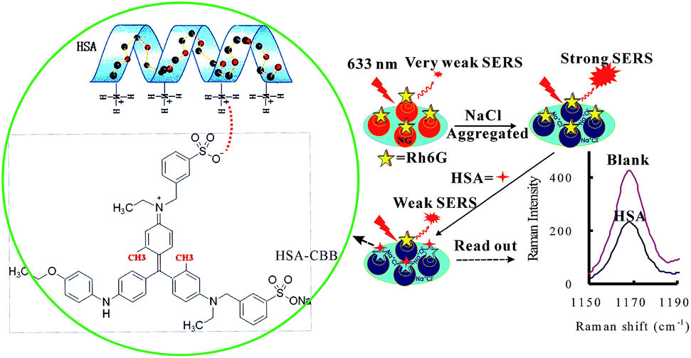
In pH 6.6 PBS, NGs were dispersed in solution and CBB showed a very weak Raman scattering signal in the NG sol substrate. When the aggregation reagent NaCl was added, NGs were aggregated into a NGA sol with high SERS activity and reproducibility. On addition of the CBB molecular probe that can be adsorbed onto the NGA surface, the CBB–NGA complex exhibited the strongest SERS peak at 1171 cm−1. When HSA was added, the SERS peak was quenched due to the interaction between HSA and CBB by means of intermolecular forces (left of Fig. 1). When the amount of HSA was increased, the SERS peak decreased linearly due to an increase in the number of HSA–CBB complexes formed. Therefore, a new simple and sensitive SERS method was proposed for the quantitative analysis of HSA (Fig. 1).
 |
| | Fig. 1 A schematic for the SERS detection of HSA with a CBB molecular probe. | |
SERS spectra
In pH 6.6 PBS containing 0.06 mol L−1 NaCl, no CBB SESR peaks were observed (Fig. S1a†). Upon addition of NGs, it were aggregated into stable NGAs as SERS sol substrate, and CBB molecules were adsorbed onto the surface of the NGAs, in which several SERS peaks appeared at 464 cm−1, 757 cm−1, 907 cm−1, 936 cm−1, 1171 cm−1, 1403 cm−1 and 1612 cm−1 (Fig. S1b†) with an enhanced factor in the range of 1.9 × 10⁵–9.6 × 10⁵ (Table S1†). The three peaks at 464 cm−1, 757 cm−1 and 1171 cm−1 were ascribed to the C–C stretch vibration, the two peaks at 907 cm−1 and 936 cm−1 were ascribed to the benzene in-plane vibration, the peak at 1403 cm−1 was ascribed to the C–N in-plane vibration, and the peak at 1612 cm−1 was ascribed to the C![[double bond, length as m-dash]](https://www.rsc.org/images/entities/char_e001.gif) N stretch. Among them, the peak at 1171 cm−1 is the strongest, and its intensity is linear to the CBB concentration in the range of 2.9 × 10⁻⁸–4.68 × 10⁻⁷ mol L−1. CBB is commonly used to spectrophotometrically analyze μg mL−1 levels of protein and is not reported in SERS detection techniques. We have found that it can be utilized in SERS quantitative analysis, based on the interaction between HSA and CBB causing the SERS peak quenching. As shown in Fig. 2, when the HSA concentration was increased, the peak decreased mostly at 1171 cm−1, and thus this peak was selected for the SERS quantitative analysis of HSA.
N stretch. Among them, the peak at 1171 cm−1 is the strongest, and its intensity is linear to the CBB concentration in the range of 2.9 × 10⁻⁸–4.68 × 10⁻⁷ mol L−1. CBB is commonly used to spectrophotometrically analyze μg mL−1 levels of protein and is not reported in SERS detection techniques. We have found that it can be utilized in SERS quantitative analysis, based on the interaction between HSA and CBB causing the SERS peak quenching. As shown in Fig. 2, when the HSA concentration was increased, the peak decreased mostly at 1171 cm−1, and thus this peak was selected for the SERS quantitative analysis of HSA.
 |
| | Fig. 2 SERS spectra of the CBB–HSA–NG system. (a) 16.74 mg L−1 Au, 6.0 mmol L−1 pH 6.6 PBS, 0.06 mol L−1 NaCl, and 5.9 × 10⁻⁷ mol L−1 CBB; (b) a and 0.1 mg L−1 HSA; (c) a and 0.25 mg L−1 HSA; (d) a and 0.50 mg L−1 HSA; (e) a and 1.0 mg L−1 HSA. | |
RRS and absorption spectrum
The RRS spectrum is obtained easily by means of a synchronous scanning technique, which is commonly used in fluorescence spectrophotometry and is a simple and sensitive tool to study nanoparticles in solution and their aggregation.36–39 In general, increased size and aggregation causes the RRS signal to increase. Small NGs exhibit a weak RRS signal and NGAs exhibit a strong RRS peak at 740 nm in pH 6.6 PBS containing 0.06 mol L−1 NaCl as the aggregation reagent (Fig. S2†). When the CBB concentration was increased, the RRS peak increased non-linearly. This showed that there are interactions between the NGA and CBB molecules, i.e., CBB molecules have adsorbed onto the surface of NGAs. In pH 6.6 PBS, NGs were not aggregated, red in color and exhibited a surface plasmon resonance (SPR) absorption peak at 520 nm (Fig. S3a†). On addition of NaCl, NGs were aggregated to NGAs that the SPR peak disappeared. When CBB was added, the absorption value increased slowly due to the contribution of CBB adsorbed on the NGA surface.
Transmission electron microscopy
According to the procedure, the TEM images of nanogold particles were recorded. As shown in Fig. 3a, the as-prepared nanogold particles were dispersed uniformly in solution with an average diameter of 10 nm. On addition of 0.06 mol L−1 NaCl, the NGs were aggregated to form large aggregates (Fig. 3b), resulting in strong RRS and SERS effects. When CBB, HSA and CBB–HSA were added, the aggregates remained in the system.
 |
| | Fig. 3 TEM images of the nanogold particle system. (a) 16.7 mg L−1 Au and pH 6.6 PBS; (b) 16.7 mg L−1 Au, pH 6.6 PBS, and 0.06 mol L−1 NaCl. | |
Selection of analytical conditions
Analytical conditions such as pH, and the concentration of NG, CBB and NaCl were considered. The effect of pH in the range of 5.0–7.5 on the ΔI value was examined. The results (Fig. S4†) show that when the pH was 6.6, the ΔI value was largest due to the interaction between HSA bearing positive charges and CBB bearing negative charges being strongest. Therefore, 60 μL of a pH 6.6 PBS containing 0.2 mol L−1 PO43− was selected for use. The SERS substrate is vital to the quantitative analysis. In the absence of NGs, the SERS signal of CBB is almost zero (Fig. S5†). When NGs were added to the system, they aggregated into highly SERS active NGAs, and the SERS signal of CBB adsorbed onto the NGA surface was enhanced greatly. When NG was increased, the ΔI value was increased due to formation of more NGAs. 16.74 mg L−1 of NG gave the biggest ΔI value and was selected for use. The molecular probes such as CBB, rhodamine 6G, methyl blue and methyl orange were examined. The results show that 5.86 × 10⁻⁷ mol L−1 of CBB exhibited the strongest sensitivity for HSA detection and was chosen for further use (Fig. S5†). In pH 6.6 PBS solution and in the absence of the aggregation reagent NaCl, NGs were found in their dispersed state, no active NGAs existed, and no SERS peaks were observed (Fig. S6†). When the concentration of NaCl increased, the SERS peaks increased due to the formation of more active NGAs. When the NaCl concentration was 60 mmol L−1, the ΔI was largest (Fig. S7†) and was selected for further use.
Influence of foreign substances
According to the procedure, the influence of 11 foreign substances on the assay of 1.0 μg mL−1 HSA was studied with a relative error (RE) of ±10%. The results show (Table 1) that 200 times glycine (Gly) and glucose (Glu), 150 times Ca(II), Mg(II), Zn(II) and Mn(II), 100 times L-cysteine (Cye), L-lysine (Lys), L-phenylalanine (Phe) and Fe(III), and 1 times BSA did not interfere with the assay of HSA. This indicated that this SERS method had good selectivity (Fig. 4).
Table 1 Influence of foreign substances (FS) on the determination of HSA
| FS |
Tolerance ratio |
RE (%) |
FS |
Tolerance ratio |
RE (%) |
| Gly |
200 |
9.8 |
Cye |
100 |
5.4 |
| Glu |
200 |
9.0 |
Lys |
100 |
4.9 |
| Ca(II) |
150 |
8.3 |
Phe |
150 |
4.3 |
| Mg(II) |
150 |
9.0 |
Fe(III) |
150 |
7.9 |
| Zn(II) |
150 |
1.9 |
BSA |
1 |
3.3 |
| Mn(II) |
150 |
2.2 |
|
|
|
 |
| | Fig. 4 Influence of foreign substances. | |
Working curve
Under the selected conditions, the ΔI values for different concentrations of HSA (CHSA) were plotted vs. CHSA to obtain the working curve (Fig. S8†). The linear range is 0.04–2.0 μg mL−1 HSA, with a regress equation of ΔI = 47.7C + 6.0, a coefficient of 0.9920 and a detection of 0.02 μg mL−1 HSA. Compared to the CBB spectrophotometry, the SERS sensitivity increased 10 times. Five determinations of 0.10, 0.50 and 1.0 μg mL−1 HSA were obtained with a relative standard deviation of 6.8%, 4.9% and 4.7%, respectively. In addition, five batches of NG colloidal solutions were prepared and were used as SERS substrate for the assay of 0.50 μg mL−1 HSA with an accuracy of 5.4%. The results indicated that this SERS method has good accuracy due to the good stability of the NGA sol.
Analysis of samples
Five serum samples were obtained from hospital and were diluted 100 times with water. The contents of HSA in serum samples were determined five times, respectively. The results are listed in Table 2 and are in agreement with those of CBB spectrophotometry (CBB-S) with relative standard deviations (RSD) of 4.6–7.4% and recoveries of 94.6–105.4%.
Table 2 Analytical results of HSA in serum samples
| Sample |
SERS method/mg mL−1 |
Recoverya |
RSD/(%, n = 5) |
CBB-S/mg mL−1 |
| 1.0 μg of HSA was added in 0.50 mL sample solution used to determine the HSA content. |
| 1 |
57.0 ± 2.6 |
95.2 |
4.6 |
60.6 |
| 2 |
61.4 ± 3.7 |
105.4 |
6.0 |
62.4 |
| 3 |
48.7 ± 3.1 |
94.6 |
6.4 |
50.2 |
| 4 |
51.2 ± 3.8 |
95.3 |
7.4 |
54.5 |
| 5 |
45.7 ± 2.9 |
104.6 |
6.3 |
44.1 |
Conclusions
Good reproducibility of the SERS substrate is vital to quantitative analysis. A simple way is to explore the good reproducibility of the SERS sol coupled with a suitable molecular probe using modern nanotechnology. In this study, a reproducible NGA sol with high SERS activity was prepared and combined with a CBB molecular probe to fabricate a SERS nanosensor for trace CBB. We have observed that HSA exhibited a strong SERS quenching effect on CBB molecular probes in the NGA substrate. Thus, a new simple and sensitive SERS method has been proposed for the quantitative analysis of HSA.
Acknowledgements
This work supported by the National Natural Science Foundation of China (no. 21165005, 21267004, 21367005, 21467001, 21465006, 21447006, and 21477025), and the Natural Science Foundation of Guangxi (no. 2013GXNSFFA019003, 2014GXNSFAA118050, and 2014GXNSFAA118059).
References
- M. Fleischmann, P. J. Hendra and A. J. McQuillan, Chem. Phys. Lett., 1974, 26, 163 CrossRef CAS.
- P. L. Stiles, J. A. Dieringer, N. C. Shah and R. P. VanDuyne, Annu. Rev. Anal. Chem., 2008, 1, 600 Search PubMed.
- D. Cialla, A. Marz, R. Bohme and F. Theil, Anal. Bioanal. Chem., 2011, 403, 27–54 CrossRef PubMed.
- D. H. Zhang, P. W. Li, Q. Zhang and W. Zhang, Biosens. Bioelectron., 2011, 26, 2877 CrossRef CAS PubMed.
- Z. Zhou, G. G. Huang, T. Katoc and Y. Ozaki, J. Raman Spectrosc., 2011, 43, 202 Search PubMed.
- S. Kalmodia, J. Harjwani, R. Rajeswari, W. Yang, C. J. Barrow, S. Ramaprabhu, S. Krishnakumar and S. V. Elchuri, Int. J. Nanomed., 2013, 8, 4327 CrossRef PubMed.
- L. M. Chen and Y. N. Liu, ACS Appl. Mater. Interfaces, 2011, 3, 3091 CAS.
- R. Liu, J. F. Liu, X. X. Zhou, M. T. Sun and G. B. Jiang, Anal. Chem., 2011, 83, 9131 CrossRef CAS PubMed.
- C. Dong, Appl. Surf. Sci., 2012, 258, 9218 CrossRef CAS PubMed.
- S. K. Bhargava, Phys. Chem. Chem. Phys., 2013, 15, 12920 RSC.
- C. Z. Dinu, Appl. Surf. Sci., 2012, 258, 9218 CrossRef PubMed.
- O. Peron, E. Rinnert, T. Toury, M. L. Chapelle and C. Compere, Analyst, 2011, 136, 1018 RSC.
- Y. H. Luo, G. Q. Wen, J. C. Dong, Q. Y. Liu, A. H. Liang and Z. L. Jiang, Sens. Actuators, B, 2014, 201, 336 CrossRef CAS PubMed.
- Q. Y. Liu, J. C. Dong, Y. H. Luo, G. Q. Wen, L. Wei, A. H. Liang and Z. L. Jiang, RSC Adv., 2014, 4, 10955 RSC.
- X. H. Zhang, C. Y. Lin, Q. Y. Liu and A. H. Liang, RSC Adv., 2014, 4, 959 RSC.
- G. Q. Wen, L. P. Zhou, T. S. Li, A. H. Liang and Z. L. Jiang, Chin. J. Chem., 2012, 30, 869 CrossRef CAS.
- Y. H. Luo, K. Li, G. Q. Wen, Q. Y. Liu, A. H. Liang and Z. L. Jiang, Plasmonics, 2012, 7, 461 CrossRef CAS.
- Q. Y. Liu, Y. Y. Wei, Y. H. Luo, A. H. Liang and Z. L. Jiang, Spectrochim. Acta, Part A, 2014, 128, 806 CrossRef CAS PubMed.
- P. G. Yin, L. Jiang, X. F. Lang, L. Guo and S. H. Yang, Biosens. Bioelectron., 2011, 26, 4828 CrossRef CAS PubMed.
- M. M. Bradford, Anal. Biochem., 1976, 72(1–2), 248 CrossRef CAS.
- Y. Nan and H. G. Li, Detect. Anal. Rare Elem., 2007, 10(12), 41 Search PubMed.
- L. L. Guo, Y. Zhu and X. W. Zhang, China Med. Eng., 2011, 19(4), 156 Search PubMed.
- F. Eertmans, V. Bogaert and B. Puype, Anal. Methods, 2011, 3, 1296 RSC.
- J. L. Fan, W. Sun, Z. K. Wang, X. J. Peng, Y. Q. Li and J. F. Cao, Chem. Commun., 2014, 50, 9573 RSC.
- Y. F. Long, C. Z. Huang and Y. F. Li, J. Phys. Chem. B, 2007, 111, 4535 CrossRef CAS PubMed.
- H. Q. Luo, N. B. Li and S. P. Liu, Biosens. Bioelectron., 2006, 21, 1186 CrossRef CAS PubMed.
- Z. Q. Yu, T. Kabashima, C. Tang, T. Shibata, K. Kitazato, N. Kobayashi, M. K. Lee and M. Kai, Anal. Biochem., 2010, 397, 197 CrossRef CAS PubMed.
- J. A. Lines, Z. Q. Yu and S. X. Chen, Biochem. Biophys. Res. Commun., 2014, 443, 308 CrossRef CAS PubMed.
- I. Willner, Nano Lett., 2004, 4, 1683–1687 CrossRef.
- K. Tonanka, T. Kabashima, T. Shibata, C. Tang, Z. Q. Yu and M. Kai, Anal. Sci., 2008, 24, 471 CrossRef.
- D. M. Barry, S. Lorna, W. Alan, F. Sabine, J. T. Nicolas, C. Chris and G. Duncan, Nat. Biotechnol., 2004, 22, 1133 CrossRef PubMed.
- C. M. Ruan, W. Wang and B. Gu, Anal. Chem., 2006, 78, 3379 CrossRef CAS PubMed.
- Y. Lei and J. Xu, J. Light Scat., 2015, 27, 1 Search PubMed.
- L. X. Chen, X. L. Fu and J. H. Li, Nanoscale, 2013, 5, 5905 RSC.
- J. H. Granger, M. C. Granger, M. A. Firpo, S. J. Mulvihill and M. D. Porter, Analyst, 2013, 138, 410 RSC.
- P. P. Fu, N. B. Li and H. Q. Luo, Analyst, 2012, 137, 1097 RSC.
- A. H. Liang, Y. Zhang, Y. Y. Fan, C. Q. Chen, G. Q. Wen, Q. Y. Liu, C. Y. Kang and Z. L. Jiang, Nanoscale, 2011, 3, 3178 RSC.
- J. C. Dong, A. H. Liang and Z. L. Jiang, RSC Adv., 2013, 3, 17703 RSC.
- G. Q. Wen, Y. H. Luo, A. H. Liang and Z. L. Jiang, Sci. Rep., 2014, 4, 3990 Search PubMed.
Footnotes |
| † Electronic supplementary information (ESI) available. See DOI: 10.1039/c4ra11778f |
| ‡ These authors contributed equally to this work. |
|
| This journal is © The Royal Society of Chemistry 2015 |
Click here to see how this site uses Cookies. View our privacy policy here.