Jinhua Li‡
a,
Ruichen Dong‡ab,
Xiaoyan Wangc,
Hua Xiong*b,
Shoufang Xuad,
Dazhong Shen*c,
Xingliang Songd and
Lingxin Chen*a
aKey Laboratory of Coastal Environmental Processes and Ecological Remediation, Shandong Provincial Key Laboratory of Coastal Environmental Processes, Yantai Institute of Coastal Zone Research, Chinese Academy of Sciences, Yantai 264003, China. E-mail: lxchen@yic.ac.cn; Fax: +86 535 2109130; Tel: +86 535 2109130
bState Key Laboratory of Food Science and Technology, Nanchang University, Nanchang 330047, China. E-mail: huaxiong100@126.com; Fax: +86 791 6634810; Tel: +86 791 6634810
cKey Lab in Molecular and Nanomaterials Probes of the Ministry of Education of China, College of Chemistry, Chemical Engineering and Materials Science, Shandong Normal University, Jinan 250014, China. E-mail: dzshen@sdnu.edu.cn; Fax: +86 531 82615258; Tel: +86 531 86180740
dSchool of Chemistry & Chemical Engineering, Linyi University, Linyi 276005, China
First published on 24th December 2014
A facile strategy was developed to prepare magnetic molecularly imprinted microspheres (MMIMs) for the selective recognition and effective removal of 17-beta-estradiol (17β-E2) by reversible addition-fragmentation chain transfer precipitation polymerization. One-pot synthesis was employed, which could simplify the imprinting process and shorten the experimental period. The resultant MMIMs displayed fast kinetics and high binding capacity, and the adsorption processes followed Langmuir–Freundlich isotherm and pseudo-second-order kinetic models. Excellent recognition selectivity toward 17β-E2 was attained over other phenolic estrogens such as 17-alpha-E2, estriol and estrone. The magnetic property of MMIMs provided fast and simple separation, and the recycling process for magnetic solid phase extraction (MSPE) was sustainable at least five times without obvious efficiency decrease. Furthermore, the MMIMs-MSPE presented satisfactory recoveries within 71.7–108.3% with the precisions of 1.1–6.0% for spiked 17β-E2 in water, soil and food samples. The developed MMIMs-based method proved to be a convenient and practical way in sample pretreatment and targeted pollutants removal.
Recently, a number of reports using versatile, robust and cost-effective molecularly imprinted polymers (MIPs)6 to specifically recognize, detect and remove 17β-E2 from polluted water and foods have been demonstrated. For example, Ma et al. prepared selective core–shell MIPs of 17β-E2 on the surface of silica nanoparticles.7 Shi et al. introduced molecularly imprinted solid phase extraction (MISPE) combined with HPLC to detect trace 17β-E2 in different dairy and meat samples.8 Noir et al. fabricated macroporous MIP/cryogel composite systems for removing trace endocrine disrupting contaminants.9 Usually, MIPs are synthesized by traditional free radical polymerization, whereas the rate of chain propagation cannot be controlled, resulting in a broad size distribution. The introduction of reversible addition-fragmentation chain transfer (RAFT) polymerization techniques into the molecular imprinting strategy has attracted significant interest, which can solve the above problem.10–14 For examples, Pan et al. described an approach to obtain water-compatible MIPs by the facile surface-grafting of functional polymer brushes via RAFT polymerization.10 Titirici et al. prepared thin film MIPs around mesoporous silica beads by adopting covalent immobilization of azo initiators and RAFT-mediated living radical polymerization.11 Our group presented atrazine MIPs for preconcentration of atrazine in food matrices by using di-thioesters based RAFT coupled to precipitation polymerization.12,13
Moreover, magnetic MIPs have received wide attentions since they can be easily isolated/collected and recycled by an external magnetic field and have been applied in many fields such as separation/purification, chemo/biosensing, and drug delivery.15–19 For instance, Li et al. synthesized a core–shell magnetic imprinted polymer for the fast and selective removal of EDCs.18 Moreover, our group successfully prepared photonic/magnetic dual-responsive MIPs for recognition of caffeine.19
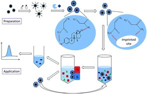
Herein, inspired by these studies, we developed an improved core–shell MIPs synthesis strategy for enhanced selective recognition and removal of 17β-E2 by combining RAFT precipitation polymerization and magnetic separation. One-pot synthesis was conducted, allowing all reagents to react together under proper conditions. Moreover, the resultant MIPs layer was grafted onto the surface of magnetic iron oxide beads by a RAFT agent for improving the polymerization efficiency. The obtained magnetic molecularly imprinted microspheres (MMIMs) were easily characterized by morphologies, structures, thermostability and magnetism, as well as static and dynamic adsorptions. Finally, the MMIMs were used as SPE sorbents and successfully applied to the extraction of 17β-E2, providing a practicable way in samples pretreatment and removal of trace targeted pollutants in environment and food.
The morphological evaluation was performed by transmission electron microscopy (TEM, JEOL-100CX-2). Infrared spectra were recorded using Fourier transform infrared spectrometer (FT/IR-4100, Thermo Nicolet Corporation, USA). Thermal gravimetric analysis (TGA) was carried out from room temperature to 800 °C with a heating rate of 10 °C min−1 under nitrogen environment by thermal gravimetric analyzer (Mettler 5 MP), presenting TGA and derivative thermogravimetry (DTG) data. Magnetic property was measured by vibrating sample magnetometer (VSM, Lake Shore 7410, Beijing, China). UV-Vis spectra were recorded using a Thermo Scientific NanoDrop 2000/2000c spectrophotometer (Thermo, USA). N2 adsorption–desorption isotherms were examined with Beishide instruments (3H-2000PS4, Beijing) for Brunauer–Emmett–Teller (BET) analysis to determine the specific surface area and pore size. The amounts of analytes were determined by HPLC-UV (Skyray LC-310, Skyray Instrument Inc., China), under the optimized conditions: sample loaded, 20 μL; mobile phase, acetonitrile–water (v/v, 7:
3); flow rate, 1.0 mL min−1; detection wavelength, 208 nm; column, 250 mm × 4.6 mm C18 at 20 °C.
![]() | ||
Fig. 1 Schematic illustration of the preparation process of MMIMs by RAFT precipitation polymerization, and MSPE application for recognition and removal of 17β-E2. |
The process of magnetic solid phase extraction (MSPE) was carried out according to a previously reported method21–23 with suitable modifications. Specifically, MMIMs of 80 mg were dispersed into 2 mL sample extract solutions spiked with 17β-E2 at three concentrations (0.1, 1.0 and 10 mg L−1). After being shaken for 3 h at room temperature, the MMIMs sorbent loaded with 17β-E2 was separated from the suspension using a magnet, and the supernatant was measured by HPLC-UV. Subsequently, the 17β-E2 adsorbed onto the MMIMs sorbent was eluted with 2 mL of methanol–acetic acid solution (9:
1, v/v). Then, the eluent was dried and re-dissolved in 2 mL acetonitrile and then determined by HPLC-UV. Finally, after the extraction process, in order to reuse the same sorbent for new extraction of the 17β-E2, the MMIMs were washed with methanol–acetic acid solution and acetonitrile several times, and then the sorbent was dried at 60 °C for new extraction. The MSPE procedure is illustrated in Fig. 1.
Fig. 2 shows the morphologies of the products at different synthetic stages. Fe3O4@SiO2 (Fig. 2A) and MMIMs (Fig. 2B) particles were monodisperse and exhibited a regular sphere morphology, which indicated the polymerization smoothly proceeded on the surface of Fe3O4@SiO2 by RAFT. By comparing Fig. 2A and B, the thickness of the imprinting shell layer could be attained within 10–30 nm (Fig. 2B). Owing to the thin imprinting layers, almost all the template molecules could be completely eluted and thereby produce the largest amounts of imprinted cavity sites, resulting in high binding capacities and fast mass transfer. However, the MMIMs-Control and MNIMs particles were not uniform in size distribution, with the average diameter ranging from 225 to 505 nm for MMIMs-Control (Fig. 2C) and from 730 to 1500 nm for MNIMs (Fig. 2D), respectively. And the non-uniform size distribution was adverse to mass transfer. When Fe3O4@SiO2 without further RAFT functionalization was used, imprinting layers could not form around the magnetic particles, but non-magnetic secondary particles formed by precipitation polymerization with much smaller diameters (100–200 nm), as shown in Fig. 2C. The secondary particles might well adhere with the magnetic particles, and thereby cause inhomogeneous size and distribution of the MMIMs-Control (Fig. 2C). For MNIMs, as seen in Fig. 2D, their sizes were quite large with a wide distribution, which was quite different from MMIMs. This was very likely owing to the absence of template in the RAFT polymerization. For the MNIMs, without the presence of template molecules, the crosslinking substances, all of which contain double bonds, could more easily form shell-layers on the surface of Fe3O4@SiO2, and thereby result in thicker shell-layers and easier aggregation, presenting non-homogeneous and much larger material (Fig. 2D).
Fig. 3A shows the FT-IR spectra of Fe3O4, Fe3O4@SiO2, Fe3O4@SiO2-RAFT, MMIMs and MNIMs particles. The polymerization process could be evaluated according to the characteristic functional groups. The characteristic absorption band of Fe3O4 was at 580 cm−1 (curve a). The absorption at 803 and 1089 cm−1 could be attributed to the stretching vibration of Si–O and Si–O–Si, respectively, proving that SiO2 was successfully covered on the Fe3O4 particles (curve b). As seen from curve c, the peak at 1629 cm−1 represented the stretching vibration of CC bond of alkene, due to the RAFT active modification on the surface of Fe3O4@SiO2. The unique absorption peak at 1728 cm−1 belonged to the stretching vibration of the C
O bond, the typical peak at 2998 and 2955 cm−1 could be ascribed to the saturated C–H bond, as well as the peaks at 1392 and 1260 cm−1 could be assigned to the C–N bond, indicating the imprinted polymer layer was grafted onto the surface of Fe3O4@SiO2 successfully, as demonstrated in curve d. In addition, the peak at 1462 cm−1 belonged to the aromatic carbon–carbon bond of DVB, which was used for the preparation of the polymer. The FT-IR spectrum of MNIMs (curve e) looks exactly the same as that of MMIMs (curve d), except for the lower intensity. All the results of FT-IR confirmed that the core–shell structured MMIMs were successfully prepared by RAFT precipitation polymerization.
The corresponding TGA results of the above five particles were displayed in Fig. 3B. As can be seen in the figure, the TGA curve of Fe3O4 was almost a straight line, indicating the particles were very pure and without any impurities (curve a). There was a slight weight loss at 250–350 °C for Fe3O4@SiO2 particles (curve b), which could be attributed to the dehydration of the SiO2 layer. As can be observed from curve c, Fe3O4@SiO2-RAFT exhibited bigger weight loss, which may be due to the pyrolysis of RAFT active groups. The TGA curves of MMIMs (curve d) and MNIMs (curve e) particles dropped rapidly at around 450 °C, suggesting that the whole imprinted and non-imprinted polymer layers on the surface of Fe3O4@SiO2 largely decomposed under high temperature. In addition, the peak temperatures of MMIMs and MNIMs were 454.36 and 451.72 °C, respectively, and the residual amounts were 25.61% and 36.30%, respectively, as can be seen from the TGA results in Table S1† and DTG curves in Fig. S1.† These results demonstrated that the MMIMs possessed higher polymerization efficiency and good thermal stability below 400 °C.
Fig. 3C shows the magnetic hysteresis loops analysis of the MMIMs, MMIM-Control and MNIMs, and the inset illustrates the dispersion and agglomeration processes of the MMIMs. It is seen that there is a similar general shape to the three curves, although the saturation magnetization value of MMIMs is low (Table S2†). The results suggested that the prepared MMIMs were magnetically responsive. As can be seen from Table S2,† the MMIMs had better magnetic induction intensity, which may be caused by the strong magnetism of independent Fe3O4@SiO2 particles that did not participate in the reaction. Consequently, the homogeneously dispersed MMIMs could go straight towards the magnet and adhere to the inner side wall of the vials when the external magnetic field was applied, and the turbid solution became clear and transparent, as evidenced in the inset of Fig. 3C, showing a fast and simple magnetic separation.
The N2 adsorption–desorption isotherms and pore diameter distribution of MMIMs, MMIMs-Control and MNIMs are shown in Fig. 4. The type IV isotherm curves with a loop were observed for MMIMs (Fig. 4A), which indicated that MMIMs had a well-defined porous structure. As can be seen in the figure, the MMIMs showed a hysteresis loop, where the desorption curve was closer but slightly leveled above the adsorption curve, representing that the MMIMs were stable with low swelling and low solvent uptake.24 The narrow pore size distribution and low average pore diameter that can be observed from the adsorption plot (Fig. 4B) suggested that the size of the cavities formed in the MMIMs matrix played an important role in the binding capacity. The structure parameters of the three microspheres obtained by BET analysis are listed in Table S3.† As can be seen in the figure, the specific surface area, cumulative pore volume and average pore diameter of MMIMs particles were 444.86 m2 g−1, 0.10 mL g−1 and 4.18 nm, respectively, while those of MMIMs-Control particles were 6.89 m2 g−1, 0.030 mL g−1 and 7.96 nm, and those of MNIMs particles were 46.18 m2 g−1, 0.038 mL g−1 and 4.35 nm, respectively. Obviously, the specific surface area and cumulative pore volume of MMIMs were much larger, while the average pore diameter was slightly smaller than that of the latter two. The large specific surface area proved that MMIMs had a uniform, regular spherical structure (Fig. 2B). Generally, MIPs have slight differences in surface area and pore volume from NIPs. Here, the significantly high values of MMIMs might be easily attributed to the template functioning somehow with the RAFT polymerization. During MMIMs preparation, when the template molecules were added, the synthesis would become a doping synthesis to some extent. This would produce thinner imprinting shell-layers with smaller diameters, along with amounts of imprinted cavities, and therefore would lead to large surface area and pore volume.
![]() | ||
Fig. 4 N2 adsorption–desorption isotherms (A) and pore diameter distribution curves (B) of MMIMs, MNIMs and MMIMs-Control. |
To evaluate the binding isotherms of MMIMs, four adsorption isotherm models, including Langmuir, Freundlich, Redlich–Peterson and Langmuir–Freundlich, were employed,24 as shown in Fig. 5B. Their corresponding equations and parameters for adsorption of 17β-E2 onto the three polymers are listed in Table S4.† Detailed descriptions for model fitting are given in the ESI.† It can be seen that for all the three polymers, the Langmuir–Freundlich isotherm model yielded the best fitting among the four models with correlation coefficients (R2) of 0.9995, representing its ability to simultaneously model both subsaturation and saturation behaviors.26 In addition, the MMIMs provided the highest concentration of binding sites per gram of polymers (Nt = 212.8 μmol g−1) and the largest median binding affinity (α = 5.677 g μmol−1), indicating an excellent imprinting effect due to the presence of a number of specific binding sites on the MMIMs.
Dynamic binding experiments were conducted to assess the mass transfer properties of the MMIMs. Fig. 6A shows the time-dependent increase in the amount of 17β-E2 adsorbed by MMIMs and MMIMs-Control. As can be seen in the figure, the MMIMs demonstrated significantly higher dynamic binding performances than that of MNIMs, which indicated that the uniform spherical structure and larger specific surface area of the MMIMs were favorable to rapid mass transfer.
As for the dynamic models of MMIMs, pseudo-first-order, pseudo-second-order, Elovich and intraparticle diffusion27–29 for 17β-E2 are displayed in Fig. 6B, and the related equations and fitting results are listed in Table S5.† Detailed descriptions for the model fitting could be found in ESI.† As can be seen in the figure, the pseudo-second-order model could better describe the time effect on the adsorption system than other kinetic models, which provided the most suitable correlation for the adsorption with the highest correlation coefficient of 0.9966. The pseudo-second-order equation can be expressed as follows:
![]() | (1) |
In order to evaluate the competitive recognition ability of the obtained MMIMs, seven PEEs (E3, 17α-E2, DS, BPA, DES, HS and E1) as structural analogues were used. As seen from Fig. 7A (up), MMIMs presented a higher binding capacity for 17β-E2 than that for those competitive PEEs. The binding capacities and selectivity factors of the MMIMs for E3, 17α-E2 and DS were close and higher than the other four PEEs, as their structures are more similar to that of 17β-E2 (Fig. 7A, below). Moreover, compared with the MMIMs-Control, the MMIMs showed obviously higher binding capacity, revealing the utilization of RAFT as an ideal strategy could greatly improve the binding capacity and mass transfer rate of the imprinted materials. However, MNIMs adsorbed much less template molecules, and there was no significant difference in binding capacity between the competitive PEEs and 17β-E2, since there were no tailor-made recognition sites formed in the MNIMs. Therefore, the obtained MMIMs could selectively recognize the delicate difference of 17β-E2 from its analogues.
In order to assess the practical applicability of MMIMs-MSPE, several real samples of water, soil and food were analyzed. Table 1 lists the removal recoveries of MMIMs applied in seawater, lake water, soil and yogurt samples spiked at three concentration levels of 17β-E2. Satisfactory recoveries were obtained, such as 76.7–108.3% with precision of 1.5–4.9% at 0.10 mg L−1. The MMIMs were demonstrated to be potentially applicable for highly efficient preconcentration and separation of 17β-E2 in real samples. Fig. S2† presents the typical HPLC-UV chromatograms. As can be seen in the figure, the matrix effects were reduced and the spiked 17β-E2 compounds were concentrated by MMIMs-MSPE, indicating that MMIMs had excellent imprinting efficiency. Moreover, the magnetic property would enable the MMIMs-MSPE more attractive. Compared to conventional SPE, the MSPE provided a simple fast magnetic separation and enrichment procedure, preventing from the complex pretreatments such as filtration and centrifugation.
Samples | Spiked concentration (mg L−1) | ||
---|---|---|---|
0.1 | 1.0 | 10.0 | |
a Experimental conditions: polymer, 80 mg; sample volume, 2 mL; incubating time, 3 h; separation, magnet; eluting, 2 mL methanol–acetic acid solution (9![]() ![]() |
|||
Seawater | 76.7b ± 4.9c | 82.7 ± 2.7 | 74.2 ± 3.4 |
Lake water | 86.4 ± 4.7 | 75.6 ± 4.4 | 71.7 ± 2.8 |
Soil | 108.3 ± 4.7 | 93.4 ± 2.6 | 105.8 ± 3.0 |
Yogurt | 88.9 ± 1.5 | 96.0 ± 1.1 | 100.3 ± 6.0 |
Footnotes |
† Electric supplementary information (ESI) available. See DOI: 10.1039/c4ra11177j |
‡ Equally contributed to this work. |
This journal is © The Royal Society of Chemistry 2015 |