Unraveling the binding interaction of Toluidine blue O with bovine hemoglobin – a multi spectroscopic and molecular modeling approach

Krishnamoorthy Shanmugaraja, Shanmugam Anandakumarb and Malaichamy Ilanchelian*a
aDepartment of Chemistry, Bharathiar University, Coimbatore – 641046, Tamil Nadu, India. E-mail: chelian73@yahoo.com; Fax: +91 422 2422 387; Tel: +91 422 2428 317
bDepartment of Bioinformatics, Bharathiar University, Coimbatore – 641046, Tamil Nadu, India

Received 24th September 2014 , Accepted 5th December 2014

First published on 5th December 2014


Abstract

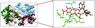
Toluidine blue O (TBO) is a cationic photosensitizer that belongs to the class of phenothiazinium dyes. It has been widely used in photodynamic therapy due to its preferential affinity towards cancer cells in vivo. The present study reports a detailed binding mechanism of photodynamic therapeutic agent TBO with bovine hemoglobin (BHb) using steady state emission, time resolved fluorescence, UV-Vis absorption, circular dichroism (CD) and three dimensional emission (3D) spectral studies. The results from the steady state emission and time resolved fluorescence studies revealed that the quenching of BHb emission by TBO is due to the formation of a ground state (BHb–TBO) complex with a substantial binding affinity value of (1.19 ± 0.30) × 105 dm3 mol−1. Moreover, TBO induced tertiary and secondary conformational changes of BHb were monitored by UV-Vis absorption, CD and 3D emission spectral studies. The most probable binding location of TBO within the cavity of BHb has been investigated by AutoDock-based blind docking simulation. The negative Gibbs energy change and docking results revealed that hydrogen bonding and hydrophobic interactions played a crucial role in the complexation of TBO with BHb.


1. Introduction

Proteins are important biomolecules that play crucial roles in sustaining life and are closely related to the origin, evolution and metabolism of life. The studies of the binding interaction of small exogenous ligands to proteins have attracted great interest in the fields of chemistry, life science, biophysics and clinical medicine for decades.1 The various specific and nonspecific interactions of drugs with the residues of proteins have important consequences to the biological function of protein.2 Many drugs bind reversibly to proteins, and this in turn alters the absorption, distribution, transport, metabolism, excretion of various drugs, cellular uptake and activity of drugs in the circulatory system. Therefore, detailed investigation of drug–protein interaction assumes significance for thorough understanding of the pharmacodynamic and pharmacokinetics behavior of the drug.3

Hemoglobin (Hb) is an iron-containing respiratory blood protein, and is mostly present in red blood cells of vertebrate erythrocytes. It is responsible for carrying oxygen from the lungs to different respiring tissues, to disperse hydrogen peroxide and to effect electron transfer to all body parts and organs.4,5 In addition, it is also involved in many clinical diseases such as anemia, leukemia and heart disease.6 Hb has the ability to reversibly bind a number of endogenous and exogenous molecules and thus act as a drug carrier for effective delivery to the required physiological site for the treatment of various diseases.7–11 The enzymatic and antioxidant activities of Hb are well documented and it is widely available in literature.12–14 Hb exists as a tetramer of globin chains, consisting of two α and two β subunits. The α-chains contain 141 amino acid residues, whereas, the β-chains contain 146 amino acid residues. Each α-chain is in contact with the β-chains.15 Each subunit has a polypeptide chain attached to an iron-containing component called heme. Bovine hemoglobin (BHb) is an important functional protein for reversible oxygen carrying and storage. BHb has a less exothermic oxygen binding ability and delivers oxygen even at low temperatures. BHb shares 90% amino acid sequence homology with human hemoglobin and is a better oxygen carrier than its human counterpart.10

Hb has long been used as the paradigm for understanding the structure–function relationships of proteins.16–20 Furthermore, the unfolding of this kind of protein is closely associated with physiological abnormalities. For example, under very acidic conditions, the cooperative oxygen binding property of Hb is decreased and the pro-oxidative activity is dramatically increased mainly due to the significant conformational changes in its structure and heme crevice.21,22 In addition, Hb can undergo structural changes very easily and hence it can be used as a good model protein to study the conformational changes. The fundamental understanding of the conformational behavior of proteins in protein–ligand system is of critical importance in the field of chemistry and biology. Thus, the study on the binding interaction of antitumour drug with Hb is imperative and of fundamental importance in terms of understanding their pharmacological actions.23

Toluidine blue O (TBO) (Fig. 1), a positively charged phenothiazine dye and a potential antitumour drug24,25 is generally used as polymerization inhibitor, complexing agent, biological sensitizer and stain.26,27 TBO is effectively used for the diagnosis and treatment of oral diseases, since it is a selective stain for oral cancer and also for various oral pathogens.28,29 TBO has also been proven as an antifungal and antibacterial drug for the inactivation of yeast and some gram-positive and gram-negative bacteria.30 Moreover, TBO has been studied for its mutagenic action,31 toxic interaction on binding with DNA,32 RNA,33 and living bull spermatozoa,34 and photoinduced inactivation of viruses.35 Ephros and Mashberg reported that the use of TBO as a mouth rinse and subsequent flushing to the environment presents potentially serious consequences that might adversely affect fish and other aquatic life.36 Therefore, it is necessary to investigate the binding mechanism of TBO with BHb, as it can provide valuable insight into the toxicity of the drug on binding to protein.


image file: c4ra11136b-f1.tif
Fig. 1 Structure of TBO.

This work aims to delineate the binding mechanism of TBO with BHb by means of steady state emission, time-resolved fluorescence, UV-Vis absorption, CD and 3D emission spectral studies. The binding parameters such as binding constant, the number of binding sites and binding force were obtained from the emission spectral studies. The effect of TBO on the conformational and secondary structural changes of BHb was studied by UV-Vis absorption, CD and 3D emission spectral studies. The exact binding location of TBO within BHb was explored using AutoDock based blind docking strategy. This report provides quantitative binding information on the complexation of photodynamic therapeutic agent TBO with BHb.

2. Experimental procedures

2.1. Materials

BHb was procured from Sigma-Aldrich, USA and purified by reported procedure.14 The purity of BHb was checked by reversed phase high performance liquid chromatography and was found to be greater than 98%. TBO dye was purchased from Sd fine chemicals, India, and purified by silica gel column chromatography using ethanol–benzene (7[thin space (1/6-em)]:[thin space (1/6-em)]3 v/v) containing 0.4% glacial acetic acid and finally recrystallized from ethanol.37–39 A stock solution of TBO was prepared at a concentration of 1.20 × 10⁻⁴ mol dm−3 using phosphate buffer solution (PBS) at pH = 7.40 and stored in dark until further use. The stock solution of BHb was prepared in PBS at pH = 7.40. All other reagents were of analytical grade and water used in this investigation was doubly distilled over alkaline potassium permanganate using an all-glass apparatus.

2.2. Steady state emission spectral measurements

The emission spectral studies were performed with JASCO FP-6600 spectrofluorometer equipped with a 1.0 cm quartz cuvette. A 10−6 mol dm−3 solution of BHb was prepared daily for experiments. Various concentrations of TBO solutions were freshly prepared by pipetting an aliquot of the stock solution into a 5 mL standard measuring flask containing 1 mL of BHb solution (10−6 mol dm−3), and finally made up to 5 mL with PBS. BHb and TBO solutions are mixed uniformly and allowed to equilibrate for 15 minutes before recording spectral data. BHb solution was excited at 295 nm and the emission wavelength was recorded between 300 to 500 nm with a scanning speed of 200 nm min−1. The emission and excitation slit widths used throughout the experiment were 5 and 10 nm, respectively. 3D emission spectra were performed under the following conditions; the emission was recorded between the wavelength range of 200 nm to 500 nm and excitation was recorded between the wavelength ranges from 200 nm to 340 nm. The excitation and emission bandwidths for 3D emission spectral studies were 10 and 6 nm, respectively.

2.3. Absorption spectral measurements

Absorption spectral measurements were performed with a JASCO V-630 UV-Visible spectrophotometer using PBS as references. Quartz cuvettes of path length 1.0 cm were used to carry out the absorption spectral studies.

2.4. Time resolved fluorescence lifetime measurements

The fluorescence lifetime measurements were performed using time-correlated single photon counting (TCSPC) technique using Horiba Jobin Yvon. The samples were excited at 295 nm using a picosecond diode (IBH NanoLED-295) in an IBH fluorocube apparatus. The emission data were collected at a magic angle (54.7°) relative to the excitation, passed through a monochromator and into a fast detector, using a Hamamatsu MCP photomultiplier (2809U). The repetition rate was 1 MHz. The instrumental response function (IRF) was determined experimentally on the basis of light signal scattered from Ludox (colloidal silica in water) and was used for subsequent deconvolution of the fluorescence signal. The fluorescence decays were deconvoluted using IBH DAS6 software in order to obtain best residuals and acceptable χ2 (0.9–1.2) values. The following equation was used to analyze the experimental time-resolved fluorescence decays (eqn (1)),40
 
image file: c4ra11136b-t1.tif(1)
where, F(t) is the fluorescence intensity at time t and ai is the pre-exponential factor representing the fractional contribution to the time resolved decay of the ith component with a lifetime τi. For multiexponential decays, the average lifetime τavg was calculated from the following equation (eqn (2)).40
 
image file: c4ra11136b-t2.tif(2)
where, τi is the fluorescence lifetime and ai are their relative amplitude with i variable from 1 to 2.

2.5. Circular dichroism measurements

Circular dichroism (CD) spectroscopy is a sensitive instrumental method to monitor the secondary structural change of protein upon interaction with ligands. CD measurements were recorded with a JASCO-810 spectropolarimeter equipped with a quartz cuvette of 0.1 cm path length. The spectra were recorded in the wavelength range of 200 nm to 260 nm and from 350 to 450 nm with a scan speed of 50 nm min−1. Each spectrum presented was the average of three scans and all observed CD spectra were baseline subtracted for buffer solution. All the measurements were carried out at room temperature (25 °C).

2.6. Molecular docking studies

Molecular docking study of BHb–TBO complexation was carried out on AutoDock 4.2 program which utilizes Lamarckian Genetic Algorithm (LGA).41 The crystallographic coordinates of TBO was obtained from the PubChem database (http://pubchem.ncbi.nlm.nih.gov). The native structure of BHb (PDB ID: 1G09) was retrieved from Brookhaven Protein Data Bank (http://www.rcsb.org/pdb). As required in the Lamarckian Genetic Algorithm, all water molecules were removed and hydrogen atoms were added followed by the calculation of Gasteiger charges. The grid size along the x-, y-, z-axes were set to 50 Å, 50 Å and 50 Å, respectively. The grid center along the x-, y-, z-axes were set as 1.07 Å, 65.021 Å and 12.094 Å, respectively. The AutoDocking parameters used were as follows: GA population size = 150, maximum number of energy evaluations = 250[thin space (1/6-em)]000 and GA crossover mode of two points. The lowest binding energy conformer was searched out of 25 different conformers for the docking simulation and the resultant was used for further analysis. The docked conformations were viewed using PyMOL (http://www.pymol.org) software package.

3. Results and discussion

3.1. Emission spectral studies

The intrinsic emission of many proteins is mainly attributed to tryptophan (Trp) and tyrosine (Tyr) residues, which are very sensitive to their microenvironment. BHb contains six Trp residues, in which two αβ dimers have three Trp residues such as α-Trp14, β-Trp15 and β-Trp37.42 Additionally, it is also comprised of five Tyr residues in each αβ dimer as α-Tyr24, α-Tyr42, α-Tyr140, β-Tyr34 and β-Tyr144.43 The intrinsic emission of BHb primarily originates from β-Trp37 that plays a key role in the quaternary state change upon ligand binding.44 The emission spectra of BHb with increasing concentrations of TBO is shown in Fig. 2(A). The emission spectrum of BHb in the absence of TBO shows an emission maximum at 334 nm (Fig. 2(A)(a)), when excited at 295 nm. In the presence of increasing concentrations of TBO, the emission intensity of BHb decreased gradually accompanied by the bathochromic shift around 2 nm from 334 to 336 nm. Recently, a similar kind of behavior was reported for the interaction of hemoglobin with malachite green dye.45 It has been proposed that the decrease in emission intensity along with slight red shift is mainly attributed to the complexation induced decrement in the hydrophobicity around β-Trp37 residue.45 Thus, in the present study, it is believed that the observed emission quenching with slight red shift in the emission maximum wavelength is mainly due to the formation of a complex between TBO with BHb, with the binding site of TBO situated near the β-Trp37 residue. Moreover, observed red shift in the emission wavelength of BHb is due to the alteration in protein conformation, which in turn leads to the changes in the microenvironment around the Trp residues.
image file: c4ra11136b-f2.tif
Fig. 2 (A) Emission spectra of BHb (3.00 × 10⁻⁶ mol dm−3) at various concentrations of TBO. [TBO]: [a] 0.00, [b] 2.40 × 10⁻⁷, [c] 4.80 × 10⁻⁷, [d] 7.20 × 10⁻⁷, [e] 9.60 × 10⁻⁷, [f] 12.00 × 10⁻⁷, [g] 14.40 × 10⁻⁷, [h] 16.80 × 10⁻⁷, [i] 19.20 × 10⁻⁷, [j] 21.60 × 10⁻⁷ and [k] 24.00 × 10⁻⁷ mol dm−3; (B) emission spectra of TBO (3.00 × 10⁻⁶ mol dm−3) at various concentrations of BHb. [BHb]: [a] 0.00, [b] 4.00 × 10⁻⁷, [c] 8.00 × 10⁻⁷, [d] 12.00 × 10⁻⁷, [e] 16.00 × 10⁻⁷, [f] 20.00 × 10⁻⁷, [g] 24.00 × 10⁻⁷, [h] 28.00 × 10⁻⁷, [i] 32.00 × 10⁻⁷ and [j] 36.00 × 10⁻⁷ mol dm−3; pH 7.40.

TBO posses substantial emission property and it exhibits strong emission maximum at 654 nm, when excited at 595 nm.37,39 Hence, the effect of BHb on the emission property of TBO was monitored and the results are represented in Fig. 2(B). Upon the addition of increasing concentrations of BHb, the emission intensity of TBO decreased gradually with slight red shift of around 2 nm. In recent past, a similar behavior has been reported for the interaction of sanguinarine with hemoglobin.5 The results from the emission spectral experiments clearly enabled us to conclude the complexation of TBO with BHb protein.

Since the existence of inner filter effect (IFE) is known to affect emission titration measurements,40,46 we took advantage of some effective methods to correct the emission intensities of the complex and reduce the IFE. The elimination of IFE of BHb is also discussed in detail in the ESI. After the removal of IFE, it is imperative to ascertain the specific emission quenching mechanism of BHb by TBO viz., collisional or binding-related quenching.47 Therefore, in the ensuing section the exact emission quenching mechanism involved in the BHb–TBO system was discussed in detail.

3.2. Emission quenching mechanism

Quenching mechanisms are usually classified as either static or dynamic quenching. To elucidate the nature of quenching mechanism, the emission quenching data were analyzed using the Stern–Volmer equation (eqn (3)).40
 
image file: c4ra11136b-t3.tif(3)
where, F0 and F represent the corrected emission intensity in the absence and presence of quencher, respectively. kq is the bimolecular quenching rate constant, ksv is the Stern–Volmer quenching constant, which measures the efficiency of quenching, τ0 is the average lifetime of the biomolecule without quencher (τ0 = 10−8 s)40 and [Q] is the concentration of the quencher.

The emission quenching data from BHb–TBO (Fig. 2(A)) system were analyzed using eqn (3) and their corresponding Stern–Volmer plot of F0/F vs. [TBO] are displayed in Fig. 3(A). As shown in Fig. 3(A), the plot exhibited a good linear relationship within the investigated concentrations of TBO. In general, a linear Stern–Volmer plot indicates the occurrence of single type of quenching, either static or dynamic quenching. The value of Stern–Volmer quenching constant ksv was obtained from the slope of the linear plot and their values are presented in Table 1. The bimolecular quenching constants kq for BHb–TBO system was calculated from the relation kq = ksv/τ0 and was found to be (1.48 ± 0.45) × 1013 dm3 mol−1 s−1. It has been well established that for a system involving static quenching process, the bimolecular quenching constant kq is far greater than the maximum scattering collision quenching constant (2.00 × 10¹⁰ dm3 mol−1 s−1).40 Thus, the emission quenching of BHb by TBO is due to formation of ground state complex via static quenching process.


image file: c4ra11136b-f3.tif
Fig. 3 (A) Stern–Volmer plot and (B) modified Stern–Volmer plot for the quenching of BHb by TBO. [BHb] = 3.00 × 10⁻⁶ mol dm−3; [TBO] = (2.40 to 24.00 × 10⁻⁷ mol dm−3); pH 7.40.
Table 1 Stern–Volmer quenching constant (ksv) and modified Stern–Volmer affinity constant (ka) of BHb–TBO system
System Eqn (3) Eqn (4)
ksva (×105 dm3 mol−1) Kqa (×1013 dm3 mol−1 s−1) Rb kaa (×105 dm3 mol−1) Rb
a The mean value of three individual experiments with standard deviation (S.D.).b R is the correlation coefficient.
BHb + TBO 1.48 ± 0.45 1.48 ± 0.45 0.9982 1.21 ± 0.38 0.9980


For a system involving the complex formation process, the affinity constant (ka) between BHb and TBO can be computed by employing the modified Stern–Volmer equation (eqn (4)).40

 
image file: c4ra11136b-t4.tif(4)
where, ΔF = (F0F) is the difference in corrected emission intensity of BHb in the absence and presence of quencher [Q], fa is the fraction of accessible fluorescence and ka is the effective quenching constant for the accessible fluorophores. The dependence of F0F on the reciprocal value of the quencher concentration [Q]−1 is linear, with a slope equal to the value of (faKa)−1 and the corresponding result are shown in Fig. 3(B). The ka value thus obtained for the complexation of TBO with BHb is listed in Table 1. This result suggests that TBO forms a strong complex with BHb and is in good agreement with literature report.38

3.3. Fluorescence lifetime measurement

Fluorescence lifetime measurements have been exploited to delve the quenching mechanism of BHb–TBO system.40 The typical nanosecond-resolved fluorescence decay profile of the BHb in the presence of TBO is displayed in Fig. 4 and the relevant decay parameters are summarized in Table 2. As shown in Fig. 4, the fluorescence decay curve of BHb is fitted well to a biexponential function with an emerging relative fluorescence lifetime values of τ1 = 1.15 ns and τ2 = 4.55 ns. After the addition of maximum concentration of TBO (24.00 × 10⁻⁷ mol dm−3) to BHb, we observed the fluorescence lifetime values of τ1 = 1.18 ns and τ2 = 4.45 ns. As Trp is known to divulge multiexponential decays, we have not tried to delegate the individual components; conversely, the average fluorescence lifetime was exploited to gain a qualitative analysis. The average fluorescence lifetime of BHb did not change significantly from 3.46 to 3.47 ns at maximum added concentration of TBO. It is well established that the static quenching mechanism is generally associated with stable fluorescence life time values, whereas, in the case of dynamic quenching the fluorescence life time values are altered significantly.40 From the above time resolved fluorescence analysis, it is apparent that the unperturbed fluorescence lifetime of BHb in the presence of TBO further reiterate the earlier observation that emission quenching of BHb is primarily static in nature and it is initiated mainly because of BHb–TBO ground state complex formation.45,48,49
image file: c4ra11136b-f4.tif
Fig. 4 Time resolved fluorescence decay of BHb in the absence and presence of TBO. [BHb] = 3.00 × 10⁻⁶ mol dm−3; [TBO] = 24.00 × 10⁻⁷ mol dm−3; the samples were excited at 295 nm and the emission monochromator was set at 335 nm; pH 7.40.
Table 2 Time resolved fluorescence decay of BHb and BHb–TBO systema
System τ1 (ns) τ2 (ns) a1 a2 τavg (ns) χ2
a τavg〉 = τ1a1 + τ2a2. The magnitude of χ2 denotes the goodness of the fit.
BHb 1.15 4.55 0.32 0.68 3.46 1.07
BHb + TBO 1.18 4.45 0.30 0.70 3.47 1.09


3.4. Binding stoichiometry of BHb–TBO complex

The binding constant and binding stoichiometry for the interaction between TBO with BHb have been estimated from the emission quenching data (Fig. 2(A)) by employing the Benesi–Hildebrand equation (eqn (5)).50
 
image file: c4ra11136b-t5.tif(5)
where, F0 is the corrected emission intensity of BHb in the absence of TBO, F is the corrected emission intensity of BHb at intermediate concentration of TBO, F1 is the corrected emission intensity of BHb at infinite concentration of TBO, [TBO] is the concentration of TBO and K is the binding constant.

As is evident from the Benesi–Hildebrand plot of TBO with BHb (Fig. 5), the plot of 1/[F0F] vs. 1/[TBO] abides by a linear regression indicating the formation of a 1[thin space (1/6-em)]:[thin space (1/6-em)]1 stoichiometry between BHb and TBO.51 The binding constant value was determined from the ratio of intercept to slope of the plot and is listed in Table 3. To confirm the formation of 1[thin space (1/6-em)]:[thin space (1/6-em)]1 binding stoichiometry and exclude the probability of 1[thin space (1/6-em)]:[thin space (1/6-em)]2 complex formation in BHb–TBO system, a modified Benesi–Hildebrand equation was employed (eqn (6)).50

 
image file: c4ra11136b-t6.tif(6)
where F0, F1, TBO and K are same as in eqn (5). A plot of 1/[F0 − F] against 1/[TBO]2 (Fig. 5 (inset)) showed deviation from linearity and cannot be described as a straight line.51 From the above analysis, it is confirmed that the complex formation of TBO with BHb is not due to 1[thin space (1/6-em)]:[thin space (1/6-em)]2 complex formation. The high binding constant value indicates that the interaction between TBO and BHb is strong and corroborates well with the results obtained from previous literature report.51


image file: c4ra11136b-f5.tif
Fig. 5 Benesi–Hildebrand plot of 1/[F0F] vs. 1/[TBO] for the binding of TBO with BHb. [BHb] = 3.00 × 10⁻⁶ mol dm−3; [TBO] = (2.40 to 24.00 × 10⁻⁷ mol dm−3); (inset: modified Benesi–Hildebrand plot of 1/[F0F] vs. 1/[TBO]2 for the binding of TBO with BHb); pH 7.40.
Table 3 The binding parameters for the interaction of TBO with BHb
System Eqn (5) Eqn (8)
Ka (×105 dm3 mol−1) Rb kba (×105 dm3 mol−1) na Rb
a The mean value of three individual experiments with standard deviation (S.D.).b R is the correlation coefficient.
BHb + TBO 1.19 ± 0.30 0.9980 1.42 ± 0.18 ≈1 (1.03 ± 0.02) 0.9981


3.5. Gibbs energy change of BHb–TBO complexation

The Gibbs energy change for the complexation of TBO with BHb can be calculated using the following equation (eqn (7)),52
 
ΔG = −2.303RT[thin space (1/6-em)]log[thin space (1/6-em)]K (7)
where, ΔG is the change in Gibbs energy, R is the universal gas constant, K is the binding constant, which can be obtained from the Benesi–Hildebrand plot (Fig. 5). The Gibbs energy change for the binding process of TBO with BHb was calculated as ΔG = −28.96 kJ mol−1. The observed negative Gibbs energy change value indicates that the complexation of BHb–TBO system was spontaneous and highly favorable. The complexation process in protein–ligand interactions involving hydrophobic and hydrogen bonding interactions are known to produce negative Gibbs energy change.52

3.6. Number of binding site for BHb–TBO complex

For static quenching process, when small molecules bind independently to a set of equivalent sites on a macromolecule, the binding constant (kb) and the number of equivalent binding sites (n) can be calculated by the modified double logarithmic regression equation (eqn (8)).53
 
image file: c4ra11136b-t7.tif(8)
where F0, F and [Q] are the same as in eqn (3). n is the number of binding sites per protein, and kb is the binding constant to a site. A plot of log[(F0F)/F] vs. log[Q] yields log[thin space (1/6-em)]kb as the intercept on the y-axis and n as the slope. A plot of log[F0F]/F vs. log[Q] (Fig. 6) produces a straight line, whose slope (n) is approximately equal to 1. The obtained n value suggests the existence of single class of binding site for TBO with BHb during their interaction.38,45,47,49,54,55 The 1[thin space (1/6-em)]:[thin space (1/6-em)]1 binding of TBO on BHb was further confirmed by the continuous variation method (a Job's plot analysis).56 In this method, the total concentration (c(BHb) + c(TBO)) was held constant while the molar fractions of BHb and TBO were varied. The BHb emission intensity was recorded (ΔF = FBHbFBHb+TBO) and plotted versus the TBO molar fraction. The plot of difference in emission intensity at 334 nm versus mole fraction of TBO (Fig. S3, ESI) crossed at χ = 0.501, which indicates the number of TBO molecules binding with BHb to be close to unity.57–59 Furthermore, the observed binding site value complements the findings of 1[thin space (1/6-em)]:[thin space (1/6-em)]1 binding stoichiometry discussed in the prior section. The computed binding constant and number of binding site are summarized in Table 3.

image file: c4ra11136b-f6.tif
Fig. 6 Plot of log[(F0F)/F] vs. log[TBO] for the BHb–TBO system. [BHb] = 3.00 × 10⁻⁶ mol dm−3; pH 7.40.

4. Conformational investigation

UV-Vis absorption, CD and 3D-emission spectral studies were performed to explore the effect of TBO on the conformational changes of BHb.

4.1. UV-Vis absorption spectral studies

The UV-Vis absorption spectra of BHb in the absence and presence of TBO is shown in Fig. 7. In the absence of TBO, BHb exhibits two absorption bands, one at 272 nm due to the phenyl group of Trp and Tyr residues and another sharp peak at 405 nm owing to the Soret absorption by the heme system.60 Upon the addition of increasing concentrations of TBO to BHb, the absorption intensity of BHb at 405 nm (Fig. 7 (inset)) showed a decrease without any shift in the maximum absorption wavelength. Wang et al. observed a similar kind of behavior for the interaction of caffeine with bovine hemoglobin.61 The observed decrease in Soret band clearly indicates that the heme is not exposed from the crevices at the exterior of the subunit and TBO is easily integrated into the hydrophobic pocket of BHb. The absorption band at 272 nm increased with notable red shift to 280 nm along with the concomitant increase in the absorption intensity of TBO at 624 nm. Therefore, the observed red shift in the peak at 272 nm is attributed to changes occurring in the microenvironment of Trp and Tyr residues of BHb.62 Consequently, the perturbation in the absorption spectral studies obviously confirmed the formation of ground state complex and the microenvironmental changes occurring at the Trp and Tyr residues of BHb upon the addition of TBO.
image file: c4ra11136b-f7.tif
Fig. 7 Absorption spectra of BHb (3.00 × 10⁻⁶ mol dm−3) at various concentrations of TBO. [TBO]: [a] 0.00, [b] 2.40 × 10⁻⁷, [c] 4.80 × 10⁻⁷, [d] 7.2 × 10⁻⁷, [e] 9.60 × 10⁻⁷, [f] 12.00 × 10⁻⁷, [g] 14.40 × 10⁻⁷, [h] 16.80 × 10⁻⁷, [i] 19.20 × 10⁻⁷, [j] 21.60 × 10⁻⁷ and [k] 24.00 × 10⁻⁷ mol dm−3; (inset: absorption spectra of BHb at 405 nm in the presence of increasing concentrations of TBO); pH 7.40.

4.2. Circular dichroism studies

To understand the effect of TBO binding on the secondary structure of BHb, CD measurements were performed. The CD spectrum of native BHb exhibits two negative peaks at 208 nm and 222 nm, which are characteristic of the α-helical structure. The band at 208 nm corresponds to π–π* transition of the α-helix, whereas, the band at 222 nm is due to n–π* transition for both the α-helix and random coil.16,45 The influence of TBO on the secondary structure of BHb was ascertained by monitoring the CD spectra of BHb in the presence of TBO (Fig. 8). The CD results were expressed in terms of the mean residue molar ellipticity [θ], in units of deg cm2 dmol−1 according to the following equation (eqn (9)).63
 
image file: c4ra11136b-t8.tif(9)
where, μ is the mean residual molecular weight (MRW) of protein, θ is observed rotation in degree, L is the path length in cm and C is the concentration of BHb in g mL−1. The MRW for peptide bond is calculated from MRW = M/(N − 1), where M is the molecular mass of the polypeptide chain (in Da) and N is the number of amino acids in chain; the number of peptide bonds is N − 1.

image file: c4ra11136b-f8.tif
Fig. 8 CD spectra of BHb in the absence and presence of TBO (inset: Soret band of CD spectral change of BHb in the absence and presence of TBO). ([BHb] = [TBO] = 3.00 × 10⁻⁶ mol dm−3); pH 7.40.

Quantitative assessment of the percentage of α-helix in BHb in the absence and presence of TBO can be estimated by using the equation (eqn (10)).64

 
image file: c4ra11136b-t9.tif(10)
where, [θ]208 is the observed mean residue ellipticity (MRE) value at 208 nm, 4000 is the MRE of the β-form and random coil conformation cross at 208 nm and 33[thin space (1/6-em)]000 is the MRE value of a pure α-helix at 208 nm.

The CD spectra of BHb showed a decrease in the intensity of both the negative bands at 208 nm and 222 nm upon the addition of TBO indicating that the α-helical content of BHb is decreased (Fig. 8). With the addition of equimolar concentration of TBO to BHb [1[thin space (1/6-em)]:[thin space (1/6-em)]1], the α-helical content of BHb showed a decrease from 81 (±2)% to 75 (±2)%. A similar kind of behavior has also been reported for the interaction of small molecules with hemoglobin.5,11,23,45,49 The Soret band of the CD spectrum of BHb in the absence and presence of TBO are displayed in (Fig. 8 (inset)). In the absence of TBO, Soret region of BHb is characterized by a large positive maximum centered at 410 nm and a small negative trough (shown in inset of Fig. 8). Upon the addition of TBO, the CD signal of positive maximum of Soret band of BHb was decreased. The changes in the Soret band further reiterate the occurrence of structural changes around the heme part of BHb upon TBO complexation.5,11,23,65 From the CD spectral analysis, it is clear that interaction of TBO with BHb brought about slight conformational changes in the secondary structure of BHb protein along with a decrease in α-helical stability.

4.3. Three-dimensional (3D) emission spectral studies

Three-dimensional (3D) emission spectroscopy has become a popular technique in recent years that provides information on ligand induced conformational changes of protein by changing the excitation and emission wavelengths simultaneously.66 A comparative study of the spectral changes in 3D emission can provide conformational and microenvironmental changes in the protein. The contour map and 3D emission spectra of BHb in the absence and presence of TBO are shown in Fig. 9(A) and (B). The corresponding characteristic parameters are listed in Table 4. As can be seen from Fig. 9(A), the 3D emission spectra of BHb shows four peaks, namely peak 1, peak 2, peak a and peak b. Peak 1 is the first-order Rayleigh scattering peak (λem = λex) and peak 2 represents the second order Rayleigh scattering peak (λem = 2λex). Peak a (λex = 280 nm, is mainly attributed to the characteristic intrinsic emission spectral behavior of Trp and Tyr residues.5 It must be noted that the absorption spectrum of BHb (Fig. 7(a)) shows an absorption maximum at 272 nm due to π–π* transition of aromatic amino acid residues viz., Trp, Tyr and Phenylalanine (Phe) that occur in the binding cavity of BHb. Peak b (λex = 230 nm) in BHb is mainly caused by the characteristic transition of n–π* of the polypeptide backbone. As depicted in Fig. 9(B), the emission intensities of peaks a and b decrease at different degrees in the presence of TBO; with the emission intensity ratio of peak a and peak b are found as 1[thin space (1/6-em)]:[thin space (1/6-em)]1.16 and 1[thin space (1/6-em)]:[thin space (1/6-em)]1.13, respectively. The observed decrease in emission intensity of peak a and peak b in combination with the absorption and CD spectral studies suggests that the binding of TBO to BHb induced slight unfolding of the polypeptide chain of BHb, which in turn resulted in the exposure of some hydrophobic regions that had been buried.
image file: c4ra11136b-f9.tif
Fig. 9 The contour map and three-dimensional emission spectra of BHb in absence (A) and presence (B) of TBO. [BHb] = 3.00 × 10⁻⁶ mol dm−3 and [TBO] = 24.00 × 10⁻⁷ mol dm−3; pH 7.40.
Table 4 3D-emission spectral characteristics of BHb and BHb–TBO system
Peak BHb BHb–TBO
Peak position λex/λem (nm/nm) Stokes shift Δλ (nm) Emission intensity (a.u.) Peak position λex/λem (nm/nm) Stokes shift Δλ (nm) Emission intensity (a.u.)
Peak a 280/341 61 688.95 280/343 63 592.76
Peak b 230/342 112 82.09 230/343 113 72.94


5. Molecular docking studies

Molecular docking of TBO with biomacromolecules provides insight into the preferred binding location and can be exploited to validate experimental observations to a great extent. In order to understand the binding interaction of TBO with BHb, molecular docking study was employed to examine the exact binding location of TBO at the active site of BHb using AutoDock tools. During the docking process, the lowest binding energy conformer was searched out of 25 different conformers for each docking simulation and the resultant conformer was used for further analysis. The best ranked result, which has the lowest Gibbs energy for BHb–TBO system is shown in Fig. 10. The docking summary of BHb–TBO complex is listed in Table 5. The docking results revealed that TBO binds within the cavity constituted by α1, α2, and β2 subunits of BHb. The TBO dye is surrounded by hydrophobic side chains and positively charged residue, namely, β2-Pro36, β2-Trp37, α1-Tyr140 and α2-Lys127, respectively. In addition, TBO is also surrounded by polar residues such as, α1-Thr134, α1-Thr137 and α1-Ser138. The TBO dye forms a hydrogen bond with α1-Thr134 residue of BHb along with a bond length of 1.946 Å. The hydrogen bond serves as an ‘anchor’, which determines the three-dimensional space position of TBO in the central cavity, and stimulates the π–π interactions of the TBO with the side chain of protein. The above results and analyses verify that TBO positions itself near the β2-Trp37 residue, and causes structural changes in BHb.45,67 It is inferred from the docking studies that the existence of hydrophobic and hydrogen bonding interactions plays a crucial role in complexation process of TBO with BHb. However, the presence of some polar residues in the vicinity of TBO molecule suggests the possibility of electrostatic interactions as well. The Gibbs energy change (ΔG) value obtained from docking simulation studies for BHb–TBO system is calculated as ΔG = −26.94 kJ mol−1. The ΔG value from docking study is slightly lower than the ΔG value obtained from the experimental results (−28.96 kJ mol−1). This apparent difference in the Gibbs energy change could be due to exclusion of the solvent and/or rigidity of some other receptor besides Trp in the molecular docking studies.68,69 The docking results thus obtained, provide a good structural basis to correlate the experimental results of BHb–TBO system.
image file: c4ra11136b-f10.tif
Fig. 10 Molecular docking of TBO with BHb. (A) Schematic representation of BHb with TBO. Each subdomain is marked with a different color (green α1; pink β1; blue α2; brown β2). TBO colored in red, (B) docking simulation shows TBO located within the BHb central cavity. (C) The amino acid residues of BHb are surrounded by TBO. Magenta dotted lines indicate the formation of Hydrogen bond.
Table 5 Docking summary of BHb with TBO
Rank Run Binding energy (kcal mol−1) Estimated inhibition constant (Ki) Cluster RMSD Reference RMSD
1 1 −6.44 19.00 μm 0.00 70.99
2 5 −6.42 19.67 μm 0.15 71.05
3 10 −6.22 27.68 μm 0.00 68.72
4 6 −6.22 27.68 μm 0.03 68.72
5 15 −6.22 27.69 μm 0.01 68.72
6 9 −6.22 27.69 μm 0.03 68.72
7 4 −6.22 27.72 μm 0.02 68.72
8 3 −6.22 27.77 μm 0.04 68.71
9 16 −6.22 27.77 μm 0.01 68.71
10 2 −6.22 27.78 μm 0.03 68.71


6. Conclusions

In conclusion, the results described herein clearly elucidate the in vitro binding interaction of TBO with BHb by spectroscopic and molecular docking studies. The results from the steady state emission and time resolved fluorescence studies revealed that the quenching of BHb by TBO is primarily initiated by static quenching mechanism. The negative Gibbs energy change value for BHb–TBO system suggests a major role for hydrogen bonding and hydrophobic interactions in stabilizing the complex. The results from absorption, CD and 3D emission spectral studies confirmed that the secondary structure and microenvironment of BHb was altered upon interaction with TBO. AutoDock based molecular docking studies revealed the probable binding location of TBO on BHb. The study provides accurate and comprehensive information pertaining to the binding mechanism of TBO with BHb. Further, this work will provide useful information in the synthesis and designing of efficient photoactive drugs.

Acknowledgements

KS acknowledges Department of Science and Technology INSPIRE fellowship (DST-INSPIRE Program), Government of India for the financial support (IF110497). MI acknowledges the University Grants Commission (UGC-MRP, Project no. 41-309/2012 (SR)), India for the financial support. Professor A. Ramu of Madurai Kamaraj University is gratefully acknowledged for allowing us to record the CD spectra. We are thankful to Professor J. Jayabharathi of Annamalai University for time resolved fluorescence spectral studies.

References

  1. S. Naveenraj and S. Anandan, J. Photochem. Photobiol., C, 2013, 14, 53–71 CrossRef CAS PubMed.
  2. M. Mahato, P. Pal, T. Kamilya, R. Sarkar, A. Chaudhuri and G. B. Talapatra, J. Phys. Chem. B, 2010, 114, 7062–7070 CrossRef CAS PubMed.
  3. F. Deng, C. Dong and Y. Liu, Mol. BioSyst., 2012, 8, 1446–1451 RSC.
  4. F. W. Scheller, N. Bistolas, S. Q. Liu, M. Janchen, M. Katterle and U. Wollenberger, Adv. Colloid Interface Sci., 2005, 116, 111–120 CrossRef CAS PubMed.
  5. S. Hazra and G. Suresh Kumar, J. Phys. Chem. B, 2014, 118, 3771–3784 CrossRef CAS PubMed.
  6. E. A. Rachmilewitz, J. Peisach, T. B. Bradley and W. E. Blumberg, Nature, 1969, 222, 248–250 CrossRef CAS.
  7. R. Mandal, R. Kalke and X. F. Li, Chem. Res. Toxicol., 2004, 17, 1391–1397 CrossRef CAS PubMed.
  8. L. Messori, C. Gabbianii, A. Casini, M. Siragusa, F. F. Vincieri and A. R. Bilia, Bioorg. Med. Chem., 2006, 14, 2972–2977 CrossRef CAS PubMed.
  9. J. C. Jang, H. Liu, W. Chen and G. L. Zou, J. Mol. Struct., 2009, 928, 72–77 CrossRef CAS PubMed.
  10. R. Wang, Y. Yin, R. Wang, Y. Xie, B. Ge, Z. Li, Z. Li, J. Shi and J. Chang, J. Lumin., 2013, 144, 79–86 CrossRef CAS PubMed.
  11. S. Hazra, M. Hossainz and G. Suresh Kumar, Mol. BioSyst., 2013, 9, 143–153 RSC.
  12. R. Gabbianelli, A. M. Santroni, D. Fedeli, A. Kantar and G. Falcioni, Biochem. Biophys. Res. Commun., 1998, 242, 560–564 CrossRef CAS PubMed.
  13. A. H. Hegde, B. Sandhya and J. Seetharamappa, Int. J. Biol. Macromol., 2013, 52, 133–138 CrossRef CAS PubMed.
  14. A. Basu and G. Suresh Kumar, Food Funct., 2014, 5, 1949–1955 CAS.
  15. A. L. Lehninger, D. L. Nelson and M. M. Cox, Principles of Biochemistry, CBS Publishers and distributors, India, 2nd edn, 1993 Search PubMed.
  16. Q. Shao, P. Wu, P. Gu, X. Xu, H. Zhang and C. Cai, J. Phys. Chem. B, 2011, 115, 8627–8637 CrossRef CAS PubMed.
  17. H. Badghisi and D. C. Liebler, Chem. Res. Toxicol., 2002, 15, 799–805 CrossRef CAS PubMed.
  18. J. Choi and M. Terazima, J. Phys. Chem. B, 2002, 106, 6587–6593 CrossRef CAS.
  19. L. B. Sagle, J. Zimmermann, P. E. Dawson and F. E. Romesberg, J. Am. Chem. Soc., 2004, 126, 3384–3385 CrossRef CAS PubMed.
  20. V. L. Robinson, B. B. Smith and A. Arnone, Biochemistry, 2003, 42, 10113–10125 CrossRef CAS PubMed.
  21. H. G. Kristinsson and H. O. Hultin, J. Agric. Food Chem., 2004, 52, 3633–3643 CrossRef CAS PubMed.
  22. H. G. Kristinsson and H. O. Hultin, J. Agric. Food Chem., 2004, 52, 5482–5490 CrossRef CAS PubMed.
  23. S. Chatterjee and G. Suresh Kumar, RSC Adv., 2014, 4, 42706–42715 RSC.
  24. J. F. Tremblay, S. Dussault, G. Viau, F. Gad, M. Boushira and R. Bissonnette, Photochem. Photobiol. Sci., 2002, 1, 852–856 CAS.
  25. P. Paul and G. Suresh Kumar, RSC Adv., 2014, 4, 25666–25674 RSC.
  26. B. Arikan and M. Tuncay, Colloids Surf., A, 2006, 273, 202–207 CrossRef CAS PubMed.
  27. P. Paul and G. Suresh Kumar, Spectrochim. Acta, Part A, 2013, 107, 303–310 CrossRef CAS PubMed.
  28. M. Wilson, T. Burns, J. Pratten and G. J. Pearson, J. Appl. Bacteriol., 1995, 78, 569–574 CrossRef CAS PubMed.
  29. C. E. Millson, M. Wilson, A. J. MacRobert and S. G. Bown, J. Photochem. Photobiol., B, 1996, 32, 59–65 CrossRef CAS.
  30. M. N. Usacheva, M. C. Teichert and M. A. Biel, Lasers Surg. Med., 2001, 29, 165–173 CrossRef CAS PubMed.
  31. A. J. Dunipace, R. Beaven, T. Noblitt, Y. Li, S. Zunt and G. Stookey, Mutat. Res., 1992, 279, 255–259 CAS.
  32. Z. Chi, R. Liu, Y. Sun, M. Wang, P. Zhang and C. Gao, J. Hazard. Mater., 2010, 175, 274–278 CrossRef CAS PubMed.
  33. L. M. Popa and L. Bosch, FEBS Lett., 1969, 4, 143–146 CrossRef CAS.
  34. C. Van Duijn, Exp. Cell Res., 1962, 26, 373–381 CrossRef CAS.
  35. C. W. Hiatt, E. Kaufman, J. J. Helprin and S. Baron, J. Immunol., 1960, 84, 480–484 CAS.
  36. A. Mashberg and H. Ephros, Oral Surg., Oral Med., Oral Pathol., 1999, 87, 527–528 CrossRef.
  37. M. Ilanchelian and R. Ramaraj, J. Fluoresc., 2011, 21, 1439–1453 CrossRef CAS PubMed.
  38. A. Selva Sharma, S. Anandakumar and M. Ilanchelian, J. Lumin., 2014, 151, 206–218 CrossRef CAS PubMed.
  39. M. Ilanchelian, C. Retna Raj and R. Ramaraj, J. Inclusion Phenom. Macrocyclic Chem., 2000, 36, 9–20 CrossRef CAS.
  40. J. R. Lakowicz, Principles of fluorescence spectroscopy, Springer Science + Business Media, 3rd edn, New York, 2006 Search PubMed.
  41. G. M. Morris, R. Huey, W. Lindstrom, M. F. Sanner, R. K. Belew, D. S. Goodsell and A. J. Olson, J. Comput. Chem., 2009, 30, 2785–2791 CrossRef CAS PubMed.
  42. S. Venkateshrao and P. T. Manoharan, Spectrochim. Acta, Part A, 2004, 60, 2523–2526 CrossRef CAS PubMed.
  43. T. C. Mueser, P. H. Rogers and A. Arnone, Biochemistry, 2000, 39, 15353–15364 CrossRef CAS PubMed.
  44. B. Alpert, D. M. Jameson and G. Weber, J. Photochem. Photobiol., A, 1980, 31, 1–4 CrossRef CAS PubMed.
  45. W. Peng, F. Ding, Y. K. Peng and Y. Sun, Mol. BioSyst., 2014, 10, 138–148 RSC.
  46. M. Van de Weert and L. Stella, J. Mol. Struct., 2011, 998, 144–150 CrossRef CAS PubMed.
  47. J. Zhang, Z. Tian, L. Liang, M. Subirade and L. Chen, J. Phys. Chem. B, 2013, 117, 14018–14028 CrossRef CAS PubMed.
  48. F. Ding, W. Peng, J. X. Diao, L. Zhang and Y. Sun, J. Agric. Food Chem., 2013, 61, 4497–4505 CrossRef CAS PubMed.
  49. K. Shanmugaraj, S. Anandakumar and M. Ilanchelian, J. Photochem. Photobiol., B, 2014, 131, 43–52 CrossRef CAS PubMed.
  50. H. Benesi and J. Hildebrand, J. Am. Chem. Soc., 1949, 71, 2703–2707 CrossRef CAS.
  51. A. Samanta, B. Kumar Paul and N. Guchhait, Biophys. Chem., 2011, 156, 128–139 CrossRef CAS PubMed.
  52. D. P. Yeggoni, M. Gokara, D. M. Manidhar, A. Rachamallu, S. Nakka, C. S. Reddy and R. Subramanyam, Mol. Pharmaceutics, 2014, 11, 1117–1131 CrossRef CAS PubMed.
  53. K. A. Connors, Binding Constants. The Measurements of Molecular Complex Stability, Wiley, New York, 1987 Search PubMed.
  54. M. Mathew, S. Sreedhanya, P. Manoj, C. T. Aravindakumar and U. K. Aravind, J. Phys. Chem. B, 2014, 118, 3832–3843 CrossRef CAS PubMed.
  55. Z. X. Chi and R. T. Liu, Biomacromolecules, 2011, 12, 203–209 CrossRef CAS PubMed.
  56. P. Job, Ann. Chim., 1928, 9, 113–203 CAS.
  57. W. Liu, F. Ding and Y. Sun, J. Solution Chem., 2011, 40, 231–246 CrossRef CAS.
  58. H. J. Zo, J. N. Wilson and J. S. Park, Dyes Pigm., 2014, 101, 38–42 CrossRef CAS PubMed.
  59. F. Ding, W. Liu, X. Zhang, L. J. Wu, L. Zhang and Y. Sun, Spectrochim. Acta, Part A, 2010, 75, 1088–1094 CrossRef PubMed.
  60. A. E. F. Nassar, J. F. Rusling and N. Nakashima, J. Am. Chem. Soc., 1996, 118, 3043–3044 CrossRef CAS.
  61. Y. Q. Wang, H. M. Zhang and Q. H. Zhou, Eur. J. Biochem., 2009, 44, 2100–2105 CAS.
  62. Y. Li, Y. Zhang, S. Sun, A. Zhang and Y. Liu, J. Photochem. Photobiol., B, 2013, 128, 12–19 CrossRef CAS PubMed.
  63. D. Chakraborty and M. Bhattacharyya, Mol. Cell. Biochem., 2000, 204, 17–20 CrossRef CAS.
  64. N. Greenfield and G. D. Fasman, Biochemistry, 1969, 8, 4108–4116 CrossRef CAS.
  65. Y. Q. Wang, H. M. Zhang and J. Cao, J. Photochem. Photobiol., B, 2014, 141, 26–35 CrossRef CAS PubMed.
  66. X. Pan, P. Qin, R. Liu and J. Wang, J. Agric. Food Chem., 2011, 59, 6650–6656 CrossRef CAS PubMed.
  67. F. Ding, Y. Sun, J. X. Diao, X. N. Li, X. L. Yang, Y. Sun and L. Zhang, J. Photochem. Photobiol., B, 2012, 106, 53–60 CrossRef CAS PubMed.
  68. K. Shanmugaraj, S. Anandakumar and M. Ilanchelian, Dyes Pigm., 2015, 112, 210–219 CrossRef CAS PubMed.
  69. S. Ghosh, S. Jana and N. Guchhait, J. Phys. Chem. B, 2012, 116, 1155–1163 CrossRef CAS PubMed.

Footnotes

This paper is dedicated to Professor Ramasamy Ramaraj, School of Chemistry, Madurai Kamaraj University, Madruai-625021, India for his excellent contribution to the fields of photochemistry and photoelectrochemistry.
Electronic supplementary information (ESI) available. See DOI: 10.1039/c4ra11136b

This journal is © The Royal Society of Chemistry 2015
Click here to see how this site uses Cookies. View our privacy policy here.