Ismaila Emahi,
Isabel M. Mulvihill and
Dana A. Baum*
Saint Louis University, Department of Chemistry, 3501 Laclede Avenue, St. Louis, Missouri, USA 63103. E-mail: dbaum1@slu.edu
First published on 22nd December 2014
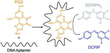
We have identified by in vitro selection DNA aptamers for the redox cofactor pyrroloquinoline quinone (PQQ). Using a spectroscopic assay, we determined PQQ maintains its redox properties when bound to the DNA aptamers. These complexes could find potential use as biocatalysts when direct electrical communication with electrode surfaces is desirable.
One challenge faced in electrochemical applications utilizing oxidoreductases is how to effectively transfer electrons from the enzyme active site to an electrode surface. In biofuel cells, NAD+- and FAD-dependent dehydrogenases typically employ mediated electron transfer (MET) processes due to their buried cofactors, which often lead to decreased catalytic activity and eventually low power density.10 Although FAD-dependent oxidases have better electron transfer ability, their direct electron transfer (DET) processes are often inefficient.11,12 PQQ-dependent dehydrogenases, on the other hand, are recognized for their effective DET processes,13–15 which is preferred in biofuel cell systems.16
One way to improve DET while providing a stable environment for the cofactor would be to bind the cofactor to a different biomolecule, such as nucleic acids, particularly DNA. Single-stranded DNA can fold into structures that allow for abilities other than coding genetic information. These functional DNA sequences are identified through an iterative process known as in vitro selection17 or Systematic Evolution of Ligands by Exponential Enrichment (SELEX).18 Aptamers are single-stranded nucleic acids that can bind to a target with affinity and specificity,19 whereas deoxyribozymes are DNA sequences with catalytic ability.20 The wide variety of targets for which aptamers have been identified suggests the versatility of aptamers and the diversity of applications for which they may be useful.21,22 Aptamers for NAD+ and FAD have been reported, although their identification was largely driven by a desire to understand how these cofactors would interact with RNA or to explore structural motifs that may have played significant roles in the hypothesized RNA world.23 Since in vitro selection can be used to isolate aptamers that will function either under physiological or non-physiological conditions, it is possible to exploit aptamers for use in bioanalytical applications. This is especially true for DNA because of its stability relative to RNA.
In some instances, aptamers have demonstrated properties other than binding. A sulforhodamine-binding DNA aptamer can catalyze the oxidation of a structurally-related fluorophore, dihydrotetramethylrosamine, albeit with weak redox activity.24 A DNA sequence originally isolated as an aptamer for hemin was later termed a deoxyribozyme or DNAzyme because this DNA–hemin complex mimics peroxidases25 and can be used in various applications,26 including biofuel cells.27 We are interested in exploring how DNA can bind to redox cofactors and potentially mimic protein oxidoreductases by catalyzing redox reactions. In this study, we report the identification of DNA aptamers for PQQ and investigate whether the redox activity of PQQ is affected when bound to these aptamers.
We utilized 4 different sets of reaction conditions to identify PQQ-aptamers (Table 1). In designing the in vitro selection conditions, we set out to investigate the effect of calcium and glycerol. Ca2+ is an important cofactor for PQQ-dependent dehydrogenases28 and may be important for maintaining PQQ's redox activity. By including glycerol, a possible biofuel, we can potentially isolate some aptamers that not only contain a binding site for glycerol, but also catalyze its oxidation.
PQQ is polyanionic under neutral to slightly acidic conditions due to its carboxylic acid groups (Fig. 1).3 Binding of PQQ to negatively charged nucleic acids may therefore present some challenges. However, the presence of Ca2+ and Mg2+ could help overcome these repulsive interactions and allow PQQ to bind DNA. Divalent metal ions, particularly Mg2+, are important for functional nucleic acids, such as aptamers, to fold into their active tertiary structures by helping shield the negative charge of the phosphodiester backbone.29 It is possible to form a structure that will orient PQQ in a way to avoid repulsion by the negatively charged DNA backbone. For example, the negatively charged triphosphate of ATP does not interact with either the DNA or the RNA aptamer for ATP, indicating an orientation that prevents repulsion.30,31
Selections were conducted by immobilizing PQQ on amine-modified magnetic beads and partitioning DNA sequences that bound to PQQ from those sequences that did not bind (Fig. S1†). The progress of the selections was monitored by measuring the amount of DNA that binds to immobilized PQQ and then elutes in PQQ-containing washes (Fig. S3†). The binding oligonucleotides from the 15th round of selection were cloned and sequenced. Sequence analysis revealed 12 unique sequences out of 44 sequenced clones (Fig. S2†). All 12 aptamers showed varying degrees of binding to immobilized PQQ, with those selected under the AD conditions in the presence of calcium showing the highest binding affinity (Fig. S4†).
In order to confirm that the aptamers were truly binding to PQQ and not to the tether used to immobilize PQQ, we also employed solution-based binding studies in which aptamers were challenged to bind to PQQ free in solution. Unlike proteins and other large molecular weight compounds that can interact with nucleic acids through various strategies including shape complementarity and electrostatic van der Waals interactions,32 the binding of small organic molecules to nucleic acids is largely dependent on hydrogen bonding and intercalation or stacking.33,34 If the binder is a chromophore like PQQ, these types of interactions can alter absorption and lead to hypochromicity. We monitored the absorption of PQQ at 330 nm in the presence of each potential aptamer, as well as an unstructured DNA and two G-quadruplex-containing control sequences (Fig. S5†). We observed no measurable change in PQQ absorbance in the presence of some of the aptamers, suggesting very weak affinity for PQQ, binding to the immobilization tether, or binding that does not involve intercalation and thus leads to no hypochromicity (data not shown). Similar results were observed with the control DNA sequences (Fig. S5†). Three of the aptamers showed significant binding to PQQ, resulting in a decrease in absorption of the complex relative to that of the PQQ alone. For these aptamers, the decrease in absorption grew more pronounced with increasing PQQ concentration (Fig. S5†).
The isolated aptamers were also studied by fluorescence anisotropy to investigate PQQ binding that does not lead to PQQ hypochromicity.35–40 Several studies on PQQ have suggested that PQQ is fluorescent only when it is hydrated at the C-5 position,41,42 which can be affected by the buffer used. We utilized HEPES buffer pH 7.5 in our selections; however this buffer interfered with the anisotropy measurements (data not shown). We investigated different buffers to identify a buffer that supported PQQ–aptamer binding and caused minimal interference in anisotropy measurements. Phosphate buffered saline (PBS) showed a slight improvement in the binding compared to HEPES, and yielded only baseline anisotropy values; therefore we used PBS for our anisotropy measurements.
Two of the aptamers stood out in terms of their binding affinity for PQQ in the above studies (Fig. S6†). These aptamers were isolated in the AD selection and were designated 15ADa9 and 15ADb11 based on naming convention within our lab. The increase in anisotropy with increasing DNA concentration indicated binding of both 15ADa9 and 15ADb11 to PQQ. Control experiments were conducted in which the aptamer was replaced with an unstructured DNA which differs from the aptamers at the 70-nucleotide random region or with G-quadruplex DNA sequences. There was no significant change in the anisotropy values obtained by titrating these control DNAs into the PQQ solution at even higher concentrations than the aptamers, indicating that the observed binding profiles for the aptamers are due to specific interactions with PQQ (Fig. S6†). Nonlinear analysis of the anisotropy data resulted in the sigmoidal binding profiles shown in Fig. 1 and provided dissociation constants (KD) of 16.4 ± 1.0 μM for 15ADa9 and 9.11 ± 1.0 μM for 15ADb11.
To determine if PQQ maintains its redox properties when bound to the DNA aptamers, we developed a spectroscopic assay based on Garcia-Castineiras et al.'s analysis of hydrogen peroxide with dichlorophenolindophenol (DCPIP).43 DCPIP is a redox active dye that undergoes a two-electron transfer process, the same as PQQ. Oxidized DCPIP produces a blue solution that becomes colorless as DCPIP is reduced, so the state of DCPIP can be monitored by measuring the absorption at 605 nm. DCPIP has a redox potential of +0.220 V at 25 °C and pH 7.0, which is compatible with PQQ's redox potential of +0.090 V. In our assay, we initially reduced DCPIP using sodium ascorbate to produce a colorless solution. We then used PQQ or our PQQ–aptamer complexes to oxidize DCPIP and monitored the increase in absorbance at 605 nm.
Initially, 1 μM of each aptamer was incubated with 100 μM PQQ in order to oxidize 25 μM of reduced DCPIP. The PQQ–DNA complex for each aptamer was able to oxidize the dye at a rate similar to that of PQQ alone (Fig. 2A). In order to confirm that the observed oxidation ability of the PQQ–aptamer complex was not the result of any excess or unbound PQQ in solution, we carried out additional assays in which equimolar amounts of DNA and PQQ were used. Again, a similar rate of oxidation of the dye was observed for the complexes as well as the PQQ alone (Fig. 2B). We did not observe a rate enhancement for the PQQ–aptamer complex compared to PQQ alone, which would have been desirable. However, we also did not observe any inhibition or decrease in rate for the PQQ–aptamer complexes relative to PQQ, which is encouraging and demonstrates the redox ability of PQQ is not affected by the presence of the aptamer. These results indicate that PQQ is not necessarily buried in a DNA structure that prevents access to the solution and allows PQQ to participate in redox reactions.
Footnote |
† Electronic supplementary information (ESI) available: Experimental details of in vitro selection and characterization experiments. See DOI: 10.1039/c4ra11052h |
This journal is © The Royal Society of Chemistry 2015 |