RGD-functionalized ultrasmall iron oxide nanoparticles for targeted T1-weighted MR imaging of gliomas†
Abstract
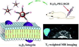
We report a convenient approach to prepare ultrasmall Fe3O4 nanoparticles (NPs) functionalized with an arginylglycylaspartic acid (RGD) peptide for in vitro and in vivo magnetic resonance (MR) imaging of gliomas. In our work, stable sodium citrate-stabilized Fe3O4 NPs were prepared by a solvothermal route. Then, the carboxylated Fe3O4 NPs stabilized with sodium citrate were conjugated with polyethylene glycol (PEG)-linked RGD. The formed ultrasmall RGD-functionalized nanoprobe (Fe3O4-PEG-RGD) was fully characterized using different techniques. We show that these Fe3O4-PEG-RGD particles with a size of 2.7 nm are water-dispersible, stable, cytocompatible and hemocompatible in a given concentration range, and display targeting specificity to glioma cells overexpressing αvβ3 integrin in vitro. With the relatively high r1 relaxivity (r1 = 1.4 mM−1 s−1), the Fe3O4-PEG-RGD particles can be used as an efficient nanoprobe for targeted T1-weighted positive MR imaging of glioma cells in vitro and the xenografted tumor model in vivo via an active RGD-mediated targeting pathway. The developed RGD-functionalized Fe3O4 NPs may hold great promise to be used as a nanoprobe for targeted T1-weighted MR imaging of different αvβ3 integrin-overexpressing cancer cells or biological systems.


 Please wait while we load your content...