DOI:
10.1039/C4RA13075H
(Paper)
RSC Adv., 2014,
4, 62076-62085
Real-time control of inertial focusing in microfluidics using dielectrophoresis (DEP)
Received
25th September 2014
, Accepted 31st October 2014
First published on 31st October 2014
Abstract
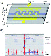
In this paper, we propose a novel hybrid microfluidic device, for the first time, which integrates and fully couples both dielectrophoresis (DEP) and inertial focusing. A DEP force is coupled with inertial lift force to adjust particle's equilibrium position in the vertical direction in real time. The focusing pattern and position of the particles along the horizontal plane can be concurrently adjusted by adjusting their vertical position with DEP forces. The magnitude of secondary flow drag dramatically increases when particles are levitated towards the centre of the channel in the vertical direction. The paper analytically and experimentally investigates the mechanism of this tuneable DEP-inertial microfluidic device. The proposed hybrid device possesses the advantages of both DEP and inertial microfluidic devices, working in a high-throughput manner, as well as having precise controllability in real-time. This DEP-inertial microfluidic device is potentially a versatile and robust platform for a feedback-controlled manipulation and separation of particles and cells.
Introduction
Microfluidic technology has achieved significant progress over the last few decades. This fascinating technology holds broad application areas, including sample preparation,1 clinical diagnosis,2 waste water treatment,3 drug screening4 and material synthesis.5 Manipulation of particles, such as biological cells, is an essential capability of microfluidics to analyse biochemical processes at the single-cell level or to separate cells according to their unique biophysical signatures.
Various techniques have already been proposed and developed to manipulate particles in microfluidics. According to the source of the manipulating force, they can be categorised as active and passive techniques. Active techniques, such as dielectrophoresis (DEP),6 magnetophoresis (MP)7 and acoustophoresis (AP),8 rely on an external force field, whereas passive techniques depend entirely on the channel geometry or intrinsic hydrodynamic forces, such as pinched flow fractionation (PFF),9 deterministic lateral displacement (DLD)10 and inertial microfluidics.11 Generally, an active technique allows for a more precise control of target particles while being very flexible for a wide range of biological particles and tuneable in real-time. However, the flow speed is always limited because particles must be exposed to the outer force field for sufficient duration to achieve effective functionality, which reduces their throughput. In contrast, a passive microfluidic device is always very robust and has a high throughput. However, the fixed geometry and design of passive devices limit their manipulative capability, making them only effective for a specific range of particle properties. This indicated that, separating the particles out of the previous design range would require a completely new device design. Ideally then, a combination of high-throughput passive techniques with precisely tuneable active ones would result in a more powerful and versatile platform.
Attempts to combine active and passive techniques in one microfluidic device have rarely been reported in the past.12–15 Ozkumur et al.13,14 reported a hybrid microfluidic device, which combined deterministic lateral displacement (DLD), inertial focusing and magnetophoresis (MP) to isolate rare circulating tumour cells (CTCs) in an antigen-dependent and -independent manner. In this device, after a sample of raw blood is introduced into the chip, the blood cells first encounter an array of circular pillars with a specific gap, in which a DLD size-based hydrodynamic force filtrate smaller red blood cells (RBCs) from white blood cells (WBCs) and CTCs with considerably larger size. Subsequently, the mixture of WBCs and CTCs flows into an asymmetric serpentine channel in which they are promptly focused along a single path by the inertial effects within the serpentine channel. At the end of the serpentine channel, a strong magnetic field is applied to deflect the cells that are magnetically labelled. Then, the CTCs can be separately collected. Benefiting from the initial debulking of RBCs by DLD and its serpentine inertial focusing section, this device allows for a throughput as high as 107 cells per second. Another impressive work was reported by Moon et al.15 who successfully separated human breast cancer cells (MCF-7) from a spiked blood sample by a combination of multiorifice flow fractionation (MOFF) and dielectrophoresis (DEP) techniques. The inertial separation by MOFF takes advantage of the high-throughput filtration of blood cells, and the serially connected DEP separator works as a precise post-processor to further enhance the efficiency and purity of the separation process. The paper claimed that 99.24% of RBCs and 94.23% of WBCs were removed, with a 162-fold increase in MCF-7 cells. In both works, the separators were actually connected in series, and no coupling between the separators exists.
To further broaden the scope and capability of microfluidic technology, we explored the possibility of combining DEP and inertial focusing in a fully coupled manner and proposed a new concept, which is called DEP-inertial microfluidics. Rather than choosing a horizontal DEP force to directly deflect particles, a vertical DEP force was used to levitate particle equilibrium positions vertically to the centre of the channel. Due to the no-slip boundary, the fluid velocity at the centre of the channel is at its maximum, which imposes a considerably stronger secondary flow drag onto the particles. This indicates that the inertial focusing pattern and position of the particles along the horizontal plane can also be tuned. The mechanism of this tuneable DEP-inertial microfluidic device was discussed, and the effects of the DEP force on each inertial focusing pattern were investigated. Our proposed hybrid device has the advantages of both DEP and inertial microfluidic devices, which works in a high-throughput manner, as well as with precise controllability. The concept reported here is expected to provide a versatile and robust platform for feedback-controlled manipulation and separation of particles and cells.
Materials and methods
A. Design and fabrication of the DEP-inertial hybrid device
The serpentine channel used in our experiments consisted of 15 symmetric zigzag periods. The width of the microchannel was 200 μm. The length and width of each U-turn were both 700 μm. The height of the channel was uniformly 40 μm. The polydimethylsiloxane (PDMS) slab embedded in the microchannel was fabricated by standard photolithography and soft replica techniques.16 On the glass slide, a 50 nm Ti/150 nm Pt electrode layer was patterned using the standard lift-off process.17 The spacing and width of the interdigitated electrodes (IDEs) were both 20 μm. The IDEs were 13.5 mm long, which left one zigzag period of microchannel uncovered by IDEs to allow for the clear observation of the particle trajectory. The PDMS slab and glass slide were bonded with the help of oxygen plasma after being carefully aligned. Fig. 1 shows a schematic image of the DEP-inertial microfluidic device.
 |
| Fig. 1 (a) Schematic of the DEP-inertial microfluidic device. (b) Distribution of inertial lift force vectors and DEP force vectors within a cross section of the DEP-inertial microfluidic device. The serpentine microchannel is embedded in the PDMS slab, and the slab is bonded with a clean glass slide where interdigitated electrodes (IDEs) are patterned. The particle suspension is delivered into the microchannel by a syringe pump. An alternating current (AC) signal is imported into the IDEs, activating the DEP force to levitate particles along the vertical z direction. The DEP force competes with the inertial lift force along the z direction, and thus alters the final vertical position of the particles. As a result, the inertial focusing process and pattern can be tuned in three dimensions. | |
B. Particle preparation
Internally dyed fluorescent polystyrene particles (10 μm in diameter, Thermo Fisher Scientific) were suspended in deionized (DI) water with 0.025% w/w. Tween 20 (SIGMA-ALDRICH Product no. P9416) with 0.1% w/v was added to prevent the particles from aggregating. Bulk conductivity and permittivity of the polystyrene beads are 2.4 × 10⁻⁴ S m−1 and 2.6εo, respectively. The conductivity and permittivity of DI water are 1.5 × 10⁻⁴ S m−1 and 78εo, respectively.18
C. Experimental setup and methods
The microfluidic device was placed on an inverted microscope (CKX41, Olympus, Japan), illuminated by a mercury arc lamp. A particle suspension was delivered into the microchannel by a syringe pump (Legato 100, KD Scientific). Fluorescent images were observed and captured by a CCD camera (Rolera Bolt, Q-imaging, Australia), and then post-processed and analysed using Q-Capture Pro 7 (Q-imaging, Australia) software. The exposure time for each frame was consistent at 100 ms. A sinusoidal signal with a frequency of 1 MHz was generated by a waveform generator (33250A, Agilent, USA) and amplified by an RF power amplifier (TIA-1000-1R8-2, Mini-Circuits, USA). The applied frequency in all the experiments was set to 1 MHz. Copper wires with one end soldered onto the IDEs pads were connected to the power amplifier to activate the electrodes.
Mechanism
A. Inertial migration
The inertial focusing of the particles in a straight channel is driven by the counteraction of two inertial effects: the shear gradient lift force FLS and the wall lift force FLW. The shear gradient lift force FLS depends on the curvature of the fluid velocity profile and its interaction with a finite size particle, whereas the wall lift force FLW is a result of a disturbance of the flow field around the suspending particles and its reflection off the wall. The equilibrium positions result from the balance between these two inertial lift forces. The net inertial lift force FL can be expressed as follows when the particle size is small compared to the channel size:11,19 |
 | (1) |
|
 | (2) |
where ρf, Um and μf are fluid density, maximum velocity, and dynamic viscosity, respectively; a is the particle diameter, and Dh is hydraulic diameter of the channel. fL(RC, z) is the lift coefficient of the net inertial lift force, which is a function of position of the particles within the cross section of channel z and channel Reynolds number RC.11 The situation of fL = 0 corresponds to the dynamic stable and unstable equilibrium positions for particles.
The introduction of channel curvature in the downstream direction introduces additional inertial forces from the fluid (secondary flow drag) and the particles (centrifugal force), which will modify and assist the inertial focusing progress.20 Normally, the particle centrifugal force is negligible unless the particle has quite different density compared to the fluid. Subsequently, the focusing pattern (e.g. position and number of dynamic equilibrium positions) is always determined by the balance between secondary flow drag and inertial lift force. When secondary flow is too weak to alter inertial focusing, the focusing pattern is still determined by the inertial lift force, and particles are normally ordered on the two sides of a channel.21 If the secondary flow is strong enough, it can modify particles as a single stream in a curved channel from the top view.20,22,23
B. The n-DEP force in interdigitated electrodes (IDEs)
Dielectrophoretic (DEP) forces occur on particles/cells when a nonuniform electrical field interacts with a field-induced electrical polarisation. A particle immersed in an alternating electrical field is subject to a time-averaged DEP force determined by the frequency-dependent polarisability of the particle relative to the medium, which is given by the following equation:24 |
FDEP = 2πεmr3Re[K(ω)]∇|Erms|2
| (3) |
where εm is the permittivity of the medium, r represents the particle radius, |Erms| is the root-mean-squared value of the applied electrical field, and K(ω) refers to the Clausius–Mossotti (CM) factor. Re indicates the real part of the factor, and K(ω) depends on the relative permittivity of the particle and the suspending medium, as well as the frequency of the external electric field. |
K(ω) = (ε*p − ε*m)/(ε*p + 2ε*m)
| (4) |
where ε* = ε − iσ/ω is the complex permittivity, σ is the electrical conductivity, and ω is the frequency of the electric field. The subscripts p and m denote the particle and suspending medium, respectively.
When the dielectric constant of a particle is larger than the medium, Re[K(ω)] > 0, the DEP is positive and particles move towards the location with the highest electric field gradient. However, when the dielectric constant of a particle is smaller than the medium, Re[K(ω)] < 0, the DEP force is negative and particles move to locations with the lowest electric field gradient.
In the present work, an IDEs array was patterned onto the bottom of the microchannel to generate a negative DEP (n-DEP) on the particles and push them upwards, Fig. 2(a). Because the DEP force in the horizontal direction is relatively small and negligible, we focused on the vertical DEP force (FDEPz) and its counteraction with the inertial lift force along the vertical plane. Without a specific notice, the DEP force in the entire manuscript indicates the DEP force component in the vertical (z) direction. Near the surfaces of the electrodes (z ≤ 10 μm), the magnitude of the DEP force oscillates along the longitudinal (x) direction. The peak points are at the edges of the electrodes, whereas the trough points are at the centre of the electrodes and the gap between them. Moreover, the closer the particle to the electrode covered surface, the higher is the magnitude of the oscillation, Fig. 2(b). Furthermore, when particles move away from the surface of the electrodes, the magnitude of the DEP force and the magnitude of oscillation sharply decreases, Fig. 2(b and c). However, in the region far from the surface of the electrodes (z ≥ 10 μm), the DEP force remains almost constant in the longitudinal direction. When fluid rapidly flows through this microchannel patterned with IDEs, e.g. RC ∼ 100 (or U ∼ 1 m s−1), particles suspended in the medium will follow the streamlines at a very high speed. The net effect on the particles of the fluctuating DEP force depends on its average magnitude.
 |
| Fig. 2 (a) The electrical field and n-DEP force distribution within a longitudinal section. The arrow vectors represent the magnitude and direction of the n-DEP force. The magnitude of the n-DEP force along the z direction is implied by the vectors' colour and corresponds to the left colour bar. The colour map surface shows the electric field distribution and corresponds to the right colour bar. (b) Longitudinal distribution of the n-DEP force along the z direction (FDEPz). (c) Vertical distribution of the n-DEP force along the z direction (FDEPz). COMSOL Multiphysics 4.3 was used to calculate the electric field, and the DEP force was calculated using eqn (3). Particle diameter is 10 μm, and Re[K(ω)] for polystyrene beads in DI water at electric frequency of 1 MHz is approximated as −0.5. | |
C. Coupling of the n-DEP force and inertial lift force along the vertical direction
If particles are close to the walls of the channel in the vertical direction z, a wall lift force FLW, generated by the wall-induced disturbance on the flow field around the particles, pushes them towards the centre of the channel. The parabolic nature of the fluid velocity profile directs the shear gradient induced lift force FLS towards the channel wall, acting against the FLW. The net inertial force FL [the dashed curve in Fig. 3(a)] exhibits two dynamic stable equilibrium positions located about 0.2 times the height of the channel away from the walls. In addition, these equilibrium positions move closer to channel walls with increasing Reynolds number.25
 |
| Fig. 3 (a) The distribution of dielectrophoresis (DEP) force FDEP and inertial lift force FL along the height of the channel z. The inertial lift force is obtained from the analytical solution from Matas et al.25,39 and the n-DEP force is obtained from the simulation results by COMSOL Multiphysics. Particle diameter is 10 μm. (b) At relative low applied voltages, two stable equilibrium positions (A and D) along z exist, which are modifications of the original inertial equilibrium positions. The direction of the net force (red arrows) on both sides of stable positions will always point to themselves because they can overcome any small disturbance. However, point B is in an unstable position because the net force along both sides is directed outwards, and a little disturbance will induce the particles to move away from point B. The blue dashed curve is the mirror image of the FL negative part on a positive coordinate. (c) At a critical voltage (Vcpp), the FDEP curve is tangential with the FL negative mirroring curve at point C, which is in a half stable (saddle) position. If particles were shifted to below (z < zC), the net force will attract particles back to position C, but when the particles were shifted to above (z > zC), the net force is directed upwards and particles will never return. (d) When the applied voltage is higher than the critical voltage Vcpp, there is only one vertical equilibrium position D at the upper half of the channel. | |
The n-DEP force is always upwards (positive z direction), shown as solid curves in Fig. 3(a), which is consistent with the direction of FL near the bottom of the channel. The particles in this region (z < 0.2 H) experience a large upward net force and rapidly migrate away from the surface of the channel. If the particles reach the region where FL is negative, three different situations occur: (i) at low voltage, the DEP force curve intersects with the negative FL mirroring curve (blue dashed line) at points A and B at the bottom half of the channel, as well as at point D, which is very close to the original inertial equilibrium position FL = 0 at the top half, Fig. 3(b). Between A and B (zA < z < zB), the net force of FL and FDEP is negative and is directed downwards. However, outside the A–B section (z < zA or z > zB), the net force is positive. Therefore, point A is a stable equilibrium position because the net force is directed at point A on both sides, which can overcome any small disturbance and draw particles back to point A. Moreover, point B is an unstable position because the net force on each side is directed outwards, thus even a little disturbance can easily induce particles to move away. (ii) At a critical voltage Vcpp, the FDEP curve is tangent with the FL mirroring curve at a point C, Fig. 3(c). This is a half stable position because the net force always points to the positive z direction whether it is below or above point C. If particles were shifted below (z < zC), the net force will attract particles back to position C. If particles were shifted above (z > zC), the net force is directed upwards and the particles will never return. Thus, very few particles are stable at point C. (iii) When the applied voltage is larger than Vcpp, there is only one node D at the top half of the channel [Fig. 3(d)], and it is common to the other two situations. This point is very close to the original inertial equilibrium positions because the DEP force exponentially decreases with increasing z and is negligible at the top half of the channel with almost no effect on the inertial equilibrium position.
We can conclude from this discussion that particles will gradually be levitated as the applied voltages (DEP force) are increased. Finally, all the particles will be focused to the equilibrium position D at the top half of the channel if the DEP force at point C can overcome the inertial lift force. It should be noted that the force of gravity [FG = (4/3)πr3(ρp − ρf)g] was omitted in this analysis because the density of particles (ρp = 1050 kg m−3) was very close to its suspension medium (ρf = 1000 kg m−3).
Because of the no-slip boundary condition, the fluid velocity is zero at the walls of the channel and at its maximum velocity at the centre of the channel. The secondary flow drag [FD = 3πμa(vfr − vpr)] is enhanced if the particles are levitated towards the vertical centre of the channel by the DEP force. Then, the particle trajectory along the horizontal plane will be modified. Here, vfr and vpr are the radial velocity of fluid and particles, respectively.23 Finally, using the DEP force to control the vertical position of the particles, their inertial focusing pattern in 3D can be modified and adjusted.
In order to test the above hypothesis, we developed a numerical model of the serpentine microchannel and released microparticles from different heights at the inlet using a particle tracking module in COMSOL Multiphysics. The results showed that the particles released near the channel vertical centre can migrate and focus considerably faster to the channel lateral centre than those released near channel walls (Fig. 4), which agreed with our hypothesis. It should be noted that in this simulation, inertial lift forces were not taken into account, because there is no explicit equation to express it; furthermore, it is not easy to implement it in a curved channel with alternating curvatures.
 |
| Fig. 4 (a) Simulated particles' trajectories released from different heights. (b) Normalized particle streak widths after traveling through different channel lengths. The particles released near the channel vertical centre focus into the channel lateral centre much faster than those released near channel walls, because fluid velocity is at maximum at channel centre, and its corresponding secondary flow drag and centrifugal force is much stronger than that near channel walls. | |
In the following sections, the pure inertial focusing pattern and its equilibrium positions in a serpentine channel were first experimentally determined without using the DEP force. Under different RC, three basic focusing patterns emerged. Next, a vertical DEP force generated by IDEs was applied onto these three inertial focusing patterns to explore its effects on inertial focusing patterns. Finally, a Vpp ∼ RC map was summarised to illustrate the dominance of force under different working conditions. The potential and outlook of proposed DEP-inertial microfluidics is also suggested briefly.
Results and discussions
A. Inertial focusing patterns in a serpentine channel under varying RC
In a previous work, we discussed the mechanism of inertial focusing in a serpentine channel23 and showed how particles with different sizes are separated according to their differential lateral positions.22 However, the inertial equilibrium positions in a vertical direction have not been shown experimentally. In order to observe the side view, a PDMS device was fabricated from three PDMS slabs using the method reported by Guan et al.,26 Fig. 5(c). The PDMS device could be placed vertically onto the microscope stage, giving a clear, side view of the serpentine channel, Fig. 5(b). The top view and side view enable the dynamic equilibrium positions of the particles to be determined easily.
 |
| Fig. 5 (a) Schematic 3D structure of one period in the serpentine channel and (b) its side view optical image; (c) the fabricated PDMS device was used to observe the serpentine channel from both top-down and side-side view in the experiments. (d–h) Inertial particle focusing patterns under varying flow conditions: (d) RC = 41.7; (e) RC = 62.5; (f) RC = 83.3; (g) RC = 104.2; (h) RC = 125. (Left) top views of fluorescent particles trajectory at the last zigzag period of serpentine; (middle) side views of fluorescent particles at the last zigzag period; and (right) the dynamic equilibrium positions of particles at cross section A-a. | |
From the top view [left in Fig. 5(d–h)], focusing streaks could be seen on the two sides of the channel at a relatively low RC (Fig. 5(d–f)), but as the RC increased, these two-sided focusing streaks migrated towards the centre of the channel, moved closer and closer together, and then merged into a single wide streak, Fig. 5(g). This was because the secondary flow drag increased considerably faster than the inertial lift force within the horizontal plane as RC increased. They gradually dominated the two-side-directed inertial lift forces and finally pinched the particles into the centre of the channel, in which the net drag forces are weaker and partly cancel. As RC continued to increase, the focusing streak became tighter until its width could reach the single diameter of a particle. The side view (middle in Fig. 5(d–h)), shows two focusing streaks near the top and bottom walls, which migrated toward the top and bottom walls as RC increased, which is in agreement with previous reports.19,20,25 The dynamic equilibrium positions within the cross section “A-a” were deduced and were illustrated schematically (green dots) in the right of Fig. 5(d–h).
In summary, as RC increased, the dynamic equilibrium positions in a serpentine channel shifted towards the centre of the channel in the lateral direction (y), as well as moving closer to the walls of the channel in the vertical direction (z).
B. Influence of n-DEP on the two-sided inertial focusing pattern: modification of the focusing pattern by levitation of particles
At a relatively low RC (41.7 to 83.3), the focusing streaks at the bottom half of the channel (blue dots) were symmetric with those at the top half (green dots) along the vertical centreline, which is observed as two streaks from a top view, Fig. 6(a). However, when a DEP force was applied to deflect the particles upwards, this symmetry was broken. The bottom two focusing streaks detached from the original overlapping streaks, Fig. 6(b and c). A very intriguing phenomenon occurred when the two focusing streaks at the bottom migrated towards the lateral centreline of the channel as the voltage increased, and then merged to become a single streak. This indicates that not only by controlling the flow conditions, RC, but also by adjusting the DEP force, the inertial focusing position and pattern can be modified. The underlying mechanism for this phenomenon is that with an increased DEP force, the particles levitated upward where the secondary flow velocity was considerably stronger, and the symmetrically oscillating secondary flow drag exerted a considerably stronger centre-directed pinch effect to focus the particles towards the centre of the channel. After reaching a certain height, the two-sided focusing streaks could even be combined as a single streak at the lateral centre of the channel.
 |
| Fig. 6 Migration of particle focusing patterns under different AC voltages of (a) Vp–p = 0 V, (b) Vp–p = 18 V, (c) Vp–p = 27 V, (d) Vp–p = 36 V and (e) Vp–p = 54 V. (Left) trajectory of fluorescent particles from top view, and (right) their corresponding postulated dynamic equilibrium positions within two typical cross sections “A-a” and “B-b”. Flow condition is RC = 62.5. | |
When the applied voltage was higher than a corresponding critical voltage Vcpp, the single focused streak collapsed because there was no stable position at the bottom half of the channel and all the particles escaped into the top two equilibrium positions, at which the DEP force was too small to have an effect. The overall trajectory of the equilibrium positions with increasing DEP force is shown as a dashed line in Fig. 6(e). Because biological cells exhibit varying induced boundary polarisations dependent on applied electric frequency and their physiological states,27 after choosing a proper frequency and suspending fluid, cells/particles can be separated by the differences in their dielectric properties, such as separation of viable and nonviable cells,28,29 blood cells and cancer cells,15,30 as well as CD34+ cells from peripheral-stem-cell harvests.31 For the inertial-DEP microfluidic device, a separation scheme could be imagined here. All the particles could be first squeezed down to the bottom half of the channel by a sheath flow after entering the microchannel, and then applying and adjusting a proper AC signal on the IDEs, the target particles experiencing a negative DEP could be levitated upwards and focused at the lateral centre, while the particles with different dielectric properties experiencing a weak DEP force or even no DEP force, could still occupy the two-sided equilibrium positions. In this manner, target particles could be isolated according to their dielectric properties, as well as particle size in our previous report.22 Therefore, the modification of inertial focusing patterns by the DEP force implies a more versatile capacity to differentiate and separate particles/cells by dielectric properties, as well as particle size in a high-throughput tuneable manner.
C. Influence of n-DEP on the inertial single-stream focusing pattern: sheathless three-dimensional focusing
At a high RC (>110), without the existence of a DEP force, the centre-directed pinch effect from secondary flow was already strong enough to focus particles along the channel centre as a single focusing streak in the horizontal plane. In the vertical direction, particles were symmetrically focused along the top and bottom walls due to the inertial lift force, Fig. 7(a). Due to the symmetry, only one focusing streak could be observed from the top view in this case. However, when a DEP force was applied in the vertical direction, the previously unobserved focusing streak from the bottom half of the channel appeared, Fig. 7(b and c). The particles continued to behave as a single focusing streak, and their lateral position became more prone to follow the secondary flow streamline due to the larger secondary flow drag. The electrical voltage was gradually increased until it exceeded the critical value Vcpp, at which point the upward DEP force became strong enough to overcome the downward inertial lift force. Finally, all the particles within the bottom half transitioned to the equilibrium position D at the top of the channel, forming a truly single focusing streak in three dimensions, Fig. 7(d). Such high-throughput sheathless three-dimensional particles focusing promises wide potential applications, such as on-chip flow cytometry,32–34 deformability cytometry,35–37 and imaging cytometry.38
 |
| Fig. 7 Migration of particle single central focusing patterns under different AC voltages of (a) Vp–p = 0 V, (b) Vp–p = 36 V, (c) Vp–p = 72 V and (d) Vp–p = 90 V. (Left) trajectory of fluorescent particles from top view, and (right) their corresponding postulated dynamic equilibrium positions within two typical cross sections “A-a” and “B-b”. Flow condition is RC = 114.6. | |
D. Influence of n-DEP on inertial transitional focusing patterns: pinching of widely distributed particles into a tight focusing streak
Without DEP, when Reynolds number RC was between 90 and 110, two-sided focusing streaks were combined as a single wide focusing streak in this flow region, which was considered as a transition between a two-sided focusing pattern and a single central focusing pattern. The particles in this pattern were distributed within a wide band near the centre of the channel, Fig. 8(a). After a vertical DEP force was introduced, and within our expectations, the widely distributed particles at the bottom half of the channel migrated together if they were levitated upwards. However, it was unexpected that particles at the top half were also pinched closer in these experiments, Fig. 8(c–e). The DEP force on the particles at the top half of the channel should be very small, because it exponentially decreases with the distance z, as previously discussed, Fig. 2(c). From our understanding, the centre-directed inertial effect (secondary flow drag) and the two-side-directed inertial lift force were in a delicate balance within this transitional region, thus a tiny DEP force could probably break this balance, and inadvertently promoted the central focusing. A single focusing streak in 3D could also be achieved when the electric voltage surpassed the corresponding critical voltage Vcpp. Using the DEP force to transform the transitional focusing pattern into a single central focusing pattern actually improved the inertial focusing performance and expanded the available working region of the original single central focusing. This result could be beneficial for complete separation of particles according to their differential lateral equilibrium positions in the serpentine channel, because a narrower focusing streak facilitates easier collection, and higher collection purity and efficiency.
 |
| Fig. 8 Migration of particle transitional focusing patterns under different AC voltages of (a) Vp–p = 0 V, (b) Vp–p = 18 V, (c) Vp–p = 54 V, (d) Vp–p = 81 V and (e) Vp–p = 99 V. (Left) trajectory of fluorescent particles from top view, and (right) their corresponding postulated dynamic equilibrium positions within two typical cross sections A-a and B-b. Flow condition is RC = 99. | |
E. Vpp–RC operation map
The critical voltage Vcpp is a very important parameter for determining the vertical position of the particles because if the applied voltage is larger than Vcpp, the particles will be pushed over to the top half of the channel where inertial lift becomes more dominant. Otherwise they will remain within the bottom half where their exact position depends on the balance of vertical inertial lift force and DEP force. A dimensionless factor δ was defined to scale with the ratio between the DEP force and inertial lift force at point C (see Fig. 3). |
 | (5) |
From the definition, Vcpp is the applied voltage when δ = 1, but if δ > 1 all the particles will be pushed up to the top half of the channel, and when δ < 1, the particles below the top half will remain in this region. It is difficult to determine the exact position of the tangent point C because the lift coefficient curve varies with channel Reynolds number RC. Therefore, it is not practical to derive an explicit analytical expression to calculate Vcpp. However, from an experimental perspective, we could roughly determine the Vcpp region. In the experiments, fewer particles travelled along the bottom half of the channel with increasing DEP force, as indicated by the decreased fluorescence intensity. More particles moved into equilibrium positions D at the top half of the channel leading to an increase in fluorescence intensity, Fig. 9(a). Point C is a half stable position [Fig. 3(c)] and the last position where a few particles remained at the bottom half of the channel. Therefore, the range of Vcpp can be estimated between the maximum voltage where the bottom particles still existed and the minimum voltage where all the bottom particles disappeared, shown as a dashed line with error bars in Fig. 9(b). From the Vpp–RC map, the critical voltage Vcpp was roughly proportional to RC because the order of applied voltage in the DEP force was the same as RC (∝Um) in the inertial lift force (eqn (5)). Of course, the dependency of the lift coefficient fL(RC, z) on RC would bring a little nonlinear nature into the Vcpp–RC relationship. Above the Vcpp–RC curve, the DEP force was strong enough to push all particles into the top half of the channel. Below this curve, the particles were levitated but still remain within the bottom half.
 |
| Fig. 9 (a) Distribution of lateral fluorescence intensity with various applied voltages. The inset is a typical fluorescent image at the last zigzag period; (b) Vpp–RC map illustrates the working range of the DEP-inertial microfluidic device. The red triangles are experimental data where particles are pushed onto the top half of the channel. The applied voltages are larger than the critical voltage. The empty rectangles are experimental data where particles under the bottom half part of the channel are focused into a single central focusing streak under the effects of the DEP force. The solid rectangles are the experimental data where the DEP force is not sufficient to alter the particle inertial focusing pattern in the bottom half part of the channel. | |
Conclusions and outlook
This work, for the first time, proposed a novel DEP-inertial microfluidic device that fully couples the DEP force and inertial lift force in a serpentine microchannel. Rather than using a horizontal DEP force, a vertical DEP force was used to adjust the particle equilibrium positions in the vertical direction. Due to the much stronger secondary flow drag imposed on particles when they were near the vertical centre of the channel, focusing patterns and positions of particles along the horizontal plane could be adjusted by their vertical position. The mechanism of this DEP-inertial microfluidic device was also investigated numerically and experimentally. The potential advantages of the DEP-inertial microfluidic device include (i) real-time controllability; (ii) three-dimensional single stream focusing; (iii) enhanced focusing quality and the ability to manipulate particles; (iv) the DEP force could be used as a force probe to investigate the mechanism of particle inertial focusing process; (v) surface-patterned microelectrodes could also possibly work as an impedance-based detector, to simultaneously enumerate and differentiate the passing microparticles; (vi) separation of particles/cells by size as well as by dielectric property; and (vii) utilisation of different focusing patterns of particles by varying the DEP forces in real-time to act as an online active sorter. In summary, this DEP-inertial device offers the possibility of combining a high-throughput manner with accurate real-time controllability, which is expected to provide a versatile and robust feedback-controlled platform for manipulating and separating particles and cells.
Acknowledgements
This work was partially supported by the University of Wollongong through a UIC grant and China Scholarship Council.
References
- J. Kim, M. Johnson, P. Hill and B. K. Gale, Integr. Biol., 2009, 1, 574–586 RSC.
- T. H. Schulte, R. L. Bardell and B. H. Weigl, Clin. Chim. Acta, 2002, 321, 1–10 CrossRef CAS.
- M. A. Shannon, P. W. Bohn, M. Elimelech, J. G. Georgiadis, B. J. Marinas and A. M. Mayes, Nature, 2008, 452, 301–310 CrossRef CAS PubMed.
- P. S. Dittrich and A. Manz, Nat. Rev. Drug Discovery, 2006, 5, 210–218 CrossRef CAS PubMed.
- J. I. Park, A. Saffari, S. Kumar, A. Günther and E. Kumacheva, Annu. Rev. Mater. Res., 2010, 40, 415–443 CrossRef CAS.
- B. Çetin and D. Li, Electrophoresis, 2011, 32, 2410–2427 CrossRef PubMed.
- T. P. Forbes and S. P. Forry, Lab Chip, 2012, 12, 1471–1479 RSC.
- Z. Wang and J. Zhe, Lab Chip, 2011, 11, 1280–1285 RSC.
- M. Yamada, M. Nakashima and M. Seki, Anal. Chem., 2004, 76, 5465–5471 CrossRef CAS PubMed.
- L. R. Huang, E. C. Cox, R. H. Austin and J. C. Sturm, Science, 2004, 304, 987–990 CrossRef CAS PubMed.
- D. Di Carlo, Lab Chip, 2009, 9, 3038–3046 RSC.
- S. Yan, J. Zhang, M. Li, G. Alici, H. Du, R. Sluyter and W. Li, Sci. Rep., 2014, 4, 5060 Search PubMed.
- E. Ozkumur, A. M. Shah, J. C. Ciciliano, B. L. Emmink, D. T. Miyamoto, E. Brachtel, M. Yu, P.-i. Chen, B. Morgan and J. Trautwein, Sci. Transl. Med., 2013, 5, 179ra47 Search PubMed.
- N. M. Karabacak, P. S. Spuhler, F. Fachin, E. J. Lim, V. Pai, E. Ozkumur, J. M. Martel, N. Kojic, K. Smith and P.-i. Chen, Nat. Protoc., 2014, 9, 694–710 CrossRef CAS PubMed.
- H. S. Moon, K. Kwon, S. I. Kim, H. Han, J. Sohn, S. Lee and H. I. Jung, Lab Chip, 2011, 11, 1118–1125 RSC.
- D. C. Duffy, J. C. McDonald, O. J. A. Schueller and G. M. Whitesides, Anal. Chem., 1998, 70, 4974–4984 CrossRef CAS PubMed.
- M. Li, S. Li, J. Wu, W. Wen, W. Li and G. Alici, Microfluid. Nanofluid., 2012, 12, 751–760 CrossRef CAS.
- S. H. Baek, W.-J. Chang, J.-Y. Baek, D. S. Yoon, R. Bashir and S. W. Lee, Anal. Chem., 2009, 81, 7737–7742 CrossRef CAS PubMed.
- E. S. Asmolov, J. Fluid Mech., 1999, 381, 63–87 CrossRef CAS.
- D. Di Carlo, D. Irimia, R. G. Tompkins and M. Toner, Proc. Natl. Acad. Sci. U. S. A., 2007, 104, 18892–18897 CrossRef CAS PubMed.
- J. Zhang, S. Yan, W. Li, G. Alici and N.-T. Nguyen, RSC Adv., 2014, 4, 33149–33159 RSC.
- J. Zhang, S. Yan, R. Sluyter, W. Li, G. Alici and N.-T. Nguyen, Sci. Rep., 2014, 4, 4527 Search PubMed.
- J. Zhang, W. Li, M. Li, G. Alici and N.-T. Nguyen, Microfluid. Nanofluid., 2013, 17, 305–316 CrossRef.
- D. J. Collins, T. Alan and A. Neild, Lab Chip, 2014, 14, 1595–1603 RSC.
- J.-P. Matas, J. F. Morris and É. Guazzelli, J. Fluid Mech., 2009, 621, 59–67 CrossRef.
- G. Guan, L. Wu, A. A. Bhagat, Z. Li, P. C. Chen, S. Chao, C. J. Ong and J. Han, Sci. Rep., 2013, 3, 1475 Search PubMed.
- H. Li and R. Bashir, Sens. Actuators, B, 2002, 86, 215–221 CrossRef CAS.
- B. H. Lapizco-Encinas, B. A. Simmons, E. B. Cummings and Y. Fintschenko, Anal. Chem., 2004, 76, 1571–1579 CrossRef CAS PubMed.
- H. Shafiee, M. B. Sano, E. A. Henslee, J. L. Caldwell and R. V. Davalos, Lab Chip, 2010, 10, 438–445 RSC.
- A. Alazzam, I. Stiharu, R. Bhat and A. N. Meguerditchian, Electrophoresis, 2011, 32, 1327–1336 CrossRef CAS PubMed.
- M. Stephens, M. Talary, R. Pethig, A. Burnett and K. Mills, Bone Marrow Transplant., 1996, 18, 777–782 CAS.
- J. Oakey, R. W. Applegate Jr, E. Arellano, D. D. Carlo, S. W. Graves and M. Toner, Anal. Chem., 2010, 82, 3862–3867 CrossRef CAS PubMed.
- K. C. Cheung, M. Di Berardino, G. Schade-Kampmann, M. Hebeisen, A. Pierzchalski, J. Bocsi, A. Mittag and A. Tárnok, Cytometry, Part A, 2010, 77, 648–666 CrossRef PubMed.
- D. C. Spencer, G. Elliott and H. Morgan, Lab Chip, 2014, 14, 3064–3073 RSC.
- T. Henry, D. R. Gossett, Y. S. Moon, M. Masaeli, M. Sohsman, Y. Ying, K. Mislick, R. P. Adams, J. Rao and D. Di Carlo, Sci. Transl. Med., 2013, 5, 212ra163 CrossRef PubMed.
- J. S. Dudani, D. R. Gossett, T. Henry and D. Di Carlo, Lab Chip, 2013, 13, 3728–3734 RSC.
- D. R. Gossett, T. Henry, S. A. Lee, Y. Ying, A. G. Lindgren, O. O. Yang, J. Rao, A. T. Clark and D. Di Carlo, Proc. Natl. Acad. Sci. U. S. A., 2012, 109, 7630–7635 CrossRef CAS PubMed.
- K. Goda, A. Ayazi, D. R. Gossett, J. Sadasivam, C. K. Lonappan, E. Sollier, A. M. Fard, S. C. Hur, J. Adam and C. Murray, Proc. Natl. Acad. Sci. U. S. A., 2012, 109, 11630–11635 CrossRef CAS PubMed.
- J.-P. Matas, J. F. Morris and É. Guazzelli, J. Fluid Mech., 2004, 515, 171–195 CrossRef.
|
This journal is © The Royal Society of Chemistry 2014 |
Click here to see how this site uses Cookies. View our privacy policy here.