HPLC–ICP-MS analysis of selenium speciation in selenium-enriched Cordyceps militaris

Mei Tiea, Baorui Lia, Yang Liua, Jie Hana, Tiebiao Suna and Huawei Li*b
aCollege of Environment, Liaoning University, Shenyang, China
bCollege of Chemistry and Life Sciences, Shenyang Normal University, Shenyang, China.. E-mail: lbr_simmer@126.com

Received 23rd October 2014 , Accepted 12th November 2014

First published on 12th November 2014


Abstract

This study examined the distribution of selenium in proteins from selenium-enriched Cordyceps militaris extract using reversed-phase high performance liquid chromatography (RP-HPLC) and size-exclusion chromatography (SEC) coupled to on-line ICP-MS specific for selenium detection. An analytical method was developed for detecting the presence of selenocysteine, selenomethionine and selenopeptides. The results showed that soluble selenium was the major form of selenium in selenium-enriched C. militaris extract. About 84.4% of the selenium was present as low-molecular-weight molecules (MW < 4 kDa), whereas 7.13% of the selenium was present in the form of high-molecular-weight molecules, probably as selenoproteins. Furthermore, analysis of acid-hydrolyzed C. militaris extract revealed a higher proportion of Se-Cys2 than Se-Met (two main selenium-conjugated amino acids) in the extract, although various forms of smaller selenium-proteins or selenium-peptides were also present in low but detectable amounts. The present analysis of selenium thus combined different chromatographic techniques for efficient and rapid separation of selenoproteins and other seleno-conjugated molecules.

Introduction

Selenium (Se) is a key trace element required in small amounts by humans and animals.1 It exists in different chemical forms, which exhibit different nutritional status, bioavailability and toxicity in the body. Compared to the inorganic form of selenium, the organic form of selenium has higher bioavailability and lower toxicity, and it exists mainly as selenoproteins, selenolipids, selenopeptides and selenoamino acids.2 Most of the biological functions of selenium are mediated by selenoproteins, and these include the biologically important antioxidants, glutathione peroxidases (GPX), thioredoxin reductases (TrxR) and iodothyronine 50-deiodianases (IDI).3–5 In selenoproteins, selenium is incorporated in the amino acid chain in the form of selenocysteine (Se-Cys) or selenomethionine (Se-Met). Se-Cys is recognized as the 21st amino acid and it plays an essential role in the active sites of several enzymes.6,7 The insertion of Se-Cys during protein synthesis is directed by UGA codon, and thus recognition elements within the mRNAs are essential for the translation of UGA to Se-Cys.8,9 It is known that Se-Met can nonspecifically insert into proteins by the usual methionine incorporation process. However, the human body cannot synthesize Se-Cys and Se-Met, so we have to ingest them from an external source. It is known that one of the safest ways to get organic selenium is to supplement cultivated plants with inorganic selenium during growth. Therefore, selenium enriched foods should be evaluated for their nutritional availability.10,11

The use of high performance liquid chromatography in the separation of biological molecules has the advantages of being economical and accurate. In terms of identifying selenium compounds, various methods of HPLC separation, such as ion-exchange chromatography, reversed-phase chromatography, and ion-pair reversed-phase chromatography, as well as size-exclusion chromatography have been used.12–14 There are many kinds of selenoproteins, and some of these proteins have similar structures, although they may have different isoelectric points, polarity and molecular masses. Therefore, working out the precise conditions for a HPLC method that would improve the separation efficiency for different selenoproteins is of great importance for researchers studying these proteins. Size-exclusion chromatography (SEC), which separates proteins on the basis of sizes has been a useful method for separating proteins from a variety of sources.15,16 However, separation of proteins based on sizes does have a limitation, and therefore, by coupling SEC to plasma mass spectrometry (ICP-MS), it may be possible to study the distribution of selenium in both high-molecular-weight (>12 kDa, likely to be proteins) and low-molecular-weight (0.36–2 kDa, small peptides or free amino acids) fractions of a cell extract. Inductively coupled-plasma mass spectrometry (ICP-MS) was regarded as an advanced technique for analyzing and detecting selenium compound in the 1980s. It combines high temperature (7000 K) and sensitive scanning of quadrupole mass spectrometry with its unique interface technology.17 Compared with traditional analytical techniques, ICP-MS has the advantages of higher sensitivity, wide linear range, greater precision of analysis, higher accuracy, good reproducibility, and high sensitivity.18,19 It is the optimal method used for trace element analysis. A combination of HPLC and ICP-MS provides a powerful and sensitive technique for on-line analysis of elemental speciation.20–22 Due to the high sensitivity of this technique the analysis can usually be carried out without sample pre-concentration steps, since these steps may influence the composition of the species in the samples.

In this study, the different forms of soluble selenium in Cordyceps militaris extract were separated and identified by HPLC–ICP-MS. Size-exclusion chromatography (SEC–HPLC) coupled to ICP-MS was employed to investigate the presence of selenium in proteins, whereas reversed phase high performance liquid chromatography (RP-HPLC) coupled ICP-MS was used for the analysis of selenoamino acids. The result showed that the combined use of HPLC and ICP-MS could provide a simple method for separating soluble selenium-containing proteins from other molecules in the crude extract.

Materials and methods

Instrumentation

An Elan 7500c inductively coupled plasma mass spectrometer (Agilent Technologies, USA) equipped with an Agilent cross-flow nebulizer was used to determine the total content of selenium as well as to detect the presence of selenium compounds. The chromatographic system consisted of an 1100 HPLC pump connected to an auto sampler (Aglient Technologies, USA). Reversed phase HPLC was performed using a C18 Zorbax (Agilent ZORBOX USA) column and a Diode array. While TSK-G3000SW gel (Japan) was used for size-exclusion chromatography (SEC) separation. The operating conditions for HPLC–ICP-MS are summarized in Table 1.
Table 1 Instrumental operation conditions
ICP-MS parameters
Forward power 1490 W
Carrier gas (Ar) flow rate 1.15 L min−1
Dwell time 0.1 s per isotope
Isotopes monitored 77Se, 82Se
[thin space (1/6-em)]
SEC–HPLC parameters
Column TSK-G3000SW (300 mm × 7.5 mm × 10 μm)
Mobile phase 30 mM Tris buffer, pH 7.2
Flow rate 0.5 mL min−1
Column temperature 20 °C
Injection volume 50 μL
[thin space (1/6-em)]
RP-HPLC parameters
Column ZOBARX-C18 (250 mm × 4.6 mm × 4 μm)
Mobile phase 95% H2O, 5% acetonitrile
Flow rate 0.8 mL min−1
Injection volume 100 μL


Reagents and standard

Stock solution of sodium selenite standard (500 mg L−1) was obtained from Nacalai Tes-que (Kyoto, Japan). Standard Se-Met and Se-Cys2 were purchased from Sigma (St. Louis, MO, USA), and each was dissolved in 3% (v/v) HCl and kept in the dark at 4 °C. Deionized water (18.2 MV cm) was generated with a NanoPure treatment system (Barnstead, Boston, MA, USA). Bovine serum (66 kDa), carbonic anhydrase (29 kDa) and cytochrome c from bovine heart (12.4 kDa) were used as protein molecular standards. These were obtained from Sigma. All other chemicals used were of analytical grades.

Cultivation and preparation of Se-enriched Cordyceps militaris

C. militaris growth medium contained K2HPO4, MgSO4, rice and sodium selenite. The initial concentration of selenium in the medium was 5 mg kg−1. The medium was dispensed into glass jars, and the jars were sealed with plastic film and sterilized at 0.1 MPa for 30 min. After sterilization, the medium was allowed to solidify by cooling the jars to room temperature. The jars were then inoculated with C. militaris and the cultures were incubated in the dark for 60 days at an initial temperature of 17 °C. At the end of incubation, C. militaris was harvested from the jars and put into a plastic bag. The bag was frozen at −85 °C 24 h, and the content of the bag was then transferred to a vacuum vessel and further dried at 2.8 × 10⁻² kPa and −50 °C for 72 h. After drying, it was homogenized to powder.

Extraction of selenium for chromatographic speciation studies

Two methods were used to prepare the sample for analysis. In the first method, 0.1 g of dried C. militaris powder was dispensed in a plastic tube and extracted with 3 mL 0.1 M Tris–HCl (pH = 7.2) for 4 h, and then centrifuged at 4000 × g per min for 10 min. The supernatant (100 μL) was loaded onto the SEC–ICP-MS system. In the second method, 0.1 g of the same power was hydrolyzed in 4 M HCl for 12 h and the sample was then subjected to selenoamino acid analysis.

Determination of total selenium and soluble selenium

C. militaris powder (0.5 g) was mixed with 2 mL HNO3 and the mixture was allowed to digest inside a microwave (model MDS 2002A) under three different sets of conditions: 0.2 MPa for 2 min, 0.6 MPa for 2 min followed by 1.5 MPa for 5 min. After cooling, the final volume of the sample was made to 25 mL with Milli-Q water. Samples (5 mL) of the extract were digested with 2 mL HNO3 inside a microwave, under the same conditions as mentioned above. After that, the total selenium and soluble selenium contents were determined by ICP-MS. Digestion performed without C. militaris powder was used as blank control.

HPLC–argon (Ar) ICP-MS

Major isotopes of selenium can be subjected to severe interference from the plasma gas Ar in the Ar-ICP-MS. The isotopes of selenium identified at m/z 77 and 82 are commonly used to monitor the ion signals in the determination of Se. However, signals at m/z 77 and 82 are not free from interference, due to strong interference by 40Ar36Ar+and 40Ar38Ar+ when the sample contains HNO3. Despite this, the ion intensity at m/z 82 still presents a better signal-to-noise ratio than the ion intensity at m/z 77, and therefore it was used for the quantification of selenium.

In order to overcome the challenge associated with separating a complex mixture of selenium species present in the extract of C. militaris by HPLC, the condition of separation needed to be optimized. Thus different flow rates and compositions of the mobile phases were tested and those that gave the best separation were adopted for all experiments. The amount of material from each peak in the chromatogram was determined by peak area measurement. The relative molecular weight of the selenium compounds were detected based on their retention times.

Results and discussion

Total selenium and soluble selenium determination

C. militaris was grown using standard medium that initially contained 5 mg kg−1 selenium in the form of selenite. After 60 days, the total selenium and soluble selenium in C. militaris were determined by ICP-MS. Compared to the control sample (0.34 μg g−1), the selenium content of Se-enriched C. militaris was 33.04 μg g−1. The selenium content of soluble selenium-containing compound was 27.07 μg g−1, which represented 81.94% of the total selenium. Thus, in selenium-enriched C. militaris, selenium existed mainly as a water-soluble form.

Chromatographic speciation studies

To study the selenium speciation of C. militaris, different HPLC–ICP-MS systems were used.

SEC–ICP-MS

The condition of the HPLC used in the separation of selenium compounds was optimized through testing the effectiveness of different flow rates and mobile-phase compositions. A flow rate ranging from 0.1 to 0.6 mL min−1 was tested with two different mobile phases (Tris–HCl buffer and phosphate buffer, both at pH 7.2), and good separation of the various peaks resolved from the C. militaris extract was achieved with both mobile phases. Slightly better separation was achieved with the lower range of the flow rate, but this also resulted in longer retention time. Thus a flow rate of 0.5 mL min−1 was chosen as the optimum flow rate, which gave reasonably good separation while maintaining acceptable column back pressure.

The elution profile of selenium-conjugated molecules in the extract of C. militaris as resolved by SEC–HPLC coupled DAD is shown in Fig. 1A. Six peaks were resolved and each with a distinct retention time, indicating they all had different molecular weights. Only the peaks indicated by II, III and IV in Fig. 1 were detected by SEC–HPLC–ICP-MS (Fig. 1B). The molecular weights and selenium contents of Peaks II, III and IV are shown in Table 2. It appeared that C. militaris extract contained three main groups of Se-containing molecules, the 133 kDa and 22 kDa species could be selenoproteins, which accounted for about 7.13% of the total soluble selenium in the extract, whereas the species represented by molecular weights of less than 4 kDa could be selenopeptides, and they accounted for 84.4% of the total soluble selenium.


image file: c4ra12975j-f1.tif
Fig. 1 Separation of C. militaris extract by HPLC. (A) SEC–HPLC–DAD; (B) SEC–HPLC–ICP-MS.
Table 2 Molecular weights and selenium contents of different Se-containing fractions in Se-enriched C. militaris
Se-containing compound II III IV
Retention time (min) 5.195 6.601 6.927
Molecular weight (kDa) 133 22 Molecular weight <4 kDa selenium compounds
Content of selenium (μg g−1) 0.988 0.943 22.801


After being preserved at 4 °C for 3 and 5 days, extract prepared from C. militaris powder was subjected to the same analysis to determine the stability of the various selenoproteins and selenopeptides detected in the previous analysis (Fig. 1 and Table 1). The results are shown in Fig. 2 and 3. Peak II and III appeared to decrease with increased storage time, suggesting that they were not very stable (Fig. 3). No obvious increase was observed for the other peaks with molecular weights lesser than those of Peaks II and III. This may suggest that the degradation of Peaks II and III resulted in species that may be too small to be resolved by the HPLC column used in the experiment.


image file: c4ra12975j-f2.tif
Fig. 2 Changes in the distribution of selenium-containing proteins and peptides in C. militaris extract as detected by SEC–HPLC–DAD upon storage at 4 °C.

image file: c4ra12975j-f3.tif
Fig. 3 Changes in the distribution of selenium-containing proteins and peptides in C. militaris extract as detected by SEC–HPLC–ICP-MS upon storage at 4 °C.

Increases in storage time at 4 °C resulted in increased amount of Peak IV when resolved by SEC–HPLC–ICP-MS (Fig. 3). This increase could in fact be the degraded products from Peak II and Peak III (which contained the selenium component), although this increase was not really detected by SEC–HPLC–DAD (Fig. 2).

RP-ICP-MS

The separation of Se-Cys2 and Se-Met by the RP-HPLC C18 Zorbax column (Agilent ZORBOX USA) was examined using different flow rates and mobile phases. The best separation was achieved with a flow rate of 0.8 mL min−1 under the mobile phase consisting of 95% water and 5% acetonitrile. Under these conditions, Se-Cys2 and Se-Met were well separated from each other (with retention time of 3.2 min for Se-Cys2 and 5.6 min for Se-Met), yielding good baseline between the two peaks, while requiring the shortest separation time (Fig. 4). Thus subsequent separation of Se-Cys2 and Se-Met was performed using these conditions.
image file: c4ra12975j-f4.tif
Fig. 4 Influence of the different mobile phases on separation of selenium-containing amino acids.

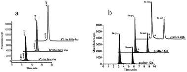
When C. militaris extract was subjected to acid hydrolysis followed by RP-HPLC–ICP-MS analysis, two peaks with the same retention times as those observed for Se-Cys2 and Se-Met were obtained (Fig. 5), suggesting that selenoproteins and selenopeptides resolved from the extract contained selenium primarily conjugated to cysteine and methionine. Furthermore, with prolonged hydrolysis, the two peaks also increased slightly with the appearance of two minor peaks, indicated by A and B in the spectra. The identities of these two minor peaks remain unknown at this stage, but they could be the products of proteolytic digestion, which probably resulted in the release of numerous proteins or peptide-bound selenium. Proteolytic digestion also increased the amount of Se-Cys2 and Se-Met released from the samples. The relative contents of Se-Cys2 and Se-Met as well as the relative contents of selenium in C. militaris extract obtained after acid hydrolysis are given in Table 3. Se-Cys2 was clearly present in a higher proportion than Se-Met.


image file: c4ra12975j-f5.tif
Fig. 5 Effect of hydrolysis on speciation of Se in Se-enriched C. militaris extract.
Table 3 Distribution of different species of selenium in C. militaris extract and its hydrolysate
  Acid hydrolyzed for 12 h Acid hydrolyzed for 24 h Acid hydrolyzed for 48 h
Se-Cys2 Se-Met Se-Cys2 Se-Met Se-Cys2 Se-Met
Concentration (μg g−1) 13.50 8.65 17.27 8.87 17.61 9.04
Relative content of selenium (%) 41.0 26.2 52.3 26.8 53.3 27.4


The percentage of selenium found in the form of Se-Cys2 or Se-Met with respect to soluble selenium in the dried samples was estimated based on the results of spiking experiments. For the selenium-enriched C. militaris extract, 67.2% of selenium was present in the combined form of Se-Cys2 and Se-Met. Upon acid hydrolysis for 24 h and 48 h, the contents of Se-Cys2 and Se-Met increased by 30% and 4.6%, respectively, over that of 12 h hydrolysis.

Conclusion

Due to the ultra trace of selenium often found in an organism, the analysis of selenium requires high sensitivity and accuracy. However, at present, the quantitative analysis of selenium still needs to be improved. For this reason, we have taken the initiative to develop an ion-pair reversed-phase chromatographic separation system for the separation of selenium compounds using size-exclusion chromatography coupled to on-line ICP-MS. The reliability of the method for the quantitative analysis of selenium-conjugated molecules was tested using C. militaris extract as source of selenium. Three different groups of products were detected and quantified, based on the molecular range of the molecules and the selenium to molecule mass ratio. By reference to standards, the presence of Se-Cys2 and Se-Met in selenium-enriched C. militaris extract was also demonstrated. The method described in this study may also be used to study the content and species of selenium-conjugated molecules in other species of organisms.

Compliance with ethics requirements

We declare that we have no financial and personal relationships with other people or organization. This article does not contain any studies with human or animal subjects.

Acknowledgements

This work was supported by Liaoning Scientific Research Fund 2011205001. Special thank is given to Dr Alan K Chang (Liaoning University) for his valuable discussion and contribution in revising the language of the manuscript.

References

  1. R. J. Shamberger and C. E. Willis, Crit. Rev. Clin. Lab. Sci., 1971, 2, 211–221 CrossRef CAS .
  2. A. Chatterjee, H. Tao, Y. Shibata and M. Morita, J. Chromatogr. A, 2003, 997, 249–257 CrossRef CAS .
  3. B. Zachara, J. Trace Elem. Electrolytes Health Dis., 1992, 6, 137–151 CAS .
  4. S. Korabu, R. Nair, C. Pawar, R. Shah, P. Jadhav and B. Chorghe, Asian J. Res. Chem., 2013, 6, 278–285 Search PubMed .
  5. E. S. Arnér, Exp. Cell Res., 2010, 316, 1296–1303 CrossRef PubMed .
  6. D. Yuan, X. Zhan and Y. Wang, Poult. Sci., 2012, 91, 936–942 CrossRef CAS PubMed .
  7. D. L. Hatfield, P. A. Tsuji, B. A. Carlson and V. N. Gladyshev, Trends Biochem. Sci., 2014, 39, 112–120 CrossRef CAS PubMed .
  8. A. A. Turanov, A. V. Lobanov, D. L. Hatfield and V. N. Gladyshev, Nucleic Acids Res., 2013, 41, 6952–6959 CrossRef CAS PubMed .
  9. R. Thyer, A. Filipovska and O. Rackham, J. Am. Chem. Soc., 2012, 135, 2–5 CrossRef PubMed .
  10. P. Whanger, J. Am. Coll. Nutr., 2002, 21, 223–232 CrossRef CAS .
  11. W. Thosaikham, K. Jitmanee, R. Sittipout, S. Maneetong, A. Chantiratikul and P. Chantiratikul, Food Chem., 2014, 145, 736–742 CrossRef CAS PubMed .
  12. J. Zheng, M. Ohata, N. Furuta and W. Kosmus, J. Chromatogr. A, 2000, 874, 55–64 CrossRef CAS .
  13. B. Chen, M. He, X. Mao, R. Cui, D. Pang and B. Hu, Talanta, 2011, 83, 724–731 CrossRef CAS PubMed .
  14. C. Casiot, J. Szpunar, R. Łobiński and M. Potin-Gautier, J. Anal. At. Spectrom., 1999, 14, 645–650 RSC .
  15. L. Ly and V. C. Wasinger, Proteomics, 2011, 11, 513–534 CrossRef CAS PubMed .
  16. G. M. Janini and T. D. Veenstra, Electrophoresis, 2002, 23, 3048–3061 CrossRef .
  17. S. A. Baker, N. J. Miller-Ihli, P. Fodor and Á. Woller, Encyclopedia of Analytical Chemistry, 2000 Search PubMed .
  18. Y. Muramatsu, S. Uchida, K. Tagami, S. Yoshida and T. Fujikawa, J. Anal. At. Spectrom., 1999, 14, 859–865 RSC .
  19. G. Jenner, H. Longerich, S. Jackson and B. Fryer, Chem. Geol., 1990, 83, 133–148 CrossRef CAS .
  20. Y. Zhao, J. Zheng, M. Yang, G. Yang, Y. Wu and F. Fu, Talanta, 2011, 84, 983–988 CrossRef CAS PubMed .
  21. S. Maneetong, S. Chookhampaeng, A. Chantiratikul, O. Chinrasri, W. Thosaikham, R. Sittipout and P. Chantiratikul, Microchem. J., 2013, 108, 87–91 CrossRef CAS PubMed .
  22. E. G. da Silva, L. R. Verola Mataveli and M. A. Zezzi Arruda, Talanta, 2013, 110, 53–57 CrossRef CAS PubMed .

This journal is © The Royal Society of Chemistry 2014
Click here to see how this site uses Cookies. View our privacy policy here.