A three-dimensional co-culture of HepG2 spheroids and fibroblasts using double-layered fibrous scaffolds incorporated with hydrogel micropatterns

Hyue Won Leea, Yun-Min Kooka, Hyun Jong Leea, Hansoo Parkb and Won-Gun Koh*a
aDepartment of Chemical and Biomolecular Engineering, Yonsei University, 50 Yonsei-ro, Seodaemun-gu, Seoul 120-749, South Korea. E-mail: wongun@yonsei.ac.kr; Fax: +82-2-312-6401; Tel: +82-2-2123-5755
bSchool of Integrative Engineering, Chung-Ang University, 84 Heukseok-ro, Dongjak-gu, Seoul 156-756, South Korea

Received 13th October 2014 , Accepted 7th November 2014

First published on 7th November 2014


Abstract

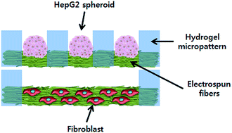
We developed a novel methodology of constructing a three-dimensional (3D) heterotypic co-culture system based on double-layered fibrous scaffolds incorporated with hydrogel micropatterns. The combination of electrospinning and hydrogel patterning generated micropatterned fibrous scaffolds consisting of poly(ethylene glycol) (PEG) hydrogel micropatterns and polycarprolactone (PCL) fibers. The thickness of the hydrogel micropatterns and fiber matrices, which were in the ranges of 100 to 250 μm and 20 to 80 μm, respectively, could be controlled by the volume of the hydrogel precursor solution and the collection time used for electrospinning. Because the resultant micropatterned fibrous scaffolds were obtained as a free-standing and bidirectionally-porous sheet, they could be stacked in a double-layered structure in which each scaffold contains different cell types for co-culture studies. As a model system, double-layered scaffolds for the co-culture of HepG2 and fibroblast cells were constructed by placing HepG2-containing scaffolds on top of fibroblast-containing scaffolds. The micropatterned fibrous scaffolds were demonstrated to be suitable for the culture of both HepG2 and fibroblasts cells. In addition, by controlling the micropattern size, HepG2 spheroids of uniform size (187.2 ± 10.7 μm) were formed in the top layer and used for the co-culture studies. According to the co-culture experiment, enhanced albumin secretion was observed from the co-cultured HepG2 cells compared with the single-cultured HepG2 cells, suggesting that micropatterned fibrous scaffolds are a promising tool that can be applied to heterotypic co-culture systems in various tissue engineering applications.


1. Introduction

Because cells exist in a three-dimensional (3D) extracellular matrix surrounded by other cells in our body, homotypic and/or heterotypic cell–cell interactions play important roles in cellular behavior such as survival, apoptosis, migration, proliferation, and differentiation.1,2 To understand various cell–cell interactions, many efforts have been made to develop in vitro co-culture systems.3–8 Co-cultures of two or more cell types have been used to create more biomimetic environments, which have demonstrated the importance of heterotypic cell–cell interactions on the regulation of cell behaviors.9–14 Initially, co-cultures were prepared by simply mixing two or more cell types and seeding them onto the same substrates. However, this method cannot precisely control the degree of cell–cell interactions. Recent progress in the field of surface chemistry in conjunction with microfabrication has enabled the spatial localization of multiple cell types relative to each other, leading to the generation of micropatterned co-culture systems.15–20 Although these micropattern systems can allow the study of heterotypic cell–cell interactions through either soluble signaling or direct cell–cell contact, there are several drawbacks that need to be overcome in these systems. First, because most micropatterned co-culture systems were developed by the sequential seeding of different cell types on a single substrate, there is a possibility for the contamination of the pure population. Furthermore, an initial cell type patterned on a substrate can be exposed to harsh chemical or physical conditions when the next cell patterns are created. Second, most micropatterned co-culture systems are still developed on a two-dimensional (2D) flat surface, whereas real tissues are inherently 3D and contain several different topographical cues that cells can sense and respond to. To resolve those problems, Bhatia group developed a microfabricated silicon substrate for cell culture with moveable interlocking parts.21 In their study, two different cell types were separately cultured on each interlocking part and then locked together with a controlled distance between the different cells for co-culture studies. However, the cells still existed on flat surfaces and formed an outspread 2D monolayer in this system. The most successful 3D co-culture systems have been achieved using cell sheet engineering, which was designed by Okano and colleagues; in these systems, one cell sheet was over layed onto the other cell sheet without using scaffold materials.22,23 Alternatively, Ito et al. used magnetic attraction as a physical approach to enhance the layered cell–cell interaction based on cell sheet engineering.24 Compared with the 2D co-culture system, in which cell–cell interaction always occurs at the same plane, the layered 3D co-culture system enables cell–cell interactions to occur at both the same and different planes. Although those layered 3D co-culture systems without artificial scaffolds are more desirable in terms of cell–cell interaction, a co-culture system using 3D scaffold materials is also necessary for conventional tissue engineering applications, which use scaffolds to reform the native structure of a tissue or organ.25–28

In this study, we developed a novel layered 3D co-culture system based on micropatterned fibrous scaffolds, which were fabricated by combining electrospinning and the hydrogel patterning technique. The incorporation of poly(ethylene glycol) (PEG) hydrogel micropatterns into fibers enabled the easy handling of a micrometer-thick fibrous scaffold that could be stacked to form a multilayer scaffold system. When different cell types are cultured in each layer, they can interact with each other due to the porous nature of fibrous scaffolds. As a model system, a double-layered, 3D co-culture system of HepG2 and fibroblast cells was constructed by placing HepG2-containing scaffolds on top of fibroblast-containing scaffolds. In addition, the hydrogel micropatterns in the top layer were designed to generate HepG2 spheroids of uniform size because it is well-known that liver-derived cells must be grown as spheroids (multi-cellular aggregates) to maintain better viability and functionality.29,30 After confirming that our scaffolds were suitable for the culture of both HepG2 and fibroblast cells, we investigated whether the co-culture of HepG2 with fibroblasts cells enhanced albumin secretion to evaluate the feasibility of this co-culture system.

2. Experimental

2.1 Materials

Poly(ethylene glycol) diacrylate (PEG-DA) (MW 575), 2-hydroxy-2-methylpropiophenone (HOMPP), polycarprolactone (PCL) (MW 80000), chloroform, methanol, dimethylsulfoxide (DMSO), ethanol, Dulbecco's Modified Eagle's Medium (DMEM), fetal bovine serum (FBS), 3-(4,5-dimethylthiazol-2-yl)-2,5,diphenyltetrazolium bromide (MTT) antibiotic/antimycotic solution, and trypsin/ethylenediaminetetra-acetate (trypsin/EDTA) were purchased from Sigma-Aldrich (Milwaukee, WI, USA). Human hepatocellular carcinoma cells (HepG2) and mouse embryonic fibroblasts (NIH-3T3) were obtained from Korean Cell Line Bank (Seoul, Korea) and American Type Culture Collection (Manassas, VA, USA), respectively. A Live/Dead Viability/Cytotoxicity Kit (L-7013), Cell Tracker Green CMFDA, Cell Tracker Orange CMTMR, and phosphate buffered saline (PBS, 0.1 M, pH 7.4) was purchased from Invitrogen (Carlsbad, CA, USA). Albumin-specific primary antibody, secondary detection antibody linked to horseradish peroxidase (HRP), and tetramethylbenzidine (TMB) were obtained from Koma Biotech Inc. (Seoul, Korea). The photomasks for photolithography were prepared using AutoCAD and were printed on transparencies using a standard laser jet printer (LaserWriter 16/600 PS, Apple Inc., Cupertino, CA, USA).

2.2 Preparation of electrospun fibrous scaffolds

The electrospinning apparatus used in this study consisted of a plastic syringe (10 mL) capped with a flat-ended 18 G metal needle, a syringe pump (KD Scientific, Holliston, MA, USA) for controlling the feeding rate, a stainless steel substrate as a collecting plate, and a high voltage power supply (NanoNC, Seoul, Korea). PCL polymer solutions (15 wt%) were prepared using a 7[thin space (1/6-em)]:[thin space (1/6-em)]1 volume ratio of chloroform:methanol. The solution was transferred to a syringe for electrospinning. To electrospin PCL fibers, a 12 kV positive voltage was applied to the solution via the needle, and a constant feeding rate of solution (3 mL hour−1) was provided by the syringe pump. The distance between the tip of the needle and the collecting plate was 20 cm. The electrospun fibers were collected on a clean aluminum foil (connected to the ground). The resulting PCL fibers were then treated with oxygen plasma (Femto Science, Kyunggi, Korea) for 5 minutes prior to the hydrogel patterning process. The radio-frequency power and pressure of plasma treatment were 40 W and 0.1 mmHg, respectively.

2.3 Incorporation of hydrogel micropatterns into fibrous scaffolds

The resultant electrospun fibers were micropatterned with PEG hydrogel using photolithography as described in our previous studies.31,32 The gel precursor solution was prepared by adding 50 μl of HOMPP as a photoinitiator to 1 mL of a 50 vol% solution of PEG-DA in water. This precursor solution was dropped onto the electrospun fibers and spread into a thin layer by covering with a photomask. The precursor solution was then exposed to 365 nm and 300 mW cm−2 ultraviolet (UV) light (EFOS Ultracure 100 ss Plus, UV spot lamp, Mississauga, Ontario, Canada) for 1 second through the photomask. After a development process with water to remove the unreacted precursor solution, a micropatterned fibrous scaffold was obtained. The morphology of the micropatterned fibrous scaffold was observed through scanning electron microscopy (SEM) (Hitachi Model S-4200 at 30 kV, Nissei Sangyo Co., Tokyo, Japan). For the cell studies, the scaffolds were sterilized in 70% v/v ethanol solution for 30 minutes and then washed five times in phosphate-buffered saline (PBS) to remove any traces of ethanol.

2.4 Cell culture and seeding

HepG2 and fibroblast cells were cultured in DMEM containing 4.5 g L−1 glucose, 10% FBS, and 1% antibiotic/antimycotic solution. The cells were then incubated at 37 °C in 5% CO2 and 95% air. To seed the cells onto the micropatterned PCL fibers, both cells were trypsinized from routine culture and centrifuged at 1200 rpm and 25 °C for 5 minutes. The supernatant was removed, and the cells were resuspended in fresh culture medium containing serum. An aliquot was obtained for cell counting in a hemocytometer to adjust the seeding density. Finally, approximately 2.0 × 10⁵ cells were seeded onto the micropatterned fibrous scaffolds. After 5 hours, the cell-containing micropatterned nanofibers were transferred to new 12-well plates to exclude the effect of the cells that adhered to the well plate.

2.5 Co-culture system

For studying the co-culture of HepG2 and fibroblasts, each cell type at the same density (2.0 × 10⁵ cells mL−1) was seeded onto different scaffolds and cultured in different well plates. After 10 hours, each scaffold was removed from the well plate and plugged into a custom-made device with four arms through the holes at the edges of a scaffold, as shown in Fig. 1. Here, we designed a multilayer co-culture system in which HepG2-seeded scaffold was located on top of the fibroblast-seeded scaffold. The co-culture was conducted in the same media conditions used for the single culture. The culture medium was refreshed every two days.
image file: c4ra12269k-f1.tif
Fig. 1 Schematic illustration and a photo of the co-culture system consisting of micropatterned fibrous scaffolds and a custom-made device capable of stacking multiple scaffolds.

2.6 Cell viability and proliferation assay

A live/dead viability/cytotoxicity fluorescence assay was used to investigate the viability of the adherent cells on the micropatterned substrate. This assay uses Calcein-AM and EthD-1 as fluorophores to distinguish the living cells from the dead cells. Calcein-AM stains live cells green, whereas EthD-1 stains dead cells red. For this assay, the micropatterned cells were incubated with 2 μm Calcein-AM and 4 μm EthD-1 in culture media for 30 minutes at 37 °C. After fixation with 2.5% glutaraldehyde, the samples were imaged using fluorescence microscopy (Carl Zeiss Inc., Thornwood, NY, USA). MTT assays were performed to investigate the in vitro proliferation of the cells cultured on the micropatterned fibrous scaffolds. Briefly, 10% v/v MTT solution (5 mg mL−1) in the culture medium was added to the cell-seeded scaffolds. The samples were incubated for 3 hours at 37 °C, and the formazan crystals transformed from MTT by mitochondrial reductase were dissolved by DMSO. The absorbance was measured at 570 nm using a microplate reader (Molecular Devices, Sunnyvale, CA, USA).

2.7 Cell imaging

For SEM imaging, the cell-attached scaffolds were fixed with 4% glutaraldehyde for 2 hours at 4 °C, washed gently with PBS for 5 minutes, dehydrated using a graded series of ethanol (25%, 50%, 70%, 80%, 90%, 95%, and 100%) and dried. The samples were platinum sputter-coated prior to SEM imaging. Cell Trackers were also used to obtain fluorescence images of the HepG2 and fibroblast cells. Prior to seeding, the HepG2 and fibroblast cells were stained with Cell Tracker Orange and Green, respectively. This reagent passes freely through cell membranes, but once inside the cell, is transformed into cell-impermeant reaction products.

2.8 Enzyme-linked immunosorbent assay for albumin

The albumin secreted by the HepG2 cells in the culture medium was quantified by enzyme-linked immunosorbent assay (ELISA). Briefly, the wells of an ELISA plate were coated with affinity-purified human albumin coating antibody for 1 hour and then blocked for 1 hour. Every two days, the culture medium was collected and added to the wells. After 1 hour incubation, HRP-conjugated human albumin detection antibody was added with TMB substrate solution, and the mixture was incubated for 20 minutes. After the reaction was stopped, the absorbance was measured at 450 nm using a microplate reader.

2.9 Statistics

The data were compared using an independent two-sample t-test and p-values less than 0.05 were considered statistically significant.

3. Results and discussion

The electrospinning of a PCL solution produced ultrathin polymer fibers with diameters of 1.0–2.5 μm, as shown in Fig. 2a. Because bare PCL fibers were too hydrophobic to allow significant infiltration of the gel precursor solution through the fiber scaffold, oxygen plasma treatment was necessary to make the fiber surface less hydrophobic. After plasma treatment, the fibrous scaffolds could be thoroughly soaked with precursor solution and fiber-incorporated hydrogel micropatterns were then successfully fabricated using photolithography with good fidelity. In this study, a photomask containing arrays of square patterns (200 × 200 μm or 1 × 1 mm) was used, and an optical image of the photomask that produced 200 × 200 μm-microwell-shaped hydrogel microstructures is shown in Fig. 2b. The design of the mask allowed only the hydrogel precursor solution below the transparent region of the photomask to be crosslinked and to become a hydrogel micropattern upon exposure to UV light while residual polymer was removed elsewhere in the pattern. Thus, the subsequent photopatterning created microwells of fibers separated by hydrogel, as shown in Fig. 2c. Because PEG hydrogels are known to resist cell adhesion, the resultant micropatterned fibrous scaffolds consisted of two different regions that interact with cells differently: one is the cell adhesion-resistant PEG hydrogel region and the other is the cell adhesion-promoting PCL fiber region. The micropatterned fibrous scaffold was easily detached from collecting plate and obtained as a free standing sheet (Fig. 2d). Usually, it is very difficult to handle micrometer-thick fibrous scaffold because they are mechanically weak and easily folded. In particular, the hydrophobic nature of most electrospun fibers made the non-woven mat form aggregates and clump after contact with water. However, incorporation of PEG hydrogel micropattern facilitated handling fibers as shown in this figure. Due to their free-standing nature, these sheets of micropatterned PCL fibers could be stacked in multiple layers.
image file: c4ra12269k-f2.tif
Fig. 2 Fabrication of micropatterned PCL fiber scaffolds by combining electrospinning and hydrogel patterning. (a) SEM image of electrospun PCL fibers (scale bar: 20 μm). (b) Optical image of photomask containing arrays of square patterns (200 × 200 μm). (c) SEM image of PCL fiber incorporated into PEG hydrogel micropatterns (scale bar: 300 μm). (d) A photo of micropatterned fibrous scaffolds.

The height of the hydrogel micropatterns and the thickness of the fiber matrices, which were in the range of 100 to 250 μm and 20–80 μm, respectively, as shown in Fig. 3a and b, could be controlled by the volume of the hydrogel precursor solution and the collection time of the electrospinning process. The SEM images in Fig. 3c show examples of scaffolds with different hydrogel heights and fiber thicknesses. The first SEM image shows a scaffold with a 200 μm-thick hydrogel and 50 μm-thick fiber matrices, whereas the second SEM image shows a scaffold with a 250 μm-thick hydrogel and 80 μm-thick fiber matrices. These cross-section images also revealed that the fibers were located at the bottom of the hydrogel patterns because hydrogel patterning was carried out in the presence of the fiber matrix on the collecting plate without any spacer.


image file: c4ra12269k-f3.tif
Fig. 3 Control of the height of the hydrogel micropatterns and the thickness of the fibrous scaffolds. (a) Dependence of the height of the hydrogel micropatterns on the volume of the precursor solution. (b) Thickness of the fibrous scaffolds as a function of the electrospinning time. (c) SEM images of micropatterned fibrous scaffolds with hydrogel micropatterns of different heights and electrospun fiber matrices of different thicknesses (scale bar: 100 μm).

The resultant micropatterned fibrous scaffolds were used for the culture of fibroblast and HepG2 cells. For HepG2, scaffolds with 200 × 200 μm patterns were used, and for fibroblasts, scaffolds with 1 × 1 mm patterns were used. The reason for using the 200 × 200 μm patterns for HepG2 cells is to obtain spheroids of uniform size (multi-cellular aggregates) because it has been well-established that liver-derived cells must be grown as spheroids to maintain better viability and functionality. Although fibrous substrates facilitate the formation of spheroids, it is very difficult to obtain spheroids with controllable and uniform size without combining micropatterning techniques. Controlling the spheroid size and the number of cells is important for optimizing spheroid culture and to obtain consistent results, which enhance the reliability of assays using cell spheroids.33,34 We expected that our micropatterned fibrous scaffolds enable us to prepare uniform HepG2 spheroids because we can confine cells only within the PCL fiber regions due to the non-adhesiveness of the PEG hydrogel toward cells. Because cell spheroids smaller than 200 μm are desirable to prevent hypoxia or malnutrition, we used 200 × 200 μm micropatterns to create a microarray of HepG2 spheroids. First, the adhesion and growth of fibroblast and HepG2 cells over a period of eight days were investigated with the MTT assay, in which the absorbance at 560 nm is proportional to the number of cells that remain viable within the micropatterned fibrous scaffolds. Fig. 4a and b indicates that both cell types remained viable and were able to proliferate within each scaffold. After four days of culture, the HepG2 cells gradually formed spheroids on each micropatterned fibrous region and the size of spheroids increased with time. As shown in Fig. 5a and b, HepG2 cells formed spheroids with high circularity only within the PCL fiber regions, and the PEG hydrogel walls served as effective barriers to cell adhesion, proliferation, and crossover. The sizes of the HepG2 spheroids formed within 200 × 200 μm patterns were 187.2 ± 10.7 μm after eight days. Fluorescent images from a live/dead assay also revealed that the HepG2 cells in the spheroids remained viable, and few dead cells were observed in the cores of the spheroids (Fig. 5c). In contrast, fibroblasts also adhered and spread only on the PCL fiber region (1 × 1 mm). Even after eight days, fibroblasts were evenly distributed within the fibrous region without forming spheroids, as shown in Fig. 5d.


image file: c4ra12269k-f4.tif
Fig. 4 Culture of fibroblast and HepG2 cells using micropatterned fibrous scaffolds. Result of MTT assay at different culture periods for (a) fibroblasts and (b) HepG2 cells.

image file: c4ra12269k-f5.tif
Fig. 5 Images of HepG2 and fibroblast cells cultured on micropatterned fibrous scaffolds. (a) Fluorescence image (from Cell Tracker Orange) and (b) SEM image of HepG2 spheroids formed within 200 × 200 μm patterns after eight days of culture. (c) Live/dead fluorescence viability assay of HepG2 spheroids after eight days of culture. (d) Fluorescence image of fibroblasts stained with Cell Tracker Green after eight days of culture. Scale bars in the figures are 200 μm.

After the hydrogel-incorporated micropatterned fibrous scaffolds were proven to be appropriate for the culture of fibroblasts and HepG2 spheroids, co-culture experiments were carried out by stacking two different micropatterned scaffolds containing HepG2 and fibroblasts, respectively. The distance between the cells in each scaffold was controlled to by changing the thickness of the hydrogel micropatterns and the thickness of the fiber matrix. In this study, micropatterned fibrous scaffolds with 100 μm-thick hydrogel micropatterns and 50 μm-thick PCL fiber matrix were used such that the distance between the HepG2 spheroids and the fibroblasts was to be approximately 100 μm to ensure that the cell–cell interactions were mainly mediated by short-range soluble factors. The HepG2-seeded scaffold was located on top of the fibroblast-seeded scaffold to allow the formation of HepG2 spheroids with greater size than the distance between the two scaffolds. The design of the double-layered co-culture system for HepG2 and fibroblast cell is shown in Fig. 6a. The effect of the stacked co-culture systems on the cellular function was determined by measuring the hepatic albumin expression level during a period of eight days. Although the albumin secretion from the fibroblasts was undetectable, the albumin secreted from the single and co-cultured hepatocytes was detected, as shown in Fig. 6b. According to this figure, a similar amount of albumin was secreted from both single and co-cultured hepatocytes at the beginning of the culture periods (up to four days), but significantly higher amounts of albumin were secreted from the co-cultured HepG2 cells from days 4 to 8, suggesting that the co-culture of HepG2 cells with fibroblasts within the stacked micropatterned scaffold system facilitated albumin secretion. Because there was no direct cell–cell contact in our system, enhanced albumin secretion was mainly due to a paracrine signaling-mediated effect. The positive effect of the co-culture on albumin secretion in this study was not as marked as in other previous studies. This result may be mainly due to the lack of direct cell–cell contact between the HepG2 and fibroblast cells in our system. Although the precise mechanism that causes the enhancement of liver-specific functions in hepatocyte co-cultures is not clear, it has been reported that cell–cell interaction via direct contact between fibroblasts and liver cells is crucial for modulating hepatocyte function, such as albumin secretion.21,24 Therefore, in the future, more efforts will be made on the application of our micropatterned fibrous scaffolds to a co-culture system that allows direct cell–cell contact.


image file: c4ra12269k-f6.tif
Fig. 6 Co-culture of HepG2 and fibroblasts using double-layered fibrous scaffolds incorporated with hydrogel micropatterns. (a) Schematic diagram of the cross-section view of the double-layered fibrous scaffolds used for the co-culture studies. (b) Cumulative amount of albumin secreted from HepG2 cells.

4. Conclusion

Micropatterned fibrous scaffolds were successfully fabricated with the combination of electrospinning and PEG hydrogel photolithography techniques for the purpose of developing novel 3D co-culture system. Because the micropatterned scaffold was obtained as a free-standing and bidirectionally-porous sheet, it could be stacked in multiple layers in which each scaffold contains different cell types for co-culture studies. As a model system, a co-culture system of HepG2 and fibroblast cells was designed by placing a HepG2-cultured scaffold on top of a fibroblast-cultured scaffold. The micropatterned scaffolds supported the adhesion and proliferation of both cell types. In addition, by controlling the micropattern size, HepG2 spheroids of uniform size were formed within the scaffold and used for the co-culture studies. Enhanced albumin secretion was observed from the co-cultured HepG2 compared with the single-cultured HepG cells, demonstrating the existence of an interaction between the two types of cells. These results suggest that the newly developed fibrous scaffolds incorporated with hydrogel is a promising tool for the construction of multilayered cell culture scaffolds consisting of heterotypic co-cultured cells that can be applied to tissue engineering and cell biology for the study of cell–cell interaction.

Acknowledgements

This work was supported by the National Research Foundation (NRF) grant funded by the Ministry of Science, ICT and Future Planning (MSIP) (2011-0022709, and 2007-0056091 “Active Polymer Center for Pattern Integration at Yonsei University“).

References

  1. A. Abbott, Nature, 2003, 424, 870–872 CrossRef CAS PubMed.
  2. S. Levenberg and R. Langer, Curr. Top. Dev. Biol., 2004, 61, 113–134 CrossRef CAS.
  3. M. Marimuthu and S. Kim, Anal. Biochem., 2011, 413, 81–89 CrossRef CAS PubMed.
  4. L. Goers, P. Freemont and K. M. Polizzi, J. R. Soc., Interface, 2014, 11, 20140065 CrossRef PubMed.
  5. K. G. Battiston, J. W. C. Cheung, D. Jain and J. P. Santerre, Biomaterials, 2014, 35, 4465–4476 CrossRef CAS PubMed.
  6. J. G. Gershovich, R. L. Dahlin, F. K. Kasper and A. G. Mikos, Tissue Eng., Part A, 2013, 19, 2565–2576 CrossRef CAS PubMed.
  7. I. Leisten, R. Kramann, M. S. V. Ferreiraa, M. Bovi, S. Neuss, P. Ziegler, W. Wagner, R. Knuchel and R. K. Schneider, Biomaterials, 2012, 33, 1736–1747 CrossRef CAS PubMed.
  8. J. E. McBane, K. H. Cai, R. S. Labow and J. P. Santerre, Acta Biomater., 2012, 8, 488–501 CrossRef CAS PubMed.
  9. H. Kaji, G. Camci-Unal, R. Langer and A. Khademhosseini, Biochim. Biophys. Acta, Gen. Subj., 2011, 1810, 239–250 CrossRef CAS PubMed.
  10. J. B. Gurdon, P. Lemaire and K. Kato, Cell, 1993, 75, 831–834 CrossRef CAS.
  11. I. K. Zervantonakis, C. R. Kothapalli, S. Chung, R. Sudo and R. D. Kamm, Biomicrofluidics, 2011, 5, 013406 CrossRef PubMed.
  12. E. J. Levorson, M. Santoro, F. K. Kasper and A. G. Mikos, Acta Biomater., 2014, 10, 1824–1835 CrossRef CAS PubMed.
  13. Y. W. Liu, L. Zhang, J. J. Wei, S. L. Yan, J. S. Yu and X. H. Li, J. Mater. Chem. B, 2014, 2, 3029–3040 RSC.
  14. C. J. Kirkpatrick, S. Fuchs and R. E. Unger, Adv. Drug Delivery Rev., 2011, 63, 291–299 CrossRef CAS PubMed.
  15. C. A. Goubko and X. D. Cao, Mat Sci Eng C Mater, 2009, 29, 1855–1868 CrossRef CAS PubMed.
  16. S. N. Bhatia, M. L. Yarmush and M. Toner, J. Biomed. Mater. Res., 1997, 34, 189–199 CrossRef CAS.
  17. D. T. Chiu, N. L. Jeon, S. Huang, R. S. Kane, C. J. Wargo, I. S. Choi, D. E. Ingber and G. M. Whitesides, Proc. Natl. Acad. Sci. U. S. A., 2000, 97, 2408–2413 CrossRef CAS PubMed.
  18. J. Fukuda, A. Khademhosseini, Y. Yeo, X. Y. Yang, J. Yeh, G. Eng, J. Blumling, C. F. Wang, D. S. Kohane and R. Langer, Biomaterials, 2006, 27, 5259–5267 CrossRef CAS PubMed.
  19. A. Folch and M. Toner, Annu. Rev. Biomed. Eng., 2000, 2, 227–256 CrossRef CAS PubMed.
  20. K. Kikuchi, K. Sumaru, J. I. Edahiro, Y. Ooshima, S. Sugiura, T. Takagi and T. Kanamori, Biotechnol. Bioeng., 2009, 103, 552–561 CrossRef CAS PubMed.
  21. E. E. Hui and S. N. Bhatia, Proc. Natl. Acad. Sci. U. S. A., 2007, 104, 5722–5726 CrossRef CAS PubMed.
  22. M. Harimoto, M. Yamato, M. Hirose, C. Takahashi, Y. Isoi, A. Kikuchi and T. Okano, J. Biomed. Mater. Res., 2002, 62, 464–470 CrossRef CAS PubMed.
  23. J. Yang, M. Yamato, C. Kohno, A. Nishimoto, H. Sekine, F. Fukai and T. Okano, Biomaterials, 2005, 26, 6415–6422 CrossRef CAS PubMed.
  24. A. Ito, H. Jitsunobu, Y. Kawabe and M. Karnihira, J. Biosci. Bioeng., 2007, 104, 371–378 CrossRef CAS PubMed.
  25. N. Di Maggio, E. Piccinini, M. Jaworski, A. Trumpp, D. J. Wendt and I. Martin, Biomaterials, 2011, 32, 321–329 CrossRef CAS PubMed.
  26. H. Jaganathan, J. Gage, F. Leonard, S. Srinivasan, G. R. Souza, B. Dave and B. Godin, Sci. Rep., 2014, 4, 6468 CrossRef PubMed.
  27. A. Kunze, M. Giugliano, A. Valero and P. Renaud, Biomaterials, 2011, 32, 2088–2098 CrossRef CAS PubMed.
  28. T. J. Liu, B. C. Lin and J. H. Qin, Lab Chip, 2010, 10, 1671–1677 RSC.
  29. N. Koide, T. Shinji, T. Tanabe, K. Asano, M. Kawaguchi, K. Sakaguchi, Y. Koide, M. Mori and T. Tsuji, Biochem. Biophys. Res. Commun., 1989, 161, 385–391 CrossRef CAS.
  30. H. F. Lu, K. N. Chua, P. C. Zhang, W. S. Lim, S. Ramakrishna, K. W. Leong and H. Q. Mao, Acta Biomater., 2005, 1, 399–410 CrossRef PubMed.
  31. H. J. Lee and W. G. Koh, ACS Appl. Mater. Interfaces, 2014, 6, 9338–9348 CAS.
  32. H. J. Lee, S. H. Nam, K. J. Son and W. G. Koh, Sens. Actuators, B, 2010, 148, 504–510 CrossRef CAS PubMed.
  33. A. Iwasaki, T. Matsumoto, G. Tazaki, H. Tsuruta, H. Egusa, H. Miyajima and T. Sohmura, Adv. Eng. Mater., 2009, 11, 801–804 CrossRef CAS.
  34. R. L. Carpenedo, S. A. Seaman and T. C. McDevitt, J. Biomed. Mater. Res., Part A, 2010, 94A, 466–475 CAS.

This journal is © The Royal Society of Chemistry 2014
Click here to see how this site uses Cookies. View our privacy policy here.