Lamellae evolution of poly(butylene succinate-co-terephthalate) copolymer induced by uniaxial stretching and subsequent heating

Zhenzhen Weiab, Ruixin Lunab, Xueqin Louab, Feng Tianc, Jinyou Lin*c, Xiuhong Lic, Jianyong Yud and Faxue Li*ab
aKey Laboratory of Textile Science & Technology, Ministry of Education, China
bCollege of Textiles, Donghua University, Shanghai 201620, China. E-mail: fxlee@dhu.edu.cn; Fax: +86-21-67792627; Tel: +86-21-67792803
cShanghai Synchrotron Radiation Facility, Shanghai Institute of Applied Physics, Chinese Academy of Sciences, Shanghai 201204, China. E-mail: Jinyoulin82@gmail.com
dModern Textile Institute, Donghua University, Shanghai 200051, China

Received 10th October 2014 , Accepted 11th November 2014

First published on 11th November 2014


Abstract

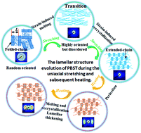
The lamellar structural evolution of biodegradable poly(butylene succinate-co-terephthalate) (PBST) random copolymer was investigated under the conditions of uniaxial stretching at 50 °C, and then heating from 50 to 150 °C at a strain of 150% by the in situ small-angle X-ray scattering (SAXS) technique. A long period referring to the lamellae while processing PBST was calculated, and a schematic for the structural evolution was proposed. It has been found that the lamellar structure experienced a remarkable transformation accompanied by the strain-induced melting of lamellae and formation of new lamellae when the strain exceeded 84% at 50 °C during the initial deformation process. After stretching by 150%, the lamellar structure remained unchanged with the perfection of lamellae in the subsequent heating process from 50 to 120 °C. Only at relatively high temperatures (120–150 °C), the long period of lamellae underwent a significant increase. Conclusions can be drawn that the lamellar structure of PBST is more sensitive to strain and only a relatively high temperature has a prominent impact on it, which is of great significance to provide targeted design guidance for the manufacturing and application of biodegradable PBST copolymer and also gives potential insights into other random and aliphatic–aromatic copolymers.


Introduction

Polymers are subjected to substantial mechanical deformation and thermal treatment during their processing, as well as in practical applications, giving rise to continuous structure changes.1–3 The mechanical properties of polymers are determined by their final structures, as well as their evolution process.4–7 Hence, revealing the structural evolution during processing with the aim of controlling the final structure is expected to regulate the mechanical properties of end products, and thus extends their applications.

The structure of crystalline polymers, viewed at scale, is normally hierarchical, i.e. from molecular chains of a few nanometers width to crystalline lamellae of dozens of nanometers thickness and spherulites of several micrometers in diameter.8,9 Among these, the lamellar structure is one of the most common structural models and possesses alternating crystalline lamellar and amorphous regions.10 Essentially, the transformation of lamellar structures arises from a variation in the macromolecular arrangement in the two regions during the processing process, resulting in the whole structural changes. Moreover, the presence and changes in crystalline and amorphous regions within the polymer enable it to exhibit different levels of rigidity and toughness.11,12 Therefore, revealing the lamellar structure developments during dynamic processes is essential both in fundamental researches and practical applications.

A wealth of researches have been conducted on lamellar structure developments during stretching, as well as heating using a host of experimental techniques such as small-angle X-ray scattering (SAXS),12–19 wide-angle X-ray diffraction,20–26 transmission electron microscopy,27,28 Fourier transform infrared microscopy,29,30 and atomic force microscopy.31,32 stretching can transform the original spherulite morphology into a highly oriented fibrillar one, in which polymeric chains are preferentially aligned along the stretching direction. On the basis of the results of SAXS experiments, strain-induced melting and recrystallization were proposed to be responsible for the morphological variation during deformation.14,18,33

Moreover, a set of uniaxial stretching experiments were carried out at different temperatures to explore the influence of temperature on polymeric structure development during stretching.34–37 The effects of temperature are mainly reflected in the initial crystallinity and chain mobility that vary with the drawing temperature. Only taking the temperature without deformation into consideration, the structural evolution of semi-crystalline polymers has also been extensively investigated during heating and annealing processes.12,13,21,23,24,38,39 Actually, the molecular mobility mechanism during heating is dependent on crystallization conditions, thermal history and other treatments to which it is subjected. Therefore, there has not been a consistent conclusion for polymers upon heating. In addition, most of the researches mentioned above focused on separately elucidating the molecular mobility mechanism during the processes of stretching and heating. In contrast, few publications have reported the structural transformation during first stretching and subsequent heating, or heating and then stretching. In fact, structural changes are of great importance to the polymers that are ordinarily subjected to tensile deformation and thermal treatment during their processing process or applications.

Poly(butylene succinate-co-terephthalate) (PBST), one type of biodegradable aliphatic–aromatic copolymer, has spurred considerable interest in the past decades. We have reported its preparation, structure, properties and application in the form of elastic fibers with high elastic recovery in previous work.40–42 Recently, unstable morphologies fundamentally ascribed to complex structure transformation in dynamic environments have been encountered in the manufacturing of PBST fibers, resulting in relatively poor mechanical properties and low productivity. As a matter of fact, some important semi-crystalline polymers including polypropylene,34,43 polyethylene,37,44 and poly(ethylene terephthalate)45 have been well documented regarding their structural developments, which have played important roles in their structure regulation and improvement of properties. Thus, it is necessary to elucidate the structural developments of PBST at the lamellar level during its deformation and/or heating for further practical application in future work. Moreover, compared with these important polymers, PBST is a type of random copolymer.46 Some investigations have been carried out on the uniaxial stretching and structural evolution of semi-crystalline block copolymers.47–49 Therefore, studies on the structural changes of PBST probably give potential insights into other random copolymers.

In this work, the in situ SAXS technique using synchrotron radiation X-rays in combination with a tensile device and a heating chamber was used to investigate the lamellar structural information during uniaxial stretching and subsequent heating. The sample was initially stretched with a constant strain of 150% at 50 °C, and then heated to 150 °C with several isothermal stages under this stable strain. The long period of PBST, as well as its evolution was extensively explored, and a schematic for its lamellar structural evolution during the uniaxial stretching and subsequent heating was proposed.

Experimental

Materials and sample preparation

Poly(butylene succinate-co-terephthalate) (PBST) copolymer was synthesized by ourselves and the specific reaction conditions and procedures were reported in our previous work.41 The chemical structure of PBST and its monomers are shown in Scheme 1. The 70 mol% butylene terephthalate unit was selected by adjusting the feeding ratio of succinic acid to terephthalic acid due to its balance between desirable biodegradability and good mechanical properties.40,41 The number average molecular weight (Mn) was 50[thin space (1/6-em)]600 g mol−1 and the polydispersity index was 1.92 through gel permeation chromatography (GPC) measurements. The endothermic peak was located at 180.4 °C in the differential scanning calorimetry (DSC) measurement and the glass transition temperature was about 13 °C, which is determined by dynamic mechanical analysis (DMA) (shown in ESI Fig. S1).
image file: c4ra12117a-s1.tif
Scheme 1 Illustration showing the chemical structure of the as-synthesized PBST.

The as-prepared PBST was melted at 200 °C in a hot press and placed at this temperature for 5 min. Latter, it was naturally cooled down to room temperature. Then, the film was cut into a dumbbell-shaped bar with dimensions of 50 mm (length) × 10 mm (neck width) × 0.5 mm (thickness).

In situ SAXS measurement

SAXS experiments were conducted at a beamline BL16B1 of the Shanghai Synchrotron Radiation Facility (SSRF, Shanghai, China). The X-ray wavelength was 0.124 nm and a Mar 165 CCD detector (2048 × 2048 pixels with a pixel size of 82.5 μm) was employed to collect two-dimensional (2D) SAXS patterns. The sample-to-detector distance was 2280 mm. The data of blank air were collected to correct for air scattering. A homemade tensile tester with a heating chamber was used for uniaxial tensile deformation during the in situ experiments. This apparatus can symmetrically stretch samples, which is beneficial for probing the same position during deformation. The schematic representation of the device is shown in Fig. 1a.
image file: c4ra12117a-f1.tif
Fig. 1 (a) Schematic representation of the in situ SAXS measurement in this work. (b) A diagram showing the uniaxial stretching and subsequent heating procedures. (c) A typical stress–strain curve of the PBST copolymer. (d) The integration sector of 2D patterns along the meridional (−15–15°) and equatorial (75–105°) directions.

The detailed experimental procedures are presented in Fig. 1b. A sample with 10 mm gauge length was first heated to 50 °C and held for 10 min, and then was stretched at a strain of 150% at a constant velocity of 20 μm s−1 at this temperature. The typical stress–strain curve of a PBST copolymer during this process was obtained as shown in Fig. 1c. The stretched sample was further heated to the preset temperatures of 80, 100 and 150 °C at a rate of 10 °C min−1 under the fixed strain. The real temperature of the sample is expected to be within ±2 °C of that of the chamber. At each temperature stage, the sample was held for 10 min before heating to a higher temperature. The 2D patterns were accumulated over periods of 20 s during the entire process.

Fit2D software was used to transfer 2D patterns into one-dimensional (1D) profiles from the integration sectors demonstrated in Fig. 1d in the form of intensity vs. q (scattering vector, q = (4π/λ)sin(θ/2), where λ is the X-ray wavelength and θ is the scattering angle). The long period (L, distance between the adjacent lamellae) was calculated from the Bragg equation L = 2π/qmax, where qmax corresponds to the peak position of the 1D SAXS profiles.

Results and discussion

Fig. 1c shows the engineering stress–strain curve of the PBST copolymer during uniaxial stretching at 50 °C. The stress first increases to its maximum value at a strain of 32% (i.e., the yield point), and then slightly decreases followed by a stress plateau. The strain-hardening phenomenon was not conspicuous for this sample due to the limited strain of 150% in this case. To elucidate the deformation behaviors of the PBST copolymer in detail, the stress–strain curve is divided into three regions, namely region I (0% ≤ strain ≤ 32%), II (32% < strain < 60%) and III (60% ≤ strain ≤ 150%).

To investigate the lamellar structure evolution of PBST induced by uniaxial stretching, the 2D SAXS patterns at different applied strains are collected as shown in Fig. 2. After naturally cooling, PBST crystallized into spherulites consisting of many chain-folded lamellae.40 The SAXS pattern of unstretched samples exhibited a single isotropic scattering ring (strain 0%), which is indicative of the existence of randomly oriented chain-folded lamellae.43,50 With the increasing strain in region I, the patterns gradually became oblate, in that the scattering maximum of SAXS patterns along the meridian shifted to a small angle, whereas that along the equator moved to a large angle. Moreover, the oblate pattern with two large arcs gradually became a two-bar-like pattern with the scattering maxima in the meridional direction (strain from 4% to 32%). As the strain increased in region II, the two bars became smeared, short and shifted towards the beam center, suggesting that destruction and fragmentation of the lamellar structure prevailed. In the early stage of region III, the two bars remained smeared (strain from 68% to 76%). When the strain reached about 84%, the two-bar-like pattern completely disappeared, indicating that the lamellar structure was possibly destroyed entirely. Above a strain of 84%, there were no obvious differences in 2D SAXS patterns. Therefore, a general understanding of the structural evolution during deformation was available from the changes in 2D SAXS patterns.


image file: c4ra12117a-f2.tif
Fig. 2 Selected 2D SAXS patterns at different strains in regions I, II and III.

To further explore the evolution involving specific parameters of the lamellar structure during uniaxial stretching, the relation of q vs. I(q) in these three regions obtained from the 2D SAXS scattering data through integrating is presented in Fig. 3, from which the long period (L) and the scattering intensity of the peak (Imax) are calculated and shown in Fig. 4. In region I (Fig. 3a and c), the changing trends of qmax along the meridian and equator are inverse, indicating that the interlamellar distances in the two directions exhibit opposite development with increasing strain. Within the strain range from 0% to 9%, in which the stress nearly linearly increased with the strain, the L value along the meridian increased from 14.2 to 15.5 nm (Fig. 4), an increase of 9.2%, close to the applied strain 9%. This increase is generally attributed to the elastic extension of chains in the amorphous region, leading to macroscopic elastic behavior of the material at a relatively low strain.18,34


image file: c4ra12117a-f3.tif
Fig. 3 Selected 1D SAXS profiles during stretching along the (a) and (b) meridional and (c) equatorial directions in regions I, II and III. The profiles of region I in (a) were vertically shifted to clearly show the tendency and avoid overlaps.

image file: c4ra12117a-f4.tif
Fig. 4 The long period and scattering intensity of the peak as a function of strain calculated from Fig. 3.

At a strain above 9%, the increase in stress was retarded and the scattering intensity more distinctly decreased, especially when the yield point was approached. The explanation that a fraction of the lamellar structure began to be destroyed after stretching by 9% was found to be reasonable for such a stress response in this case; exploring the following phenomenon in Fig. 4, the Imax at 32% strain decreased quite substantially from that of the original sample. The thickness change of the sample should not be taken into consideration for the decreased intensity during the very small deformation, while strain-induced melting should be the reason for the prominent decrease. The result agreed very well with the previous report by Hsiao et al.14 In addition, the long period along the equator displayed in Fig. 4 gradually decreased with the strain, as expected from the deformation along the meridional uniaxial stretching.

In the yielding region (region II), the behavior of strain-induced melting became more evident. As shown in Fig. 3b, the intensity distribution curves changed to monotonic decrease with the strain in this region, and it was difficult to point out the values of qmax, indicating that the periodic structure became increasingly unclear due to the melting of the original lamellar structure. Most of the molecular chains escaped from the original chain-folded lamellae into the amorphous region, causing little electron density difference, and thus an inconspicuous periodic structure. The value of the long period became indistinct but had a tendency to increase due to the destruction of some lamellae, resulting in an increase in the average distance between the remaining lamellae.

Fig. 3c shows the 1D SAXS profiles integrated from the equatorial direction at different strains in region II. The peak moved to a high q value, suggesting that some lamellae perpendicularly aligned to the stretching direction were still preserved, and its long period decreased with the increasing strain. This decrease could be attributed to the insertion of fragmentary lamellae and/or reorientation of lamellae to the stretching direction.18 Furthermore, the above results demonstrated that the strain-induced melting process was directionally dependent. Therefore, the lamellae along the stretching direction were melted first and faster than the lamellae perpendicularly aligned to the stretching direction.

It has to be pointed out that the four-point patterns occurring in many other semi-crystalline polymers under deformation in the yielding region14,17,18,35 were not observed here. The presence of four-point patterns suggested the fragmentation of lamellae and those fragmented lamellae were tilted against the stretching direction and/or had a checkerboard arrangement to minimize the stress concentration at the lamellae.12,14,18 No four-point patterns were observed here, which could be explained as follows: first, the relatively weak scattering intensity of the tilted lamellae was covered by the two bars; second, there were still lamellae aligned perpendicularly to the stretching direction, and it was unnecessary for other fragmented lamellae to tilt against the stress concentration. As a result, the slightly negative slope in the yielding region was reasonable due to the presence of lamellae aligned perpendicularly to the stretching direction.

In region III, above a strain of 84%, there were no obvious differences in 2D SAXS patterns, possibly due to the very weak intensity, but a new peak corresponding to a higher q value compared with the initial peak appeared in the 1D SAXS profiles (Fig. 3b), indicating that a new lamellar structure, possibly with extended chains, was formed with further deformation (i.e. strain-induced crystallization) and aligned along the stretching direction.

The intensity of this peak slightly increased as the strain increased, while its position stayed nearly fixed. Some researchers found that the smaller long period in the newly formed crystals (i.e. thinner lamellae) than the original lamellae was attributed to the stretching temperature being lower than the crystallization temperature of the original sample.17,51 This is consistent with our observation, since the stretching temperature was 50 °C while the crystallization temperature of the PBST sample was about 140 °C (seen in Fig. S1).

Within the strain range from 60% to 84%, the scattering intensity along the equator (Fig. 3c) gradually changed to a monotonic decrease, similar to the intensity variation along the meridian in region II (Fig. 3b), indicating that the amount of lamellae perpendicular to the stretching direction was decreasing due to their melting and orientation to the stretching direction.18,34 When the strain reached about 84%, these lamellae were completely melted and oriented along the stretching direction, resulting in almost unchanged 1D profiles at the equator with further stretching. If the newly oriented lamellae did not melt before the strain of 84%, there should be two peaks including the oriented and newly formed lamellae, which appeared with the increasing strain. It was no coincidence that the two types of lamellae were identical in their interlamellar distance because the orientation along the stretching direction would not bring about such a large change in long period (decrease from 19.4 to 10.5 nm).52 However, there was only one peak observed in the meridian, which remained constant as the strain was above 84%, indicating that the newly oriented lamellae also entirely melted before a strain of 84%. In other words, strain-induced melting for those lamellae not aligned along the stretching direction was accompanied by their orientation process. At a strain of about 84%, a transitional structure (disordered but with highly oriented molecular chains) was formed. Next, the newly oriented chains together with the original chains along the stretching direction crystallized during further deformation, leading to the formation of new lamellae.

The scattering intensity gradually decreased along both the meridian and the equator as the strain increased with the exception of the range in which new lamellae were formed (Fig. 4). The decreased and quite weak intensity at the beginning of deformation could result from the small electron density difference between crystal phases and amorphous phases during melting and the reorientation of lamellae, while the reduced thickness of the sample at large deformation should be primarily responsible for this.43 It should be noted that the 2D SAXS patterns showed no scattering streak in the equatorial direction near the beam stop when the applied strain was above the yield point (strain from 48% to 84% in Fig. 2). It could be an explanation that no micro-voids were formed during the stretching period, possibly due to the slow deformation rate and good ductility of PBST.35,43 However, with increasing strain, especially in the later stage of deformation, another relatively weak scattering streak in the equatorial direction appeared and looked stronger with increasing strain. Judging by its weak and changing intensity, the streak probably contributed to the formation of a fibrillar structure.

To understand the effects of heating on the lamellar structure of PBST copolymer under relatively small strain, after thermal annealing at 50 °C under a strain of 150%, PBST was further heated to 150 °C intermittently as shown in Fig. 1b, and some corresponding 2D SAXS patterns are presented in Fig. 5. The two-bar-like patterns along the meridian gradually became noticeable, showing the lamellar structure again prevailed during the heating process. When the temperature approached 100 °C, the two bars changed into two lobes, whose contours enlarged with the increasing temperature. The streak on the equator was nearly unchanged in the process of heating. The general representation of the two-bar-like pattern indicates that the lamellae are more regularly spaced and of equal thickness, while the two-lobe-like pattern signifies the non-uniform thickness and distance between the lamellae.44 Thus, from the transformation of these patterns, a general understanding about the lamellar changes during heating can be obtained.


image file: c4ra12117a-f5.tif
Fig. 5 Selected 2D SAXS patterns of PBST during heating under 150% strain.

The detailed effects of temperature on the development of the lamellar structure of stretched PBST (strain 150%) can be understood according to Fig. 6 and 7, which present selected 1D SAXS profiles at temperatures from 50 to 100 °C and 100 to 150 °C, respectively. For simplicity, we divided the temperature range into two sections: relatively low-temperature region (50–100 °C) and high-temperature region (100–150 °C). When the sample was under strain during heating, stress relaxation of molecular chains should be taken into consideration. Fig. S2 displays the stress relaxation curve of PBST under a strain of 150% as a function of time. As expected, the stress sharply decreased at the relatively low temperature with increasing time and gradually reached a roughly constant value after about 30 min. Consequently, the influence of stress relaxation of molecular chains should not be ignored in the relatively low-temperature region, whereas that in the second region is supposed to be overlooked.


image file: c4ra12117a-f6.tif
Fig. 6 Selected 1D SAXS profiles along the meridian in the temperature range from 50 to 100 °C.

image file: c4ra12117a-f7.tif
Fig. 7 Selected 1D SAXS profiles along the meridian in the temperature range from 100 to 150 °C.

In the low-temperature region as shown in Fig. 6, as the sample was thermally annealed at 50 °C, the new peak formed in the late stage of deformation still existed and no further changes in its position and intensity were detected with the annealing time (shown as the overlapped profiles in Fig. 6a). The similar feature can also be observed during the isothermal period at 80 °C in Fig. 6c. These findings indicated that no changes in lamellar structure occurred when the sample was held at a relatively low temperature. In comparison with Fig. 7a and c, in the isothermal periods at 100 and 150 °C, the qmax remained unchanged but the scattering intensity increased, suggesting that a higher temperature was more conducive to perfect the lamellae than a relatively low temperature for the same isothermal time. It is possible that an increase in scattering intensity can be realized if we prolong the annealing time at the low temperature according to the well-known time-temperature equivalence principle.

During the heating from 50–80 °C and 80–100 °C processes displayed in Fig. 6b and d, respectively, the scattering intensity increased with an almost invariable qmax. The rise in scattering intensity resulted from the increase in electron density difference, which was probably due to the different thermal expansion of the crystal and amorphous regions of the lamellae, perfection of lamellae, and melting of small crystals during the heating process.38 On the other hand, the constant long period (corresponding to the invariable qmax) can be ascribed to the balance between the relaxation of stretched amorphous chains inducing a long-period decrease, and the melting of small lamellae and increasing chain mobility causing a long-period increase in the thickness of the amorphous region.

In the high-temperature region (Fig. 7b), the qmax value was initially unchanged with increasing scattering intensity. Nevertheless, as the temperature increased to 120 °C, the qmax was continuously shifted to the left side and the scattering intensity also exhibited a prominent increase. In this period, the increased long period can be attributed to two main reasons as follows: first, the melting of small and thinner lamellae due to high molecular mobility leads to a thickness increase in the amorphous region; second, the recrystallization of partially melted crystals causes a thickening of the crystalline region in the lamellar structure.38

The changes occurring in this period were so significant that it was necessary to find out which one of the two factors or whether both of them played the dominant role in increasing the long period. This can be verified by exploring the specific thickness variations of the crystal and amorphous regions in this process, assuming the two-phase model of lamellar nanostructure. The one-dimensional correlation function γ(r) in the following equation can be applied to estimate the morphological variables:

image file: c4ra12117a-t1.tif
where r is the location measured along a trajectory normal to the lamellar surfaces. The resulting correlation function curves are presented in Fig. 8a. The inset of Fig. 8a shows how the long period (L) and crystalline thickness (lc) are determined. Generally, it is not possible to decide whether it is the amorphous or the crystalline thickness that is derived from the correlation function without prior knowledge of the crystallinity.12 However, the crystallinity of the sample used in the present study was 30–40% according to the results of WAXD measurement (seen in Fig. S3), ensuring the assignment of the smaller value obtained from the correlation function to the thickness of the crystalline region (lc). The amorphous thickness (la) can be obtained by subtracting the crystalline thickness from the long period (la = Llc).


image file: c4ra12117a-f8.tif
Fig. 8 (a) Correlation function calculated from the 1D SAXS profiles in Fig. 7b. (b) Values of long period L, crystal thickness lc, amorphous thickness la and inner crystallinity (lc/L) calculated from the correlation functions using a two-phase model. The inset in (a) demonstrates how the values are determined.

The calculated values of L, lc and la as a function of temperature are given in Fig. 8b. At the beginning of heating, all the values remained nearly unchanged, possibly because the molecular chains were in a metastable state after the thermal treatment in the foregoing low-temperature region. However, L, lc and la simultaneously increased at higher temperatures (over 120 °C). The increase in la should be attributed to the melting of small and thinner lamellae, while the increase in lc was mainly due to the recrystallization of partially melted crystals during the heating process, together leading to the increasing long period. Moreover, it could be found that the recrystallization of molecular chains still occurs at high temperature (about 20–30 °C lower than the melting temperature of PBST) in which the chain mobility was very high. The lamellar thickening at different temperatures was asynchronous, and thus irregular crystals were formed, corresponding to the results shown in the 2D SAXS patterns.

The inner crystallinity of the lamellae as defined by the ratio of the crystal thickness to the long period is also presented in Fig. 8b. The inner crystallinity obtained by SAXS represents the volume fraction of crystallites within the lamellae stacks.16 It was distinctly seen that the inner crystallinity roughly remained unchanged with the rising temperature despite a small fluctuation, indicative of the constant volume fraction of crystallites within the lamellar structure and independence on the morphology of the lamellae. The constant inner crystallinity was mainly attributed to the coexistence of the melting and recrystallization of the crystals in the lamellae, which was in accordance with the thickness increase of the crystalline and amorphous regions. The formation of perfect lamellae occurring during the melting and recrystallization could be the explanation of the increased scattering intensity.

Fig. 9 presents a schematic illustration of the lamellar structural evolution of PBST random copolymer during in situ stretching and heating on the basis of SAXS examination. Within the strain range from 0% to 9%, in which the stress nearly linearly increases with the strain, the increase in long period is mainly due to the elastic elongation of molecular chains in the amorphous region. Above a strain of 9%, strain-induced melting occurs to cause a nonlinear change in the stress–strain curve. The melting behavior of lamellae becomes dominant in the yielding region (32–60%), in which the stress–strain curve displays a slightly negative slope. When the strain reaches 84%, the lamellar structure changes into a disordered structure with highly oriented chains, indicating that the strain-induced melting is terminated and no lamellae exist. Moreover, before the strain of 84%, the non -oriented lamellae gradually aligned along the stretching direction accompanied by their destruction. As the strain further increases (above 84%), new and thinner lamellae with extended chains are formed and the population of the strain-induced crystals rises. During the heating period from 50 to 120 °C, the long period changes little while the scattering intensity increases, meaning that the lamellar thickness is nearly unchanged but lamellae are perfected at this relatively low heating temperature. However, when the temperature is above 120 °C, the lamellar structure is significantly altered with the increasing thickness of both crystal and amorphous phases, which should be attributed to the lamellar thickening caused by melting, recrystallization and high chain mobility in this high-temperature region.


image file: c4ra12117a-f9.tif
Fig. 9 A schematic representing the lamellae evolution of PBST copolymer during the uniaxial stretching and subsequent heating.

Different from other polymers reported, the lamellar structure changes of PBST random copolymer under uniaxial stretching exhibit a transitional state without lamellae despite experiencing similar strain-induced melting and recrystallization. The evolution of the lamellar structure of PBST random copolymer should be mainly attributed to synchronous changes in the amorphous and crystalline regions. The lamellar structural evolution of PBST copolymer could provide possible explanation on the structural changes of other random copolymers, especially those with a complicated chain arrangement and uncertainty regarding crystalline units.

Conclusions

In summary, we have presented the lamellar evolution of PBST during uniaxial stretching and subsequent heating using in situ SAXS with thermo-mechanical coupled equipment. In the initial deformation process, the lamellar structure experienced remarkable transformation, accompanied by the strain-induced melting of lamellae and the formation of new lamellae above a strain of about 84% at 50 °C. During the subsequent heating process, the lamellar structure remained unchanged when heated from 50 to 120 °C. Only at relatively high temperatures (120–150 °C), the long period of lamellae significantly increase with the increase in thickness of both amorphous and crystal phases, which was attributed to melting and recrystallization at high temperature. Hence, it is concluded that the lamellar structure of PBST is more sensitive to strain and only relatively high temperature has prominent impacts on it, which is vital for designing the manufacturing and application of PBST copolymer and sheds light on the investigation of other random copolymers.

Acknowledgements

This work has been financially supported by National Natural Science Foundation of China (no. 51303200 and 11305249), the Chinese Universities Scientific Fund (14D310116), Shanghai Natural Science Fund (14ZR1400300), and National Basic Research Program of China (no. 2011CB606104 and 2011CB605604). Additionally, the beamline BL08U1A of SSRF is appreciated.

Notes and references

  1. A. Peterlin, Colloid Polym. Sci., 1987, 265, 357–382 CAS.
  2. D. Wang, C. Shao, B. Zhao, L. Bai, X. Wang, T. Yan, J. Li, G. Pan and L. Li, Macromolecules, 2010, 43, 2406–2412 CrossRef CAS.
  3. K. Cui, L. Meng, N. Tian, W. Zhou, Y. Liu, Z. Wang, J. He and L. Li, Macromolecules, 2012, 45, 5477–5486 CrossRef CAS.
  4. M. Failla and L. Mandelkern, Macromolecules, 1993, 26, 7167–7175 CrossRef CAS.
  5. M. Kennedy, A. Peacock and L. Mandelkern, Macromolecules, 1994, 27, 5297–5310 CrossRef CAS.
  6. Z. Bartczak, Macromolecules, 2005, 38, 7702–7713 CrossRef.
  7. N. Tian, W. Zhou, K. Cui, Y. Liu, Y. Fang, X. Wang, L. Liu and L. Li, Macromolecules, 2011, 44, 7704–7712 CrossRef CAS.
  8. J. Ruokolainen, G. t. Brinke and O. Ikkala, Adv. Mater., 1999, 11, 777–780 CrossRef CAS.
  9. P. H. Nam, P. Maiti, M. Okamoto, T. Kotaka, N. Hasegawa and A. Usuki, Polymer, 2001, 42, 9633–9640 CrossRef CAS.
  10. G. R. Strobl and G. R. Strobl, The physics of polymers, Springer, 1997 Search PubMed.
  11. Y. Men, J. Rieger and G. Strobl, Phys. Rev. Lett., 2003, 91, 095502 CrossRef.
  12. Z. Jiang, Y. Tang, J. Rieger, H. F. Enderle, D. Lilge, S. V. Roth, R. Gehrke, Z. Wu, Z. Li and X. Li, Eur. Polym. J., 2010, 46, 1866–1877 CrossRef CAS PubMed.
  13. A. M. Jonas, T. Russell and D. Yoon, Colloid Polym. Sci., 1994, 272, 1344–1351 CAS.
  14. L. Z. Liu, B. S. Hsiao, B. X. Fu, S. Ran, S. Toki, B. Chu, A. H. Tsou and P. K. Agarwal, Macromolecules, 2003, 36, 1920–1929 CrossRef CAS.
  15. T. Miyazaki, A. Hoshiko, M. Akasaka, T. Shintani and S. Sakurai, Macromolecules, 2006, 39, 2921–2929 CrossRef CAS.
  16. Y. Tang, Z. Jiang, Y. Men, L. An, H. F. Enderle, D. Lilge, S. V. Roth, R. Gehrke and J. Rieger, Polymer, 2007, 48, 5125–5132 CrossRef CAS PubMed.
  17. Z. Jiang, Y. Tang, J. Rieger, H.-F. Enderle, D. Lilge, S. V. Roth, R. Gehrke, W. Heckmann and Y. Men, Macromolecules, 2010, 43, 4727–4732 CrossRef CAS.
  18. T. Kamal, T. J. Shin and S. Y. Park, Macromolecules, 2012, 45, 8752–8759 CrossRef CAS.
  19. L. Li and W. H. de Jeu, Macromolecules, 2004, 37, 5646–5652 CrossRef CAS.
  20. S. Ran, X. Zong, D. Fang, B. S. Hsiao, B. Chu and R. A. Phillips, Macromolecules, 2001, 34, 2569–2578 CrossRef CAS.
  21. T. Konishi, K. Nishida and T. Kanaya, Macromolecules, 2006, 39, 8035–8040 CrossRef CAS.
  22. D. Kawakami, C. Burger, S. Ran, C. Avila-Orta, I. Sics, B. Chu, S.-M. Chiao, B. S. Hsiao and T. Kikutani, Macromolecules, 2008, 41, 2859–2867 CrossRef CAS.
  23. A. Sanz, A. Nogales, T. A. Ezquerra, M. Soccio, A. Munari and N. Lotti, Macromolecules, 2009, 43, 671–679 CrossRef.
  24. J. Zhao, Z. Wang, Y. Niu, B. S. Hsiao and S. Piccarolo, J. Phys. Chem. B, 2011, 116, 147–153 CrossRef PubMed.
  25. J. Cai, B. S. Hsiao and R. A. Gross, Macromolecules, 2011, 44, 3874–3883 CrossRef CAS.
  26. H. R. Yang, J. Lei, L. Li, Q. Fu and Z. M. Li, Macromolecules, 2012, 45, 6600–6610 CrossRef CAS.
  27. J. Li, W. Cheung and C. Chan, Polymer, 1999, 40, 3641–3656 CrossRef CAS.
  28. J. Li, W. Cheung and C. Chan, Polymer, 1999, 40, 2089–2102 CrossRef CAS.
  29. G. Lamberti, V. La Carrubba, S. Piccarolo and V. Brucato, Polym. Bull., 2003, 50, 413–420 CrossRef CAS PubMed.
  30. G. Lamberti, G. W. Peters and B. A. Schrauwen, Polym. Bull., 2003, 50, 405–411 CrossRef CAS.
  31. V. Ferreiro and G. Coulon, J. Polym. Sci., Part B: Polym. Phys., 2004, 42, 687–701 CrossRef CAS.
  32. Y. Koike and M. Cakmak, Macromolecules, 2004, 37, 2171–2181 CrossRef CAS.
  33. S. Ran, X. Zong, D. Fang, B. S. Hsiao, B. Chu, P. Cunniff and R. Phillips, J. Mater. Sci., 2001, 36, 3071–3077 CrossRef CAS.
  34. Z. Cai, Y. Zhang, J. Li, F. Xue, Y. Shang, X. He, J. Feng, Z. Wu and S. Jiang, Polymer, 2012, 53, 1593–1601 CrossRef CAS PubMed.
  35. Y. Mao, X. Li, C. Burger, B. S. Hsiao and A. H. Tsou, Polymer, 2013, 54, 1432–1439 CrossRef CAS PubMed.
  36. B. Xiong, O. Lame, J. Chenal, C. Rochas, R. Seguela and G. Vigier, Polymer, 2013, 54, 5408–5418 CrossRef CAS PubMed.
  37. B. Xiong, O. Lame, J.-M. Chenal, C. Rochas, R. Seguela and G. Vigier, Polymer, 2014, 55, 1223–1227 CrossRef CAS PubMed.
  38. K. Krüger and H. Zachmann, Macromolecules, 1993, 26, 5202–5208 CrossRef.
  39. Y. Mao, F. Zuo, J. K. Keum, B. S. Hsiao, D. W. Thurman and A. H. Tsou, J. Polym. Sci., Part B: Polym. Phys., 2010, 48, 26–32 CrossRef CAS.
  40. F. X. Li, S. L. Luo, J. Zhang and J. Y. Yu, J. Therm. Anal. Calorim., 2013, 113, 915–921 CrossRef CAS PubMed.
  41. S. L. Luo, F. X. Li, J. Y. Yu and A. Cao, J. Appl. Polym. Sci., 2010, 115, 2203–2211 CrossRef CAS.
  42. F. X. Li, S. L. Luo, J. Zhang and J. Y. Yu, J. Therm. Anal. Calorim., 2013, 113, 915–921 CrossRef CAS PubMed.
  43. F. Zuo, J. K. Keum, X. Chen, B. S. Hsiao, H. Chen, S. Y. Lai, R. Wevers and J. Li, Polymer, 2007, 48, 6867–6880 CrossRef CAS PubMed.
  44. E. Heeley, T. Gough, D. Hughes, W. Bras, J. Rieger and A. Ryan, Polymer, 2013, 54, 6580–6588 CrossRef CAS PubMed.
  45. Z. G. Wang, Z. Y. Xia, Z. Q. Yu, E. Q. Chen, H. J. Sue, C. C. Han and B. S. Hsiao, Macromolecules, 2006, 39, 2930–2939 CrossRef.
  46. F. Li, X. Xu, Q. Hao, Q. Li, J. Yu and A. Cao, J. Polym. Sci., Part B: Polym. Phys., 2006, 44, 1635–1644 CrossRef CAS.
  47. I. Pereira and R. Oréfice, J. Mater. Sci., 2010, 45, 511–522 CrossRef PubMed.
  48. I. Pereira and R. Oréfice, Macromol. Symp., 2011, 299/300, 190–198 CrossRef CAS.
  49. M. Bothe and T. Pretsch, J. Mater. Chem. A., 2013, 1, 14491–14497 CAS.
  50. A. Nogales, I. Sics, T. A. Ezquerra, Z. Denchev, F. J. Balta Calleja and B. S. Hsiao, Macromolecules, 2003, 36, 4827–4832 CrossRef CAS.
  51. A. Peterlin and F. Balta-Calleja, Kolloid-Zeitschrift und Zeitschrift für Polymere, 1970, 242, 1093–1102 CrossRef CAS.
  52. A. Peterlin, J. Mater. Sci., 1971, 6, 490–508 CrossRef CAS.

Footnote

Electronic supplementary information (ESI) available: Thermal analysis curves of PBST copolymer (Fig. S1), the stress relaxation curve of PBST copolymer during the heating process as a function of time (Fig. S2), peak fitting of XRD curve of PBST copolymer using the software JADE 5.0 and its corresponding result for crystallinity calculated using the equation Xc = area of crystals peak/total area (Fig. S3). See DOI: 10.1039/c4ra12117a

This journal is © The Royal Society of Chemistry 2014
Click here to see how this site uses Cookies. View our privacy policy here.