Yan Ji and
Ying Qian*
School of Chemistry and Chemical Engineering, Southeast University, Nanjing, 211189, China. E-mail: yingqian@seu.edu.cn; jiyan98@163.com
First published on 1st October 2014
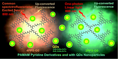
Organic dye two-photon absorption-induced frequency up-converted fluorescence (UCF) excited using a laser has been used in biological imaging and optical physics. The laser requires higher-level equipment that limits the application of UCF dyes. The experimental observation that the poly-amidoamine (PAMAM) pyridine derivatives 2PPS-G0 and 4PPS-G0 and those mixed with quantum dots (QDs) nanoparticles realized UCF using a common spectrofluorometer. The low-energy linear one-photon light source can also excite some organic molecules or QDs to give UCF emission, which replaces the need of a laser. The UCF related mechanism: second harmonic generation; two-photon absorption induced, and linear excited-states shifts (LESS) mechanisms were discussed to explain the linear one-photon light source excited UCF.
Organic dyes multi-photon (two-/three-photon) up-converted processes have been studied using laser pulses. In addition, the multi-photon excitation based laser scanning microscopy realized frequency-up-conversion imaging,10 which has been used in bio-imaging, chemical sensors, and optical physics.11
Semiconductor nanoparticles are known as quantum dots (QDs).12 QDs are characterized by large Stokes shifts, broad absorption bands, and narrow, size-dependent emission bands without a significant red tail. Multiple color QDs can be excited using a single laser excitation wavelength.13 The size-dependent emission of QDs is a result of the quantum confinement effect (QCE).14 The two-photon processes of QDs have been investigated using laser instruments.15
These experiments observed UCF emission from PAMAM pyridine organic dyes (2-pyridine poly-amidoamine Schiff-base generation 0) 2PPS-G0, and (4-pyridine poly-amidoamine Schiff-base generation 0) 4PPS-G0, and the ones mixed with quantum dots (QDs) nanoparticles (Scheme 1). The tests were carried out using a FluoroMax-4 spectrofluorometer, a common linear light source, which was excited at a wavelength of 800 nm. This experimental finding realized UCF using organic dyes and the ones mixed with QDs excited using a one-photon light source spectrofluorometer, without the need for a laser has the potential to extend the application of UCF materials. To date, there was no report on the UCF of organic dyes and the ones mixed with QDs using a common linear light source.
![]() | ||
Scheme 1 The structures of the dendrimers 2PPS-G0 and 4PPS-G0 added with CdS, CdSe, CdTe, ZnS, ZnSe and ZnTe quantum dots nanoparticles. |
Fig. 2 shows the UCF emission spectra of 2PPS-G0, 4PPS-G0, and those mixed with added CdS nanoparticles in ethanol ranging from 420 nm to 750 nm. The UCF peaks of 2PPS-G0 and 2PPS-G0 + CdS were at about 500 nm. The UCF peaks of 4PPS-G0 and 4PPS-G0 + CdS were at about 600 nm. The UCF of CdS has a sharp peak at about 533 nm. The UCF peaks of 2PPS-G0 + CdS and 4PPS-G0 + CdS also have sharp peaks at about 533 nm. But the UCF peaks of 2PPS-G0 and 4PPS-G0 have no peaks at about 533 nm. This means the peaks at 533 nm were characteristic for the CdS nanoparticles. Another phenomenon that can be noticed is that the UCF of 2PPS-G0 and 4PPS-G0 were enhanced after adding the CdS nanoparticles. These results show that the CdS nanoparticles have an interaction with the two dendrimers. Fig. 1 (CH2Cl2) and Fig. 2 (ethanol) were in two different solvents. The solvent effects make the spectra possess some difference in the observed emission wavelengths. The polarity, refractive index, and dielectric constant of the solvent all effect the dyes to give different optical physical properties.
2PPS-G0 and 4PPS-G0 have a poly-(amidoamine) (PAMAM) dendrimer16 core and pyridine triphenylamine surface groups. These two dendrimers have PAMAM flexible chains with many amines (tertiary amine, amide, aryl-amine, and imine), which can complex metal ions, package nanoparticles, or deliver drugs. The PAMAM shows emission fluorescence after storage or oxidation in air,17 which extends the application of these two dendrimers in fluorescence areas. The N atom of the pyridine forms ions under excitation, which are connected with triphenylamine by double bonds. The pyridine N ions attract electrons to be an “accepter” and the triphenylamine gives electrons to be a “donor”, which form a push–pull electronic conjugated dipole unit. This dipole unit displays intramolecular charge transfer (ICT) phenomenon, which can improve the non-linear optical properties (such as two-photon absorption) of the molecules.
Fig. 3 shows the up-converted fluorescence emission at an excitation wavelength of 800 nm using a common spectrofluorometer, which shows that the QDs gave UCF using a linear light source. Some quantum dots18 can give multi-photon absorption induced frequency up-converted emission. The observed experiment using QDs mixed with 2PPS-G0 and 4PPS-G0 can enhance UCF emission. The actions between QDs and the dyes in solution were a result of fluorescence resonance energy transfer (FRET).19 Fig. 3 shows that the QDs have an emission ranging from 300 nm to 500 nm. The UV spectra in the inset of Fig. 2 show that 2PPS-G0 and 4PPS-G0 have absorption at about 400 nm. The overlap of QDs emission and the absorbance of the two dendrimers show that there exist FRET phenomena in the QDs + dyes system. The energy transferred between the QDs nanoparticles and pyridine triphenylamine groups at close distances (about < 10 nm) in the solutions, which increased the UCF emission of the QDs + dyes system.
The fluorescence spectra of gradient concentrations of QDs (CdS, CdSe, CdTe, ZnS, ZnSe, ZnTe) added 2PPS-G0 and 4PPS-G0 was investigated (Fig. 4, S-Fig. 14, and S-Fig. 15†). The QDs added 2PPS-G0 and 4PPS-G0 gave an emission wavelength at 570 nm and 580 nm. The emission wavelength of 2PPS-G0 was at 570 nm, and that of 4PPS-G0 was at 580 nm. These two emission wavelengths were not the key emission peak areas of the QDs. Fig. 4 shows that the 2PPS-G0 and 4PPS-G0 fluorescence were enhanced after addition of the QDs. The emission wavelength changed little.
Fig. 5 shows the fluorescence lifetime and fluorescence decay curves, which show the fluorescence life time of 2PPS-G0 (2.40 ns) and 4PPS-G0 (3.19 ns), changed little after the addition of CdS (2.97 ns and 3.69 ns) and ZnS (2.40 ns and 2.38 ns) QDs. Fig. 4 shows the fluorescence peak positions maintained at about 570 nm (2PPS-G0) and 580 nm (4PPS-G0) upon addition of the QDs. Fig. 4 and 5 show that the fluorescence centers were not changed after addition of the QDs. It may be that the QDs have FRET on 2PPS-G0 and 4PPS-G0, and then enhance the fluorescence emission intensity of the two dendrimers.
![]() | ||
Fig. 5 The fluorescence lifetime (τ, unit nanoseconds) and fluorescence decay curves of 2PPS-G0 and 4PPS-G0 (2.5 × 10⁻⁶ mol L−1), and the ones mixed with CdS and ZnS (1 × 10⁻⁵ mol L−1 in CH2Cl2). |
Another interesting phenomenon is observed in Fig. 4. The fluorescence of 2PPS-G0 mixed with CdS, decreased with an increasing concentration of CdS (Fig. 4a). In addition, the fluorescence of 4PPS-G0 mixed with CdTe increased with an increasing concentration of CdTe (Fig. 4b). S-Fig. 14 and 15† show that 2PPS-G0 and 4PPS-G0 with different concentrations of QDs gave similar situations but not in a linear relationship. The reason why the two dendrimers mixed with the two types of QDs have inverse situations is not clear. These may be areas for further research.
Firstly, the UCF mechanism of QDs may be the second-harmonic generation. Fig. 6 shows the UCF spectra of 2PPS-G0 + CdS, 4PPS-G0 + CdS, and CdS excited at 800 nm (ranging from 200 nm to 850 nm). There were four main sharp peaks at 266 nm, 400 nm, 533 nm, and 800 nm. The 800 nm peaks were the scattering of the excited light source. The UCF emission peaks at 266 nm, 400 nm, and 533 nm were produced for the second-harmonic generation mechanism, which were initially for third-harmonic generation, second-harmonic generation, and one and half-harmonic generation, respectively. The second-harmonic generation UCF was the characteristic peak of the CdS quantum dots, which was also produced by the CdS mixed with the 2PPS-G0 and 4PPS-G0 dendrimers.
Secondly, the 2PPS-G0 and 4PPS-G0 dendrimers mixed with CdS also have UCF peaks at 500 nm and 600 nm. These peaks were the characteristic UCF of the 2PPS-G0 and 4PPS-G0, which were attributed to a two-photon induced UCF mechanism. The UV-vis absorbance spectra in Fig. 1b shows there was no linear absorbance at 800 nm, which means there exists a non-linear absorbance to produce the fluorescence emission under the excitation wavelength at 800 nm. It can be concluded that these two dendrimers excited at a wavelength of 800 nm produce a non-linear absorbance and gave UCF emission peaks at wavelengths from 500 nm to 600 nm. These phenomena were attributed to a two-photon absorption-induced UCF mechanism.
The two dendrimers mixed with CdS nanoparticles have UCF peaks at 266 nm, 400 nm, and 533 nm, which were attributed to the second-harmonic generation mechanism. Fig. 5 shows the UCF emission of the QDs + dyes system was a combination of a second-harmonic generation mechanism and two-photon absorption-induced mechanism.
Thirdly, the mechanism proposed for this linear light source excited up-converted fluorescence was a linear excited states shifts (LESS) up-converted fluorescence emission mechanism (Fig. 7). This mechanism shows that the 2PPS-G0 or 4PPS-G0 was excited under 400 nm to LUMO-x and gave a fluorescence emission at 500 nm or 600 nm (Fig. 2b). The 800 nm linear light source excited the molecules to LUMO-y, the LUMO-y shifts to LUMO-x, and then also can give a fluorescence emission at 500 nm or 600 nm (Fig. 2a). The excited states shifts may be triplet–triplet or triplet–singlet. The LESS mechanism can explain the UCF emission excited by a linear one-photon light source.
UCF dyes have potential and a broad range of applications.20 The experimental meaning of this finding was UCF can be investigated using a common linear spectrofluorometer without the need of laser instruments. Both this finding and these materials may improve low-intensity IR imaging, chemical sensors,21 biological imaging, biological fluorescence probes for cells, and drugs delivery trackers. The finding may further reduce the photo-damage found in optical dynamical therapy or cell imaging using a linear lower-energy excited light source. The related chemical groups will be designed and synthesized in other molecules and can extend, even improve the application values in UCF and multi-photon non-linear optical areas, which will cause interest.
The linear light excitation sources (800 nm) on the two dendrimer and QDs gave high energy up-converted fluorescence emission, which realized long wavelength excitation and short wavelength emission using a low energy light source without the need of a laser. CdS gave up-converted emission peaks at 266 nm, 400 nm, and 533 nm from third-harmonic generation, second-harmonic generation, and one and half-harmonic generation, respectively, which obey the second-harmonic generation mechanism. The up conversion spectra of 2PPS-G0 and 4PPS-G0 have emission peaks at 400 nm. This 400 nm emission was excited by a wavelength of 800 nm, which was double the frequency peak and can be explained by a two-photon absorption and linear excited states shifts (LESS) mechanism or a combination of them both. The LESS mechanism shows the 800 nm excited states shift to the 400 nm excited states, and then produce the up conversion emission. The up-conversion phenomena excited by a wavelength of 800 nm using a linear source was observed. These possible mechanisms may act on their own or as a combination.
The CdTe (ZnTe) quantum dots were prepared by jetting NaHTe CH2Cl2 solutions into Cd(OAc)2 and (Zn(OAc)2) CH2Cl2 solutions.
Footnote |
† Electronic supplementary information (ESI) available: The structures and synthesis of 2PPS-G0 and 4PPS-G0 were listed in S-Fig. 1 to S-Fig. 5. S-Fig. 6 to S-Fig. 8 was UV and fluorescence spectra. The S-Fig. 9 to S-Fig. 12 lists the 1H NMR and HRMS of 2PPS-G0 or 4PPS-G0. The MS of PAMAM is listed in S-Fig. 13, UCF of dyes + QDs in S-Fig. 14 and 15. See DOI: 10.1039/c4ra11149d |
This journal is © The Royal Society of Chemistry 2014 |