I. Jerković*a,
C. I. G. Tuberosob,
P. M. Kuśc,
Z. Marijanovićd and
M. Kranjaca
aDepartment of Organic Chemistry, Faculty of Chemistry and Technology, University of Split, N. Tesle 10/V, HR-21000 Split, Croatia. E-mail: igor@ktf-split.hr; Fax: +385 21 329 461
bDepartment of Life and Environmental Sciences, University of Cagliari, via Ospedale 72, IT-09124 Cagliari, Italy
cDepartment of Pharmacognosy, Wrocław Medical University, ul. Borowska 211a, PL-50-556 Wrocław, Poland
dDepartment of Food Technology, Marko Marulić Polytechnic in Knin, Petra Krešimira IV 30, HR-22300 Knin, Croatia
First published on 7th November 2014
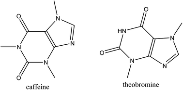
Coffea spp. honey was screened by UV/VIS, HS-SPME/GC-MS/FID, USE/GC-MS/FID and HPLC-DAD. The direct HPLC-DAD methodology overcame the major limitations of the other methods used. The obtained results constitute a breakthrough since dominant xanthine derivatives were found (theobromine and caffeine with HPLC-DAD and caffeine by USE/GC-MS/FID). Phenylacetaldehyde was the major headspace compound.
The presence of methyxanthine derivatives in the nectar and pollen of several plant species (Coffea, Camellia, Theobroma, Herrania, Cola, Ilex, Paullinia and Citrus spp.)4 suggests that honey bees may encounter methylxanthines when foraging. Methylxanthines content in the flowers and nectars of Citrus spp. was investigated in detail.4 Caffeine dominated in C. paradisi and C. maxima flowers (21 and 77 nmol g−1, respectively) accompanied by theophylline (4 and 15 nmol g−1, respectively), while theobromine and paraxanthine occurred in traces. Similarly, C. sinensis flowers contained caffeine (182 nmol g−1) and theophylline (46 nmol g−1). The nectar of C. paradise, C. maxima and C. limon contained caffeine (60–487 nmol mL−1), theobromine (0–22 nmol mL−1), theophylline (0–55 nmol mL−1) and paraxanthine (0–12 nmol mL−1). Caffeine was identified also in the nectar of three species of Coffea and it was more common in the nectar of C. canephora than in that of C. arabica or C. liberica.5 In addition, methylxanthines have been already reported in Citrus spp. honey.4,6–8 Free flying honey bees consumed saccharose solutions containing a range of caffeine concentrations similar to those found in citrus nectar (12.5–100 ppm, or 0.07–0.5 mM) and showed a preference for saccharose solutions containing 25 ppm (ca. 0.13 mM) caffeine over the solutions of saccharose alone.9 Caffeine exhibited no effect on acquisition, but increased long term memory retention in an appetitive visual learning task in the honey bees as well as increased the performance of bees in a delayed match to sample assay.10
In continuation of our ongoing investigation, the present research target is Coffea spp. honey. In general, available literature is missing data on Coffea spp. honey characterization. Therefore the goals of this study are: (a) detail screening of the samples by headspace solid-phase microextraction and ultrasonic solvent extraction followed by GC-MS/FID analyses with emphasis on headspace, volatile and semivolatile compounds; (b) targeted HPLC-DAD analysis of the samples with emphasis on methylxanthine derivatives, and (c) indication of the potential chemical markers of the honey botanical origin and pointing out the suitable methods for their identification.
Two representative samples of Coffea spp. honey of different geographical origin (Co1 and Co2) were investigated by complementary screening methodologies. Measured CIE L*a*b*C*abh°ab chromaticity coordinates correspond to medium dark colour with the parameters (L* = 67.3, a* = 10.5 and b* = 68.0) similar to heather honey.11 The investigated samples showed differences in L* values (Table 1) that could be caused by some aging of Co1 sample which was indicated by the presence of 5-hydroxymethylfurfural, Table 4. Total phenols content in Co1 and Co2 was high (622.7 and 688.1 mg GAE kg−1). It was similar to amount of phenolics found in heather honey (from 599 mg to 762 mg GAE kg−1).12 The antioxidant capacity of the samples was also determined: 0.5 mmol TEAC kg−1 (DPPH assay) and 2.7 or 2.8 mmol Fe2+ kg−1 (FRAP assay). The results are similar to those found for Dalmatian sage (0.4 TEAC kg−1, 2.5 mmol Fe2+ kg−1) and heather (0.6 TEAC kg−1, 2.1 mmol Fe2+ kg−1) honeys.13,11
Parameter | Co1 | Co2 |
---|---|---|
AV ± SD | AV ± SD | |
a Lightness.b Indicates red for positive value and green for negative value.c Indicates yellow for positive value and blue for negative value.d Chroma.e Hue, deg.f Expressed as mg kg−1 of gallic acid equivalent (GAE).g DPPH value is expressed as millimolar concentration of TEAC, obtained from a dilution of Trolox having an equivalent antiradical capacity as the honey solution.h FRAP value is expressed as millimolar concentration of Fe2+, obtained from a dilution of FeSO4 having an equivalent antioxidant capacity as the honey solution. | ||
L*a | 50.1 ± 0.0 | 72.0 ± 0.0 |
a*b | 9.4 ± 0.0 | 8.7 ± 0.0 |
b*c | 49.3 ± 0.0 | 61.5 ± 0.0 |
C*abd | 50.2 ± 0.0 | 62.1 ± 0.0 |
h°abe | 93.8 ± 0.0 | 81.9 ± 0.0 |
Total phenolsf (mg GAE kg−1) | 622.7 ± 38.0 | 688.1 ± 42.1 |
DPPHg (mmol TEAC kg−1) | 0.5 ± 0.0 | 0.5 ± 0.0 |
FRAPh (mmol Fe2+ kg−1) | 2.8 ± 0.1 | 2.7 ± 0.0 |
The applied chemical screening methodologies indicate striking differences in the obtained chemical profiles (particularly volatiles and methylxanthine derivatives distribution depending on the volatility/solubility and on the isolation/analysis protocols).
Phenylacetaldehyde was the major headspace compound in Co1 sample (36.6%) and also important in Co2 sample (13.5%), Table 2. However, it is not specific as the honey chemical marker.14 Minor percentages of other benzene derivatives were found only in Co1 (Table 2). Terpenes, particularly linalool derivatives, were also abundant in the samples: trans-linalool oxide (8.7; 20.5%), cis-linalool oxide (4.2; 12.3%) and hotrienol (4.0; 42.5%). Linalool oxide isomers were found with different headspace proportions in several honey types, e.g. Amorpha fruticosa L., oak honeydew or Thymus spp.15 3-Hydroxy-4-phenylbutan-2-one was found in both samples (dominating in Co1) and in a range of different honeys.14 α-Pinene was previously identified only in few honey types such as sunflower or Turkish pine honeydew.15
No. | Compound | RI | Area percentages (%) | |
---|---|---|---|---|
Co1 | Co2 | |||
a RI = retention indices on HP-5MS column, * = tentatively identified, ** = correct isomer not determined. | ||||
1 | Dimethyl sulfide | <900 | 0.3 | — |
2 | 2-Furancarboxaldehyde (furfural) | <900 | 5.0 | — |
3 | α-Pinene | 941 | 1.7 | 1.3 |
4 | Isobutyl isovalerate* | 947 | 6.0 | — |
5 | Benzaldehyde | 965 | 2.2 | 0.1 |
6 | β-Pinene | 984 | 2.8 | — |
7 | Benzyl alcohol | 1037 | 0.8 | — |
8 | Phenylacetaldehyde | 1048 | 36.6 | 13.5 |
9 | trans-β-Ocimene | 1054 | — | 1.8 |
10 | trans-Linalool oxide | 1076 | 8.7 | 20.5 |
11 | cis-Linalool oxide | 1091 | 4.2 | 12.3 |
12 | α-Terpinolene | 1094 | 1.0 | — |
13 | Nonanal | 1102 | 1.0 | 0.8 |
14 | Hotrienol | 1106 | 4.0 | 42.5 |
15 | 2-Phenylethanol | 1116 | 2.0 | 0.2 |
16 | 4-Ketoisophorone | 1147 | 1.2 | — |
17 | Nerol oxide | 1162 | 1.2 | — |
18 | Epoxylinalool** | 1178 | 2.7 | — |
19 | Decanal | 1207 | — | 1.6 |
20 | 3-Hydroxy-4-phenyl-butan-2-one | 1354 | 6.8 | 1.0 |
Total identified (%) | 88.2 | 95.6 |
Ultrasonic solvent extraction of Co1 and Co2 with solvent I and II revealed caffeine as predominant compound, Table 3. The percentage of caffeine was higher in solvent II (89.1%; 90.5%) than in solvent I (59.9%; 51.0%). Besides caffeine, small percentages of theobromine were found, Table 3. First of all, it should be pointed out that both methylxanthine derivatives were not identified in Coffea spp. headspace that is reasonable to explain with their lower volatility. Further comparison with the results of HPLC-DAD analysis also indicates striking differences. The explanation of these differences is triggered by the solubility distinction of methylxanthine derivatives. Namely, all xanthine derivatives except caffeine are known to exhibit poor water solubility in contrast to their parent molecule purine.1 With respect to lipophility, xanthine derivates have different properties.16 They dissolve slightly in organic liquids like chloroform. Thus they are hydrophilic and do not penetrate into organic phase of binary solvent. Caffeine shows higher lipophility than theobromine and therefore caffeine was extracted by USE, while theobromine was present in the extracts only at lower percentages. (Z)-Octadec-9-en-1-ol and octadecan-1-ol and other higher aliphatic compounds derived probably from the comb environment.17 A C13-norisoprenoid 4-hydroxy-3,5,6-trimethyl-4-(3-oxo-but-1-enyl)-cyclohex-2-en-1-one was found in the extracts (solvent I and II) of both samples with lower percentages (Table 3), but not in the headspace due to lower volatility.
No. | Compound | RI | Area percentages (%) solvent I | Area percentages (%) solvent II | ||
---|---|---|---|---|---|---|
Co1 | Co2 | Co1 | Co2 | |||
a RI = retention indices on HP-5MS column, * = tentatively identified. | ||||||
1 | 1,4-Xylene | <900 | 0.2 | 0.1 | — | — |
2 | Isobuthyl isovalerate* | 947 | 0.6 | — | 0.1 | — |
3 | Hotrienol | 1106 | — | 0.1 | — | — |
4 | 3,7-Dimethylocta-1,5-diene-3,7-diol (terpendiol I) | 1191 | 0.4 | — | 0.1 | — |
5 | 5-Hydroxymethylfurfural | 1230 | — | 0.1 | ||
6 | 3-Hydroxy-4-phenylbutan-2-one | 1354 | 4.3 | — | 1.2 | — |
7 | 3,5,5-Trimethyl-4-(3-oxo-but-1-enyl)-cyclohex-2-en-1-one (3-oxo-α-ionone) | 1663 | 2.3 | — | — | — |
8 | 4-Hydroxy-3,5,6-trimethyl-4-(3-oxo-but-1-enyl)-cyclohex-2-en-1-one | 1808 | 3.0 | 1.3 | 0.8 | 1.1 |
9 | Caffeine | 1856 | 59.9 | 51.0 | 89.1 | 90.5 |
10 | Hexadecan-1-ol | 1882 | 6.3 | 3.6 | — | — |
11 | Theobromine | 1925 | 1.1 | 1.2 | 2.9 | 2.2 |
12 | Hexadecanoic acid | 1963 | 0.2 | 1.1 | — | — |
13 | (Z)-Octadec-9-en-1-ol | 2060 | 12.3 | 25.1 | 3.3 | 3.3 |
14 | Octadecan-1-ol | 2084 | 4.0 | 7.7 | 1.0 | 1.1 |
Total identified (%) | 94.6 | 91.2 | 98.6 | 98.2 |
Targeted HPLC-DAD analysis revealed the presence of several relevant compounds, the most interesting being methylxanthine derivatives (Table 4). The most abundant was theobromine (93.11 mg kg−1; 85.30 mg kg−1) followed by caffeine (83.59 mg kg−1; 67.25 mg kg−1). Representative chromatograms are presented in Fig. 1. The caffeine presence was expected on the basis of USE/GC-MS analysis, while detected high quantity of theobromine was unexpected. In addition, traces of theophylline were detected. Small quantities of theobromine were previously reported by Kretschmar & Baumann4 in Citrus spp. honey ranging from 1–6 nmol g−1 and in the corresponding nectar (0–22 nmol mL−1). Therefore, theobromine can be pointed out as more specific chemical marker of Coffea spp. honey. Caffeine was previously found in Citrus spp. honey from various countries in small quantities in comparison with the results of Co1 and Co2. Caffeine is present in Citrus spp. honeys at about 1–10 mg kg−1.6 Later, similar findings regarding the presence of caffeine in citrus honey, flowers and nectar were reported by Vacca & Fenu (1996) and Vacca et al. (1997) with average caffeine concentrations in citrus and orange honeys of 1.790 and 4.930 mg kg−1 respectively.7,8 Kretschmar & Baumann (1999) reported caffeine concentrations in citrus honey between 0.039 and 6.00 mg kg−1.4 Caffeine was found in 80% of Palestinian multifloral honey samples from different geographic regions with concentrations ranged between 0.094 and 3.583 mg kg−1, with a mean value of 1.567 mg kg−1.18 The honey caffeine levels are usually much less than those in nectar. Some studies have reported that caffeine in nectar undergoes up to 90% reduction in fresh honey samples as compared with the nectar.4 All xanthines are products of purine metabolism. The final substance of this process is caffeine and it is formed in several steps. The first of them is conversion of purine nucleotide to xanthosine. There are at least four possible pathways for the production of xanthosine:1 purine nucleotide biosynthesis de novo, S-adenosyl-L-methionine cycle, adenine nucleotide pool and guanine nucleotide pool. Current evidence obtained from biochemical and molecular genetic studies using coffee leaves indicate that the major route of caffeine biosynthesis is xanthosine → 7-methylxanthosine → 7-methylxanthine → theobromine → caffeine pathway.19–21
RT | Compound | LOD | LOQ | Quantity (mg kg−1) | |
---|---|---|---|---|---|
Co1 | Co2 | ||||
AV ± SD | AV ± SD | ||||
a nd: not detected (below the limit of detection), tr: traces (below the limit of quantification). | |||||
5.2 | Kojic acid | 0.12 | 0.36 | 8.87 ± 0.53 | 20.30 ± 1.61 |
6.1 | Tyrosine | 0.09 | 0.27 | 359.43 ± 25.16 | 452.00 ± 28.64 |
7.0 | Phenylalanine | 0.07 | 0.22 | 363.49 ± 22.69 | 691.91 ± 50.04 |
7.3 | 5-Hydroxymethylfurfural | 0.04 | 0.12 | 7.95 ± 0.52 | nd |
8.4 | Theobromine | 0.08 | 0.25 | 93.11 ± 4.40 | 85.30 ± 3.88 |
9.5 | Theophylline | 0.10 | 0.29 | tr | 1.43 ± 0.09 |
11.6 | Caffeine | 0.07 | 0.21 | 83.59 ± 5.96 | 67.25 ± 4.71 |
20.4 | Lumichrome | 0.09 | 0.28 | nd | 22.42 ± 1.97 |
![]() | ||
Fig. 1 Representative HPLC-DAD chromatograms of the Coffea spp. honey samples solution (1![]() ![]() |
Among other quantified compounds by HPLC-DAD, kojic acid (8.87 mg kg−1 (Co1) and 20.30 mg kg−1 (Co2)) is also interesting for Coffea spp. honey characterization. Kojic acid (5-hydroxy-2-(hydroxymethyl)-4-pyrone) was identified in New Zealand manuka honey for the first time (0.13–125 mg kg−1) as one of the compounds useful for distinguishing manuka honey from other honey types.22 It is a degradation product of carbohydrates, mainly glucose, via gluconolactone and 3-ketogluconic acid lactone.22 Lumichrome was only found in Co2, and this can represent an index of an interfering nectar source (Table 4).
Targeted HPLC-DAD analysis also quantified tyrosine (359.43 mg kg−1 (Co1) and 452.0 mg kg−1 (Co2)) and phenylalanine 363.49 mg kg−1 (Co1) and 691.91 mg kg−1 (Co2). It can be seen that content of tyrosine was similar in both samples, while the content of phenylalanine was almost double in Co2. The variety of amino acids in different honey types occurring with different proportions allows their use as chemical markers of the honey botanical origin.23 For example phenylalanine and tyrosine were quantified in the honeys from rosemary (284 mg kg−1; 112 mg kg−1), eucalyptus (107 mg kg−1; 34.8 mg kg−1), lavender (18.1 mg kg−1; 299 mg kg−1), thyme (410 mg kg−1; 171 mg kg−1) and orange (122 mg kg−1; 31.5 mg kg−1).24 Since several amino acids are precursors of volatile organic compounds, attempts to find useful relationships between amino acid composition and characteristic honey aromas have been made.25 Lower amount of phenylalanine was accompanied by higher percentage of phenylacetaldehyde in Co1 which may support such hypothesis.
Representative samples of Coffea spp. honey were obtained in 2013. from the plantation of Coffea spp. plants from two geographic region: Guatemala (Co1) and Columbia (Co2). The measurements of CIE L*a*b*C*abh°ab chromatic coordinates were performed on undiluted, fluid and transparent honey samples in 10 mm optical polystyrene cuvettes (Kartell 01937) using an UV/VIS spectrophotometer Varian series Cary 50 Scan (Varian, Leinì, TO, Italy) and data were processed with Cary Win UV Colour Application V. 2.00 method.26 The total phenolic content was determined with a modified Folin–Ciocalteu method as reported by Kuś et al.,11 and the content was expressed as mg kg−1 of gallic acid equivalent (GAE), using a calibration curve of a freshly prepared GAE standard solution (10–200 mg L−1, r = 0.9998). The total antioxidant activity was evaluated using the ferric reducing antioxidant assay (FRAP).11 The quantitative analysis was performed according to the external standard method (FeSO4, 0.1–2 mmol, r = 0.9998), and the results were expressed as mmol kg−1 of Fe2+. The free radical scavenging activity was determined with the DPPH assay.11 A Trolox calibration curve in the range 0.02–0.50 mmol kg−1 was prepared (r = 0.9998), and data were expressed in Trolox equivalent antioxidant capacity (TEAC, mmol kg−1). For the three determinations, honey samples were diluted with H2O 1:
5 (w/v) and the absorbance was read on a 10 mm optical polystyrene cuvette using a Varian Cary 50 spectrophotometer, against a blank.
HS-SPME was performed in duplicate using the manual holder for SPME fibre covered with the layer of divinylbenzene/carboxene/polydimethylsiloxane/(DVB/CAR/PDMS) obtained from Supelco Co. (Bellefonte, PA, USA). Applied HS-SPME procedure was described in detail previously.27 USE was performed in duplicate in an ultrasonic bath (Transsonic Typ 310/H, Germany) by indirect sonication, at the frequency of 35 kHz at 25 ± 3 °C as described in detail previously.27 The mixture pentane–Et2O 1:
2 (v/v; solvent I) and dichloromethane (solvent II) were used for the extraction. The obtained extracts were concentrated up to 0.2 mL by careful distillation with Danish-Kuderna apparatus, and 1 μL was used for GC/FID and GC/MS analyses.
GC-FID analysis was carried out on an Agilent Technologies (Palo Alto, CA, USA) gas chromatograph model 7890A equipped with flame ionization detector. The chromatographic separations were performed on a 30 m capillary column HP-5MS (5% phenyl-methylpolysiloxane, Agilent J & W GC column) with coating thickness 0.25 mm. GC conditions were as follows: injected volume 1 μL with split ratio 1:
50; oven programmed at 70 °C for 2 min, then increased at a rate of 3 °C min−1 to 200 °C and held isothermal for 15 min; 250 °C injector temp.; 300 °C detector temp.; He carrier gas velocity 1 mL min−1. The GC-MS analyses were carried out with Agilent gas chromatograph model 7890A fitted with a mass-selective detector model 5975C (Agilent Technologies, Palo Alto, CA, USA). Mass detector conditions were set up as follows: electron impact (EI) ionization mode at 70 eV; the mass range m/z 30–300; ion source temp. was 280 °C. The compounds separation was achieved with the same conditions as for GC-FID. The individual peaks were identified by comparison of their retention indices (relative to C9–C25 n-alkanes) with those of available authentic samples and literature data28 and by comparing their mass spectra with Wiley 275 MS library (Wiley, New York, USA) and NIST08 (D-Gaithersburg) database. The percentage composition was calculated from the GC peak areas using the normalization method (without correction factors). All the analyses were performed in duplicate.
Detection and quantitative analyses were carried out using a HPLC-DAD method.13 An HPLC-DAD Varian system ProStar (Varian, Palo Alto, CA, USA) was employed, fitted with a pump module 230, an autosampler module 410, and a Thermo Separation diode array detector SpectroSystem UV 6000lp (Thermo Separation, San Jose, CA, USA). Separation was obtained with a Phenomenex Kinetex C18 column (150 × 4.60 mm, 5 μm, Chemtek Analitica, Casalecchio di Reno, I-Bologna) using 0.2 M H3PO4 (solvent A), and MeCN (solvent B) at a constant flow rate of 1.0 mL min−1, mixed in linear gradients as follows: t = 0 A:
B (100
:
0, v/v), reaching 90
:
10 (v/v) in 5 min, then 75
:
25 (v/v) in 15 min, 50
:
50 (v/v) in 10 min, 20
:
80 (v/v) in 10 min, and finally at 100% B until 50 min. The injection volume was 10 μL. Chromatograms were acquired at λ = 280 nm (5-hydroxymethylfurfural, kojic acid, methylxanthines, and lumichrome) and λ = 210 nm (tyrosine and phenylalanine), and spectra were elaborated with a ChromQuest V. 4.0 data system (ThermoQuest, Rodano, I-Milan). The honey sample was diluted with ultrapure H2O 1
:
10 (w/v), and then filtered through Econofilter RC membrane (0.45 μm, ∅ 25 mm, Agilent Technologies, I-Milan). Standard solutions were prepared in MeOH (5-hydroxymethylfurfural and kojic acid) or 0.1 M HCl–MeOH 50
:
50 (v/v) (tyrosine, phenylalanine and methylxanthines) and working standard solutions in ultrapure H2O. The method was validated in agreement with the International Conference on Harmonisation of Technical Requirements for Registration of Pharmaceuticals for Human Use (ICH) guidance note which describes validation of analytical methods13 by determining linearity, limits of detection (LOD) and limits of quantification (LOQ). Calibration curves were plotted according to the external standard method, correlating the peak area with the concentration, and the calibration curves were constructed by means of the least-squares method. This method allowed the simultaneous analysis of several polar compounds by direct injection in HPLC without any purification or derivatization procedure.
This journal is © The Royal Society of Chemistry 2014 |