Study of the post separation pH adjustment by a microchip for the analysis of aminoglycoside antibiotics

Xiangying Menga, Xingmei Suob, Beiyuan Fana, Yongliang Yuc and Yongsheng Ding*a
aCollege of Life Sciences, University of Chinese Academy of Sciences, 19A Yuquanlu Road, Beijing 100049, China. E-mail: dingysh@ucas.ac.cn; Fax: +86-10-88256079; Tel: +86-10-88256392
bSchool of Information Engineering, Minzu University of China, Beijing 100081, China
cSchool of Physics, University of Chinese Academy of Sciences, Beijing 100049, China

Received 17th September 2014 , Accepted 17th October 2014

First published on 17th October 2014


Abstract

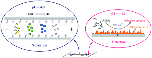
Recently, we developed a simple microchip to simultaneously accommodate acidic conditions for the separation and alkaline conditions for the electrochemical detection of aminoglycoside antibiotics [Electrophoresis, DOI: 10.1002/elps.201200309]. With two branch channels connected near the end of the separation channel, the alkaline solution was hydrostatically introduced into a Z-shaped mixing channel and combined with the acidic stream from the separation channel. As a result, the pH of the mixed solution was adjusted to the desired value for the electrochemical detection of aminoglycoside antibiotics. In this manuscript, the principle and related parameters of the pH adjustment on the microchip were investigated both in theory and practice. With the guidance of the principle of the post separation pH adjustment, we applied the functional microchip to the analysis of six aminoglycoside antibiotics in a biological sample with satisfactory analytical performances. Specifically, these compounds were electrophoretically separated in 5 mM sodium acetate (pH = 4) and 0.6 mM CTAB, and through the post separation pH adjustment, electrochemically detected in alkaline conditions (pH > 12) at a Cu–Sn–Cr alloy electrode. Additionally, this microchip may provide a possible use for the post separation reagent addition for enzyme-assisted electrochemical detection.


Introduction

Aminoglycosides (AMGs) are effective drugs that are widely used in therapy against bacterial infections, but their clinical use has been restricted due to toxic side effects, especially to the kidneys and the ear. On the other hand, their low cost means that they are often widely used in animal husbandry leading to potential residues in the food chain, which probably increase bacterial resistance against AMG antibiotics in the human body. Since these compounds are characterized by two or more amino sugars linked by glycosidic bonds to an aminocyclitol component, in general, they are short of chromophores and show high polarities. Due to these reasons, the detection of AMGs using standard HPLC or GC instrumentation generally requires pre- or post-column derivatization.1–5 Besides these derivatization methods, electrochemical detection is usually coupled with chromatographic techniques for the determination of AMGs without derivatization. However, in most cases, the use of electrochemical detection for the analysis of carbohydrates requires strongly alkaline conditions (pH > 12), which are unfavorable to the separation of AMGs by CE or non-ion exchange column chromatography.6–9 Although methods based on LC-MS/MS are the most powerful for the screening and confirmation of AMGs, they require a high cost to operate and highly qualified personnel.10–13 Consequently, the necessity still exists for rapid, facile, and low cost screening methods both in the laboratory and at on-site locations. Up to now, a few methods of screening AMGs have been reported with relatively high sensitivity, such as surface plasmon resonance (SPR)-based biosensors14 and aptamer-based sensors.15,16 However, most of them depend on costly antibodies or RNA aptamers without the separation of multiple drugs.

Microfluidic technology has evolved over the past two decades, accompanying a wealth of inventions and applications which have extensively attracted attention from a variety of scientific communities. The recent achievements and applications of microfluidic technology have been well documented in several review articles.17–20 Distinctively, a microchip coupled with electrochemical detection shows several merits, such as the miniaturization of analytical systems and the simplicity of use. Recently, we have developed a simple microfluidic chip containing two branch channels connected to a separation channel near the detection point, in which the alkaline solutions mix with the acidic stream toward an alloy modified electrode for the electrochemical detection of AMGs.21 To take better advantage of this microchip, here we did further studies on the post separation pH adjustment both in theory and practice. In addition, the electroplating conditions of the alloy electrode were re-investigated to improve the stability of the electrode. The experiments show that the microchip with the re-modified electrode can be kept in use for more than a week. The applicability of the method has been demonstrated by analyzing six aminoglycoside antibiotics in a bovine serum sample. Such a straightforward approach is a prototype of the “post-column” addition based on the microfluidic platform and also properly applied to other post separation adjustments or reactions with various reagents (e.g. enzymes) to accommodate separation and detection in the different conditions.

Experimental section

Chemicals and solutions

SU-8 2035 photoresist and SU-8 developer were purchased from MicroChem (Newton, MA, USA). Sylgard 184 silicone elastomer and the curing agent were obtained from Dow Corning (Midland, MI, USA). Cetyltrimethylammonium bromide (CTAB) was purchased from Sigma-Aldrich (Shanghai, China). Analytical grade sodium acetate, copper sulfate, tin chloride, chromium chloride, acetic acid, sulfuric acid, hydrochloric acid, and sodium hydroxide were obtained from Beijing Chemical Reagent Company (Beijing, China) and used without further purification. The six AMG standards, spectinomycin (SPE), streptomycin (STR), amikacin (AMI), kanamycin A (KAN A), paromomycin (PAR), and neomycin (NEO), were obtained from the National Institutes for Food and Drug Control (China). Running buffers were prepared by diluting stock solutions of sodium acetate and CTAB with deionized water to the desired concentrations. The pH of the buffer solution was adjusted by small additions of either 0.1 M NaOH or 0.1 M HCl. Stock solutions of the AMG antibiotics were prepared in distilled water and diluted with the running buffers. All aqueous solutions were prepared using 18.2 MΩ cm resistance water (Elga Labwater, UK) and were stored at 4 °C when not in use.

Fabrication of the microfluidic chip

The fabrication of the microfluidic chips was performed as described elsewhere.22,23 Briefly, a 100 mm diameter silicon wafer (Tianjin Semiconductor Institute of Technology, Tianjin, China) was cleaned with piranha solution (3 parts of 98% H2SO4 with 1 part of 30% H2O2; CAUTION: piranha solution is a powerful oxidizing agent that reacts violently with organic compounds and should be handled with extreme care) and thoroughly rinsed with deionized water. The wafer was then dried at 200 °C for 30 min. Next, the wafer was coated with SU-8 2035 negative photoresist using a spin coater (Laurell Technologies, PA, USA) by dispensing approximately 3 mL of the photoresist onto the wafer. A spread cycle of 500 rpm for 10 s followed by 1500 rpm for 30 s was performed, followed by two pre-exposure baking steps at 65 and 95 °C for 5 and 30 min, respectively. A digitally produced mask containing the channel pattern was placed on the coated wafer, exposed to light via a near-UV flood source (Optical Associates Inc., CA, USA) for 30 s and then subjected to a post exposure baking sequence. A positive relief was developed by placing the wafer in SU-8 developer (propylene glycol methyl ether acetate) for 15 min, rinsing with isopropanol and drying under a nitrogen stream. The height of the positive patterns on the molding master is equal to the channel depth created in the PDMS layer. It was measured roughly by cutting a piece of PDMS with the cross section of the channel under a microscope with a microscopic length scale. Two PDMS layers were fabricated by pouring a degassed mixture of Sylgard 184 silicone elastomer and a curing agent (10[thin space (1/6-em)]:[thin space (1/6-em)]1) onto either a molding master or a blank wafer, followed by curing at 65 °C for at least 2 h. The cured PDMS was separated from the mold and reservoirs were made at the end of each channel using a 5 mm circular punch. A platinum wire coated with a Cu–Sn–Cr alloy was then aligned at the end of the separation channel in a perpendicular channel designed for the working electrode. Next, the two PDMS layers were placed in a plasma cleaner (Harrick Plasma, NY, USA), oxidized for 30 s and immediately brought into conformal contact to form an irreversible seal. The extremities of the electrode channel were sealed with two drops of super glue. Finally, an electrical connection to the working electrode was made using silver paint (SPI Supplies, PA, USA) and a copper wire. A schematic drawing of the microfluidic chip is illustrated in Fig. 1.
image file: c4ra10597d-f1.tif
Fig. 1 The scheme and photograph of the microfluidic chip. Channels: 50 μm wide, 50 μm deep. Injector (double-T) volume: 1.2 nL, separation channel: 57 mm, total length of Z-shaped mixing channel: 2 mm, each double-T arm: 5 mm, each branch-like channel: 8 mm. Solution reservoirs: RB, running buffer reservoir; S, sample reservoir; SW, sample waste reservoir; BW, buffer waste reservoir; A1/A2, alkaline reservoirs.

Instrumentation

A high voltage sequencer (LabSmith Inc., CA, USA) with an adjustable voltage range of −3000 to +3000 V, was used for all of the electrophoresis experiments. Before the experiments, the microchannels were preconditioned by sequentially rinsing them with 0.1 M NaOH, deionized water and a running buffer. The microfluidic chip and its operation are described in detail as follows. The separation channel was 57 mm long. A double-T injector, with a 500 μm gap between the side channels and defining a 1.2 nL sample plug, was used for all experiments. The amount of buffer dispensed in each reservoir (RB, S, SW and BW) was 60 μL, except for two alkaline solution reservoirs (A1 and A2) in which 120 μL of 0.3 M NaOH were dispensed in order to hydrostatically push the alkaline solution into the mixing channel. The solution in each reservoir was refreshed after 5 to 6 run times to avoid evaporation. The 2 mm length mixing channel was designed as Z-shaped to enhance the mixing of the solution. During the sample injection, the potentials of −400 V and +100 V were applied to the reservoirs S and SW, respectively, while the reservoir RB was floating. During the separation step, −1000 V was applied to the reservoir RB at mean time, while the reservoirs S and SW were floating. During the above proceedings, the waste reservoir (BW) was always grounded. The present design allows the isolation of the detector from the separation current through the end-column configuration. Electrical connections were made to the microfluidic devices with platinum electrodes placed into the reservoirs at the end of each channel (except for the reservoirs A1 and A2). Electrochemical experiments were performed with a CHI 810C (Chenghua Instruments Co., Shanghai, China), using a three-electrode setup. An Ag/AgCl (3.0 M KCl) reference electrode and a platinum wire were used as the reference and auxiliary electrodes, respectively. A 5 cm length platinum wire (25 μm diameter) was used as the substrate electrode (effective length = 3 cm), on which the electrodeposition was carried out at −0.3 V (vs. Ag/AgCl) for 150 s in an electroplating bath containing 50 mM CuSO4, 0.2 mM SnCl2, 1.0 mM CrCl3, 2.0 mM EDTA and 10 mM H2SO4. The coated electrodes were also applied for cyclic voltammetry and amperometric it curve experiments.

Sample preparation

0.5 mL of bovine serum was spiked with AMG antibiotics at three concentration levels (20 μM, 50 μM and 100 μM), vortexed with 0.5 mL TCA (20% (w/v)), ultrasonicated for 10 min, and then centrifuged at 12[thin space (1/6-em)]000 rpm for 10 min. After removing the protein sediment, the supernatant was transferred to another centrifuge tube, and then 10 mM EDTA was added. The extraction of AMGs from the solution was conducted using a solid phase extraction (SPE) column (Anpelclean MCX, 60 mg, Shanghai, China). The SPE procedure is detailed as follows: loading the solution on the preconditioned column at a flow rate of 0.2 mL min−1, then washing with water until the effluent was neutral, finally eluting with 6 mL of 10% (v/v) ammonia in methanol at a flow rate of 0.2 mL min−1. The collected eluent was evaporated under a nitrogen stream in a water bath at 60 °C and the residue was reconstituted in 0.5 mL of the running buffer for injection.

Results and discussion

Theoretical description of the post separation adjustment of the pH

To better understand the behavior of the post separation adjustment of the pH based on our designed microchip, the theoretical study is described below. For a laminar flow governed by the Poiseuille equation, the volumetric flow rate is proportional to the pressure difference and the fourth power of the radius of the channel, and inversely proportional to the viscosity and the length of the channel. The equation is given as follows:
 
Q = πΔPr4/(8μL) (1)
where Q is the volumetric flow rate; ΔP is the pressure drop; L is the length of the channel; μ is the viscosity of the fluid; r is the radius of the channel; and π is the mathematical constant Pi. During the separation step, the pH of the mixed solution arriving at the detection electrode depends on the amount of alkaline solution flowing into the Z-shaped mixing channel, which is controlled by the pressure drop between the reservoirs A1/A2 and BW. In this microchip, the pressure drop is deduced from the difference between the heights of the solutions in the reservoirs A1/A2 and BW.
 
ΔP = ρg(hAhBW) (2)
where ρ is the density of water, g is the gravitational constant, and hA and hBW are the heights of the solutions in the reservoirs A1/A2 and BW.

For a cylindrical reservoir of the microfluidic chip,

 
h = VR2 (3)
where h is the height of the solution in the reservoir, V is the volume of the solution in the reservoir, and R is the radius of the reservoir.

Combining eqn (1)–(3), the Poiseuille equation governing the flow rate of the solution in the branch channels of the microchip can be written as follows:

 
Qb = ρg(VAVBW)r4/(8μLR2) (4)
where Qb is the volumetric flow rate in the branch channel, and VA and VBW are the volumes of the solutions in the reservoirs A1/A2 and BW, respectively. For the post separation adjustment of the pH in this experiment, the alkaline solutions in the reservoirs A1 and A2 start to flow into the mixing channel when the pressure drop is formed between the reservoirs A and BW (VAVBW > 0). In order to sustain an appropriate and steady pH value for the electrochemical detection, a hydrodynamic equilibrium is required to form in the mixing channel, where an active electroosmotic flow (EOF) from the upstream separation channel combines two hydrostatically driven flows from the branch channels toward the detection point. With thorough mixing, the pH of the mixed solution at the detection point can be calculated by the following equation:
 
pH = 14 + lg[2CNaOHQb/(Qm + 2Qb)] (5)
where Qm is the volumetric flow rate of the EOF in the upstream separation channel and CNaOH is the concentration of NaOH applied to the reservoirs A1 and A2. Because the volumetric flow rate from the separation channel is determined by the EOF, Qm can be obtained from the following equation:
 
Qm = πr2veof (6)
where veof is the velocity of the EOF. In this experiment, the actual value of the EOF is about 1.0 mm s−1 at −1000 V separation potential.

Combining eqn (4)–(6), the pH of the mixed solution at the end of the separation channel can be approximately calculated according to the volume difference between the reservoirs A and BW and the concentration of NaOH in the reservoir A. At a certain velocity of the EOF and a given concentration of NaOH, a series of theoretical curves between the pH and the volume difference (VAVBW) are plotted in Fig. 2A. All of the plots show the same tendency for the pH of the mixed solution to increase steeply as the volume difference (VAVBW) initially increases from 2.5 to 25 μL; then the increment of the pH becomes relatively flat when the volume difference is larger than 25 μL. However, when a larger volume difference is applied, the larger dilution percent formed is likely to decrease the response of the analyte. The dilution percent can be defined by the following formula:

 
Dilu.% = 100 × Qm/(Qm + 2Qb) (7)


image file: c4ra10597d-f2.tif
Fig. 2 The plots (A) of the pH of the mixed solution vs. the volume difference (lower x-axis) and the dilution percent of the analytes (upper x-axis); a computational simulation of the pH distribution in the mixing channel (B).

Additionally, we apply the Comsol 3.2 (student version) software to simulate the pH distribution in the mixing channel and virtually estimate the pH of the solution surrounding the detection point. Fig. 2B shows that the designed microchip can generate a high pH near the detection point under the given conditions of Qb = 0.1 × Qm and CNaOH = 0.1 M.

Experimental verification of the post separation adjustment of the pH

With guidance from the above theoretical description, the effects of the different concentrations of NaOH in the reservoir A (1/2) and the volume difference of the solution (VA–BW) on the response of the AMGs were investigated. Fig. 3A shows the electrochemical responses of the analytes under the different concentrations of NaOH at a constant volume difference (60 μL). Compared to 0.1 M and 0.5 M NaOH, the peak currents of the analytes were largest in 0.3 M NaOH. This was in part consistent with the higher concentration of NaOH leading to a higher pH to facilitate the electrochemical oxidization of the AMG. On the other hand, the peak currents of the AMGs in 0.5 M NaOH were smaller than those in 0.3 M NaOH. This can be explained by the fact that the excessively high pH of 13 of the 0.5 M NaOH results in a high background current that diminishes the peak currents of the AMGs. Thus, 0.3 M NaOH was selected in the following experiments. In order to obtain larger and more stable peak currents of the analytes, the percent of dilution and optimal pH need to be balanced at a certain concentration of NaOH. For the given concentration of NaOH, the choice of a larger volume difference means that more alkaline solution flows into the mixing channel resulting in a larger percent of dilution and higher pH, whereas applying a smaller volume difference results in a smaller pH and percent of dilution. The effect of the volume difference in 0.3 M NaOH on the electrochemical response of the AMGs is given in Fig. 3B. At a volume difference of 60 μL, the peak currents were obviously largest. This phenomenon is well in accordance with a larger volume difference resulting in a larger dilution percent, and a smaller volume difference resulting in a lower pH. Finally, 60 μL was selected as the optimal volume difference in the following experiments. The above experimental results show clear evidence for the theoretical description.
image file: c4ra10597d-f3.tif
Fig. 3 Electropherograms of the AMGs under post separation pH adjustment with different concentrations of NaOH at V(A–BW) = 60 μL (A) and with different V(A–BW) at 0.3 M NaOH (B). Other conditions: running buffer = 5 mM sodium acetate (pH = 4) with 0.6 mM CTAB, separation voltage = −1000 V, injection at −100 V/+400 V for 10 s, detection potential = 0.65 V. The peaks of the analytes (50 μM each): 1 = SPE, 2 = STR, 3 = AMI, 4 = KAN A, 5 = PAR, 6 = NEO.

Electrochemical characteristics of the alloy working electrode

Electrochemical detections of carbohydrates have extensively been studied at different metal electrodes. Conclusively, pulsed electrochemical detection (PED) needs to be applied on noble metal electrodes (e.g. Au and Pt),24,25 while constant potential amperometry needs to be applied on copper or nickel electrodes.26,27 However, both detection modes need a metallic oxide catalyst under strongly alkaline conditions (generally pH > 12). In our preliminary study, both gold and copper wires were adopted as the working electrodes for PED and constant potential amperometry, respectively. However, unavoidable hindrances were incurred during the application of the detection potential on these electrodes, such as low sensitivity, bubble formation on the gold electrode or severe corrosion to the copper electrode, etc. To overcome these problems, platinum wire was selected as a substrate, on which the multiple metal elements (Cu–Sn–Cr) were co-electrodeposited to form distinctive nanostructures with a large surface-to-volume ratio (Fig. 4A) and show corrosion-resistant characteristics (at least for one week of use). The electrochemical behavior of the electrodeposited wire was characterized by conducting cyclic voltammetry (CV) and amperometric it experiments. As can be observed in Fig. 4B, the CV data indicate that an oxidative peak of AMI on the modified electrode came out when the pH increased from 11 to 13. More details about the catalytically electrochemical oxidation of the AMGs at a copper based electrode were discussed in our previous work.21 On the other hand, the amperometric it data in Fig. 4C exhibit that the alloy modified electrode provides an obviously large sensitivity compared to the copper and platinum electrodes. The improvement of the sensitivity of the modified electrode is mainly due to the significant nanostructures formed on the alloy electrode.
image file: c4ra10597d-f4.tif
Fig. 4 Characteristics of the alloy modified electrode: SEM of the Cu–Sn–Cr alloy coated Pt wire (A); cyclic voltammograms (B) of AMI (50 μM) with a scan rate of 100 mV s−1 under the pH 11 (a), 12 (b), and 13 (c); amperometric it curves (C) of AMI (50 μM each step) under pH 13 at the platinum (a), copper (b), and Cu–Sn–Cr alloy modified (c) electrodes. Electrolyte: 5 mM sodium acetate with 0.6 mM CTAB.

The separation of the AMGs

The electrophoretic separation of the selected AMGs is likely to be obtained under an acidic running buffer due to the protonation of their amino groups. However, the dissociation of silanols on the inner surface of the PDMS microchannel will be suppressed under acidic conditions, thus the EOF becomes near to zero. To improve the separation, CTAB is chosen to modify the surface of the separation channel through the formation of a positively charged layer on the surface to reverse the direction of the EOF. Under the anodic mode, the reversed EOF is opposite to the direction of the electrophoretic mobility of the positively charged analytes, leading to the relatively long migration time that is of benefit to the separation of the AMGs. As can be seen in Fig. 5A, the reversed EOF became constant when the CTAB concentrations were larger than 0.6 mM because of the saturation of the CTAB absorption on the surface. The separation order of the selected AMGs depends on the number of amino groups in the molecule. A longer migration time corresponds to more amino groups while a shorter migration time is related to less amino groups (the molecular structures of the six AMGs are shown in Fig. S1 of the ESI). The effect of the pH of the buffer on the separation depends on the degree of protonation of the amino groups (shown in Fig. 5B); when the pH was 5.0, the three peaks of the AMGs (AMI, KAN A, and PAR) merged into a single peak. In contrast, when decreasing the pH to 4.0, the baseline separation of the six AMGs was achieved within 5 min. Thus pH = 4.0 was selected as the optimal pH of the running buffer in this experiment. Generally, a higher separation voltage produces a larger EOF that makes the migration time shorter and the peak height larger. It is worth noting in Fig. 5C that the peak currents of the AMGs became smaller with an increasing separation voltage; on the other hand, the noise became obviously large at −800 V separation voltage. These phenomena can be explained by the fact that higher separation voltages (e.g. −1400 V) increased the EOF and caused the larger volume of acidic buffer flowing into the mixing channel to decrease the pH of the mixed solution resulting in the smaller peak currents, whereas a lower separation voltage (e.g. −800 V) decreased the EOF and caused the larger volume of alkaline solution to flow from the branch channels into the mixing channel and resulted in the greater amount of noise at the higher pH condition. Finally, −1000 V was selected as the optimal separation voltage. The effect of the concentrations of sodium acetate between 2.0 and 10 mM on the separation and detection was not obvious (the data are not shown here). However, when the concentration of sodium acetate is larger than 10 mM, bubbles are formed in the separation channel and the larger buffer capacity requires the more alkaline solution for pH adjustment. Thus, the concentration of sodium acetate was selected as 5 mM.
image file: c4ra10597d-f5.tif
Fig. 5 Electropherograms of the six AMGs under different separation conditions: CTAB (A); pH (B); separation voltage (C). Other conditions were the same as in Fig. 3. The peaks of the analytes (50 μM each): 1 = SPE, 2 = STR, 3 = AMI, 4 = KAN A, 5 = PAR, 6 = NEO.

Analytical performance

Under the optimal conditions (combining 5.0 mM sodium acetate, pH = 4.0 and 0.6 mM CTAB as the running buffer, −1000 V as the separation voltage, 60 μL as the volume difference between the reservoirs A and BW, 10 s as the injection time, and 0.65 V as the detection potential), the response of the detector was analyzed as a function of the concentration for the six AMGs. Linear relationships between the concentration and the peak current were obtained for SPE, STR, AMI, KAN A, PAR, and NEO in the range 1.0 to 100 μM with R2 > 0.99. The limits of detection were between 0.2 to 2.0 μM at a signal-to-noise ratio of 3. In order to demonstrate the stability of the modified electrode, repetitive injections (n = 30) were performed. The RSDs in the peak current and migration time were less than 6.2% and 3.0%, respectively. The above analytical parameters (linear range, slope, correlation coefficient, LODs, and stability) for each analyte were summarized in Table S1 of the ESI. To further evaluate the accuracy and performance of the method, recovery experiments under the optimum conditions were conducted with the six AMGs spiked in serum samples at three concentration levels of 20 μM, 50 μM and 100 μM. As can be seen in Fig. 6, a standard mixture of the AMGs (curve a), a spiked serum sample (curve b), and a blank serum sample extract (curve c) were obtained under the optimum conditions. The average recoveries (n = 3) were 84.2 ± 4.1%, 68.2 ± 5.1%, 73.6 ± 3.9%, 88.9 ± 4.4%, 86.2 ± 4.2% and 79.6 ± 4.9% for SPE, STR, AMI, KAN A, PAR, and NEO, respectively.
image file: c4ra10597d-f6.tif
Fig. 6 Electropherograms corresponding to a mixture of standards (a), a spiking bovine serum sample (b), and a blank bovine serum sample (c). Other conditions were the same as in Fig. 3. The peak of the analytes (50 μM each): 1 = SPE, 2 = STR, 3 = AMI, 4 = KAN A, 5 = PAR, 6 = NEO.

Conclusions

This manuscript described in detail the post separation pH adjustment on a microchip and its utility in the analysis of AMGs. Specifically, the present methodology adjusted the pH after the separation of the AMGs under the acidic buffer and then applied the electrochemical detection of the AMGs under alkaline conditions. With the optimized conditions, the six AMGs were baseline separated within 5 min and detected at the relatively low micromolar level. The method offers a simple, sensitive, and portable method for the analysis of AMGs in a biological sample without derivatization. Additionally, this type of microchip provides a simple use for the post separation reagent addition for other applications.

Conflict of interest

The authors declare no competing financial interest.

Acknowledgements

This project was supported by National Natural Sciences Foundation of China (21075135) and the Chinese Academy of Sciences (no. KSCX2-EW-J-29).

References

  1. M. Zhou, G. Wei, Y. Liu, Y. Sun, S. Xiao, L. Lu, C. Liu and D. Zhong, J. Chromatogr. B: Anal. Technol. Biomed. Life Sci., 2003, 798, 43–48 CrossRef CAS PubMed.
  2. P. Vinas, N. Balsalobre and M. Hernandez-Cordoba, Talanta, 2007, 72, 808–812 CrossRef CAS PubMed.
  3. C. Z. Yu, Y. Z. He, G. N. Fu, H. Y. Xie and W. E. Gan, J. Chromatogr. B: Anal. Technol. Biomed. Life Sci., 2009, 877, 333–338 CrossRef CAS PubMed.
  4. D. A. Stead, J. Chromatogr. A, 2000, 747, 69–93 CrossRef CAS.
  5. T. A. McGlinchey, P. A. Rafter, F. Regan and G. P. McMahon, Anal. Chim. Acta, 2008, 624, 1–15 CrossRef CAS PubMed.
  6. P. D. Voegel and R. P. Baldwin, Electroanalysis, 1997, 9, 1145–1151 CrossRef CAS.
  7. W. C. Yang, A. M. Yu and H. Y. Chen, J. Chromatogr. A, 2001, 905, 309–318 CrossRef CAS.
  8. G. Brajanoski, J. Hoogmartens, K. Allegaert and E. Adams, J. Chromatogr. B: Anal. Technol. Biomed. Life Sci., 2008, 867, 149–152 CrossRef CAS PubMed.
  9. V. P. Hanko, J. S. Rohrer, H. H. Liu, C. Zheng, S. Zhang, X. Liu and X. Tang, J. Pharm. Biomed. Anal., 2008, 47, 828–833 CrossRef CAS PubMed.
  10. D. A. Bohm, C. S. Stachel and P. Gowik, Anal. Chim. Acta, 2010, 672, 103–106 CrossRef CAS PubMed.
  11. C. Chiaochan, U. Koesukwiwat, S. Yudthavorasit and N. Leepipatpiboon, Anal. Chim. Acta, 2010, 682, 117–129 CrossRef CAS PubMed.
  12. A. Kaufmann, P. Butcher and K. Maden, Anal. Chim. Acta, 2012, 711, 46–53 CrossRef CAS PubMed.
  13. F. L. van Holthoon, M. L. Essers, P. J. Mulder, S. L. Stead, M. Caldow, H. M. Ashwin and M. Sharman, Anal. Chim. Acta, 2009, 637, 135–143 CrossRef CAS PubMed.
  14. S. Rebe Raz, M. G. E. G. Bremer, W. Haasnoot and W. Norde, Anal. Chem., 2009, 81, 7743–7749 CrossRef CAS PubMed.
  15. A. A. Rowe, E. A. Miller and K. W. Plaxco, Anal. Chem., 2010, 82, 7090–7095 CrossRef CAS PubMed.
  16. N. Derbyshire, S. J. White, D. H. J. Bunka, L. Song, S. Stead, J. Tarbin, M. Sharman, D. Zhou and P. G. Stockley, Anal. Chem., 2012, 84, 6595–6602 CrossRef CAS PubMed.
  17. D. Gao, H. Liu, Y. Jiang and J.-M. Lin, Lab Chip, 2013, 13, 3309–3322 RSC.
  18. E. Primiceri, M. S. Chiriaco, R. Rinaldi and G. Maruccio, Lab Chip, 2013, 13, 3789–3802 RSC.
  19. B.-B. Xu, Y.-L. Zhang, H. Xia, W.-F. Dong, H. Ding and H.-B. Sun, Lab Chip, 2013, 13, 1677–1690 RSC.
  20. L. Yu, S. R. Ng, Y. Xu, H. Dong, Y. J. Wang and C. M. Li, Lab Chip, 2013, 13, 3163–3182 RSC.
  21. Y. Ding, L. Bai, X. Suo and X. Meng, Electrophoresis, 2012, 33, 3245–3253 CrossRef CAS PubMed.
  22. C. D. Garcia and C. S. Henry, Anal. Chem., 2003, 75, 4778–4783 CrossRef CAS.
  23. Y. Ding, A. Ayon and C. D. Garcia, Anal. Chim. Acta, 2007, 584, 244–251 CrossRef CAS PubMed.
  24. G. Brajanoski, J. Hoogmartens, K. Allegaert and E. Adams, J. Chromatogr. B: Anal. Technol. Biomed. Life Sci., 2008, 867, 149–152 CrossRef CAS PubMed.
  25. V. Manyanga, K. Kreft, B. Divjak, J. Hoogmartens and E. Adams, J. Chromatogr. A, 2008, 1189, 347–354 CrossRef CAS PubMed.
  26. F. Cao, S. Guo, H. Ma, G. Yang, S. Yang and J. Gong, Talanta, 2011, 86, 214–220 CrossRef CAS PubMed.
  27. X. Zhang, A. Gu, G. Wang, Y. Huang, H. Ji and B. Fang, Analyst, 2011, 136, 5175–5180 RSC.

Footnote

Electronic supplementary information (ESI) available. See DOI: 10.1039/c4ra10597d

This journal is © The Royal Society of Chemistry 2014
Click here to see how this site uses Cookies. View our privacy policy here.