DOI:
10.1039/C4RA10487K
(Paper)
RSC Adv., 2014,
4, 57505-57521
Organotin(IV) carboxylate derivatives as a new addition to anticancer and antileishmanial agents: design, physicochemical characterization and interaction with Salmon sperm DNA†
Received
15th September 2014
, Accepted 22nd October 2014
First published on 23rd October 2014
Abstract
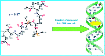
This article demonstrates the synthesis and characterization of novel organotin(IV) carboxylate complexes and their medicinal applications. Metal complexes and organometallic compounds have growing importance in medicine, particularly in oncology. Organotin derivatives have caught much attention during the last two decades for their potential biocidal activities. In recent years several organotin compounds have been synthesized, some with interesting cytotoxic properties. Here we will focus on the relevance of organotin derivatives as very promising potential candidates in anticancer therapy. Ten new organotin(IV) complexes were synthesized and characterized by using FT-IR, NMR (1H, 13C and 119Sn), elemental analysis, mass spectrometry and single crystal X-ray techniques. The results of infrared spectroscopy of the sodium salt and complexes showed that the coordination took place through the oxygen atoms of the carboxylate group. Crystallographic data for complexes 1 and 3 showed that the tin has a distorted trigonal bipyramidal geometry with the three alkyl groups in the trigonal plane and the two oxygen atoms in the equatorial plane. In the case of complex 4 the geometry is tetrahedral due to the steric hindrance of the bulky phenyl groups. The compounds were screened for in vitro antiproliferative activity against lung carcinoma (H-157) and kidney fibroblast (BHK-21) cell lines and revealed significant anticancer activity. They were also tested for antileishmanial activity against the promastigote form of leishmania major and exhibited IC50 value 0.98 ± 0.06 μM (Compound 10) as compared to amphotericin B (IC50 value 0.29 ± 0.05 μM). The synthesized compounds act as a potent intercalator of SS-DNA and insert themselves into the nitrogenous bases of the DNA resulting in hypochromism and bathochromic shift.
1. Introduction
The use of metal complexes as chemotherapeutic agents in the treatment of illness, which is a major public health concern, appears as a very attractive alternative. The discovery of cis-platin for the treatment of testicular and ovarian cancer attracted researchers’ attention to other metal-based antineoplastic agents. Metal-based compounds are of particular interest due to their physical and chemical properties. Properties such as ligand exchange rates, redox properties, oxidation states, coordination affinities, solubility, biodisponibility, and biodistribution could be modified in order to enhance the therapeutic effect while significantly decreasing the side effects.1 One approach that could produce successful results involves the metal coordination of ligands with well-known biological activity. In this way, the designed metal-based compound combines ligands with important biological activity and pharmacologically active metals in a single chemical moiety. This strategy could produce enhanced efficiency and reduced toxic or side effects while lowering the therapeutic doses and/or overcoming drug resistance mechanisms. Additionally the metal could act as a carrier and/or stabilizer of the drug until it is able to reach the target. At the same time, the organic ligand with well-known biological activity could transport and protect the metal, then avoiding side reactions on its route to the potential targets. The metal–ligand combined effects may result in a significant improvement in the activity of the resulting coordination compounds.1–3
Cancer chemotherapy based on metal complexes has gained momentum after the serendipitous discovery of cis-platin, and remains a front line treatment for most aggressive solid tumors.4 Therefore, novel treatments are urgently needed for cancer therapy. The fight against cancer is the main and primary target concerning this research. The initial efforts in the evaluation of platinum-based anticancer drugs have been shifted to non-platinum metal-based agents.5 Thus, an intensive study of other metals (Ti, Ga, Ge, Pd, Au, Co, Ru and Sn) is being carried out and is helping to improve the problems associated with the use of platinum compounds as anticancer drugs.6 Recent studies have shown very promising in vitro antitumor properties of organotin compounds against a wide panel of tumor cell lines of human origin.7 In some cases, organotin(IV) derivatives have also shown acceptable antiproliferative in vivo activity as new chemotherapy agents.8
Leishmaniasis has been defined by the World Health Organization as a group of diseases that severely affects 12 million people residing in the warm areas of the world.9 In most cases, patients cannot survive if proper treatment is not provided during development of this sand-fly mediated parasitic disease. Several antileishmanial agents have already been reported10,11 but none of these proved to be the ultimate choice of drug due to varying degrees of efficacy and toxicity. Among these, however, pentavalent antimonials are recognized to be the most useful drug for treatment of visceral leishmaniasis caused by Leishmania donovani.12 Discovery of antimony salt resistant pathogenic strains has made the situation worse.13 Miltefosine, an orally active phosphocholine analogue, also appeared to be effective in the treatment of the disease.14 However, there is still a need to identify new chemotherapeutic agents for effective therapy of the visceral form of leishmaniasis, commonly known as kala-azar.15
In continuation of our previous research work, we report here the synthesis and biological applications of organotin(IV) carboxylate derivatives of N-[(2-methoxy-5-nitrophenyl)]-4-oxo-4-[oxy]butanamide. All the synthesized compounds were successfully characterized by FT-IR, multi NMR (1H, 13C, 119Sn), elemental analysis (CHN), mass spectrometry and single crystal X-ray analysis. They were tested for in vitro antiproliferative and antileishmanial activities and got excellent results.
2. Experimental section
2.1. Materials and methods
Reagents Me3SnCl, n-Bu3SnCl, Ph3SnCl, Cy3SnCl, Me2SnCl2, n-Bu2SnCl2, tert-Bu2SnCl2, Ph2SnCl2, n-Oct2SnO, 2-methoxy-5-nitroaniline, succinic anhydride were obtained from Aldrich (USA) and were used without further purification. All the solvents purchased from E. Merck (Germany) were dried before use according to the literature procedures.16 Sodium salt of Salmon sperm DNA (SS-DNA) (Arcos) was used as received. The melting points were determined in a capillary tube using a Gallenkamp (UK) electrothermal melting point apparatus. IR spectra in the range 4000–100 cm−1 were obtained on a Thermo Nicolet-6700 FT-IR Spectrophotometer equipped with a DTGS (deuterated triglycine sulphate) detector. Elemental analysis was carried out using a CE-440 Elemental Analyzer (Exeter Analytical, Inc) and the experimentally found values are given in parentheses in the experimental part. 1H, 13C and 119Sn NMR were recorded on a 400 MHz JEOL ECS instrument, using DMSO as an internal reference [1H (DMSO-d6) = 2.50 ppm and 13C (DMSO-d6) = 39.5 ppm]. For 119Sn NMR the measurement was recorded at a working frequency of 37.718749 MHz. Chemical shifts are given in ppm and coupling constant (J) values are given in Hz. The multiplicities of the signals in 1H NMR are given with chemical shifts; (s = singlet, d = doublet, t = triplet, q = quartet, m = multiplet, dd = doublet of a doublet). The absorption spectra were measured on a Shimadzu 1800 UV-visible Spectrophotometer. X-ray data for complexes 3 and 4 were collected at 150 (2) K on a Bruker Apex II CCD diffractometer. Details are given in Table 2. All the non-hydrogen atoms were refined using anisotropic atomic displacement parameters, and hydrogen atoms bonded to carbon were inserted at calculated positions using a riding model. Hydrogen atoms bonded to O or N were located from difference maps and their coordinates refined. SHELXS-9717 was used to solve and SHELX201218 to refine the structures. The mass spectra were recorded on a Thermo Scientific executive (orbitrap) utilizing an Advion TriVersa ™NanoMate sample introduction system. The m/z values were evaluated assuming that H = 1, C = 12, N = 14, O = 16, Cl = 35, and Sn = 120.
2.2. Synthesis
Synthesis of sodium salt of ligand: sodium N-[(2-methoxy-5-nitrophenyl)]-4-oxo-4-[oxy]butanamide (NaL). An aqueous solution of sodium hydrogen carbonate (NaHCO3) was added to a suspension of the ligand, N-[(2-methoxy-5-nitrophenyl)]-4-oxo-4-[oxy]butanamide, in distilled water. The mixture was stirred at room temperature to get a clear solution, which was then rotary evaporated to get the desired sodium salt of the ligand.19 The chemical reaction is shown in Scheme 1.
 |
| Scheme 1 Systematic route for the synthesis of NaL and organotin(IV) complexes. | |
IR (4000–400 cm−1): 3357 ν (NH); 1680 ν (amide C
O); 1537 ν (COOasym); 1259 ν (COOsym); 278 (Δν).
Synthesis of organotin(IV) complexes. Organotin(IV) carboxylates were synthesized by refluxing a mixture of R3SnCl (5 mmol)/R2SnCl2 (2.5 mmol) and the sodium salt of ligand NaL (5 mmol) in dry toluene for 8 h (Scheme 1).19 The refluxed solution was kept overnight at room temperature. The NaCl precipitate was removed by filtration and the solvent was removed under reduced pressure. The product was purified by recrystallization from chloroform at room temperature. The numbering of the ligand, N-[(2-methoxy-5-nitrophenyl)]-4-oxo-4-[oxy]butanamide (HL), and alkyl groups attached to Sn is given in Scheme 2.
 |
| Scheme 2 Numbering pattern for HL and organic moieties attached to Sn atom. | |
Trimethylstannyl 4-(4-methoxy-2-nitrophenylamino)-4-oxobutanoate (1). Yield: 85%: m.p. 172–174 °C: mol. wt.: 431.0: anal. calc. for C14H20N2O6Sn: C, 39.0 (38.8); H, 4.7 (4.8); N, 6.3 (5.9): IR (4000–400 cm−1): 3358 ν (NH); 1691 ν (amide C
O); 1526 ν (COOasym); 1366 ν (COOsym); 160 (Δν); 550 ν (Sn–C); 461 ν (Sn–O): 1H NMR (DMSO-d6, 400 MHz) δ (ppm): 2.55 (t, 2H, H2, 3J[1H, 1H] = 6.8 Hz); 2.32 (t, 2H, H3, 3J[1H, 1H] = 6.8 Hz); 9.47 (s, 1H NH); 8.98 (d, 1H, H6, 4J[1H, 1H] = 2.8 Hz); 7.98 (dd, 1H, H8, 4J[1H, 1H] = 2.8 Hz); 7.21 (d, 1H, H9, 3J[1H, 1H] = 9.2 Hz); 3.94 (s, 3H, H11); 0.32 (s, 3H, Hα, 2J[119Sn–1Hα] = 69 Hz): 13C NMR (DMSO-d6, 100 MHz) δ (ppm): 172.5 (C1); 33.1 (C2); 31.5 (C3); 176.4 (C4); 128.6 (C5); 111.4 (C6); 140.8 (C7); 115.8 (C8); 120.4 (C9); 154.6 (C10); 57.2 (C11); 0.7 (Cα, 1J[119Sn–13Cα = 520 Hz): 119Sn NMR (DMSO-d6, 400 MHz) δ (ppm): −10.8: ESI-MS, m/z (%): [C14H20N2O6SnNa]+, m/z = 455 (51.4); [C14H20N2O6Sn]+, m/z = 432 (28); [C11H11O6N2]+, m/z = 267 (3); [C10H11N2O4]+, m/z = 223 (2); [C3H9Sn]+, m/z = 165 (100); [C2H6Sn]+, m/z = 150 (2); [CH3Sn]+, m/z = 135 (32); [Sn]+, m/z = 120 (13); [C13H17N2O6Sn]+, m/z = 417 (5); [C12H17N2O4Sn]+, m/z = 373 (24); [C11H14N2O4Sn]+, m/z = 358 (31); [C10H11N2O4Sn]+, m/z = 343 (70).
Triethylstannyl 4-(4-methoxy-2-nitrophenylamino)-4-oxobutanoate (2). Yield: 80%: m.p. 124–126 °C: mol. wt.: 473.1: anal. calc. for C17H26N2O6Sn: 43.0 (43.0); H, 5.5 (5.5); N, 5.9 (5.7): IR (4000–400 cm−1): 3419 ν (NH); 1734 ν (amide C
O); 1520 ν (COOasym); 1365 ν (COOsym); 155 (Δν); 546 ν (Sn–C); 486 ν (Sn–O): 1H NMR (DMSO-d6, 400 MHz) δ (ppm): 2.54 (t, 2H, H2, 3J[1H, 1H] = 6.8 Hz); 2.33 (t, 2H, H3, 3J[1H, 1H] = 6.8 Hz); 9.50 (s, 1H NH); 8.97 (d, 1H, H6, 3J[1H, 1H] = 9.2 Hz); 7.98 (dd, 1H, H8, 4J[1H, 1H] = 2.8 Hz); 7.23 (d, 1H, H9, 4J[1H–1H] = 2.8 Hz); 3.93 (s, 3H, H11); 1.02 (q, 2H, Hα, 2J[119/117Sn–1Hα] = 76, 74 Hz); 1.20 (t, 3H, Hβ, 3J[1H, 1H] = 8.0 Hz): 13C NMR (DMSO-d6, 100 MHz) δ (ppm): 172.4 (C1); 33.0 (C2); 31.2 (C3); 176.4 (C4); 128.5 (C5); 111.4 (C6); 140.8 (C7); 115.8 (C8); 120.3 (C9); 154.5 (C10); 57.1 (C11); 11.1 (Cα, 1J[119Sn–13Cα] = 580 Hz); 10.5 (Cβ, 2J[119Sn–13Cβ] = 34 Hz): 119Sn NMR (DMSO-d6, 400 MHz) δ (ppm): −16.2: ESI-MS, m/z (%): [C17H26N2O6SnNa]+, m/z = 497 (30); [C17H26N2O6Sn]+, m/z = 432 (15); [C11H11N2O6]+, m/z = 267 (34); [C10H11N2O4]+, m/z = 223 (13); [C6H15Sn]+, m/z = 207 (100); [C4H10Sn]+, m/z = 178 (35); [C2H5Sn]+, m/z = 149 (40); [Sn]+, m/z = 120 (35); [C15H21N2O6Sn]+, m/z = 445 (16); [C14H21N2O4Sn]+, m/z = 401 (11); [C12H16N2O4Sn]+, m/z = 372 (13); [C12H16N2O4]+, m/z = 252 (15).
Tributylstannyl 4-(4-methoxy-2-nitrophenylamino)-4-oxobutanoate (3). Yield: 85%: m.p. 115–117 °C: mol. wt.: 557.3: anal. calc. for C23H38N2O6Sn: C, 49.6 (48.1); H, 6.9 (7.3); N, 5.0 (4.9): IR (4000–400 cm−1): 3358 ν (NH); 1738 ν (amide C
O); 1526 ν (COOasym); 1373 ν (COOsym); 153 (Δν); 540 ν (Sn–C); 489 ν (Sn–O): 1H NMR (DMSO-d6, 400 MHz) δ (ppm): 2.56 (t, 2H, H2, 3J(1H, 1H) = 6.4 Hz); 2.34 (t, 2H, H3, 3J[1H, 1H] = 6.4 Hz); 9.43 (s, 1H NH); 9.03 (d, 1H, H6, 4J[1H, 1H] = 2.8 Hz); 7.95 (dd, 1H, H8, 4J[1H, 1H] = 2.8 Hz); 7.20 (d, 1H, H9, 3J[1H, 1H] = 9.2 Hz); 3.93 (s, 3H, H11); 0.99 (t, 2H, Hα, 3J[1H, 1H] = 8.0 Hz); 1.53 (m, 2H, Hβ); 1.26 (m, 2H, Hγ); 0.79 (t, 3H, Hδ, 3J[1H, 1H] = 7.2 Hz): 13C NMR (DMSO-d6, 100 MHz) δ (ppm): 172.4 (C1); 33.2 (C2); 31.4 (C3); 176.4 (C4); 128.8 (C5); 111.2 (C6); 140.8 (C7); 115.6 (C8); 120.2 (C9); 154.4 (C10); 57.1 (C11); 21.4 (Cα, [119Sn–13Cα] = 384 Hz); 28.2 (Cβ, [119Sn–13Cβ] = 27 Hz); 27.0 (Cγ, [119Sn–13Cγ] = 75 Hz); 14.1 (Cδ): 119Sn NMR (DMSO-d6, 400 MHz) δ (ppm): −17.3: ESI-MS, m/z (%): [C23H38N2O6SnNa]+, m/z = 581 (5); [C23H38N2O6Sn]+, m/z = 558 (15); [C11H11N2O6]+, m/z = 267 (5); [C10H11N2O4]+, m/z = 223 (10); [C12H27Sn]+, m/z = 291 (100); [C8H18Sn]+, m/z = 234 (35); [C4H9Sn]+, m/z = 177 (25); [Sn]+, m/z = 120 (10); [C19H29N2O6Sn]+, m/z = 501 (11); [C18H29N2O4Sn]+, m/z = 457 (41); [C14H20N2O4Sn]+, m/z = 400 (12); [C14H20N2O4Sn]+, m/z = 343 (23).
Triphenylstannyl 4-(4-methoxy-2-nitrophenylamino)-4-oxobutanoate (4). Yield: 85%: m.p. 161–163 °C: mol. wt.: 617.2: anal. calc. for C29H26N2O6Sn: C, 56.4 (55.6); H, 4.3 (4.2); N, 4.5 (4.0): IR (4000–100 cm−1): 3306 ν (NH); 1739 ν (amide C
O); 1536 ν (COOasym); 1324 ν (COOsym); 212 (Δν); 541 ν (Sn–C); 439 ν (Sn–O): 1H NMR (DMSO-d6, 400 MHz) δ (ppm): 2.53 (t, 2H, H2, 3J[1H, 1H] = 6.4 Hz); 2.35 (t, 2H, H3, 3J[1H, 1H] = 6.4 Hz); 9.42 (s, 1H NH); 9.01 (d, 1H, H6, 4J[1H, 1H] = 2.8 Hz); 7.98 (dd, 1H, H8, 4J[1H, 1H] = 3.2 Hz); 7.19 (d, 1H, H9, 3J[1H, 1H] = 9.2 Hz); 3.98 (s, 3H, H11); 7.45 (d, 1H, Hβ, 3J[1H, 1H] = 6.4 Hz); 7.81 (m, 1H, Hγ); 7.34 (m, 1H, Hδ): 13C NMR (DMSO-d6, 100 MHz) δ (ppm): 172.2 (C1); 32.9 (C2); 31.5 (C3); 176.2 (C4); 128.9 (C5); 111.3 (C6); 140.8 (C7); 115.7 (C8); 120.3 (C9); 154.4 (C10); 57.1 (C11); 143.7 (Cα, [119Sn–13Cβ] = 580 Hz); 136.7 (Cβ, [119Sn–13Cβ] = 45 Hz); 128.7 (Cγ, [119Sn–13Cγ] = 61 Hz); 129.2 (Cδ): 119Sn NMR (DMSO-d6, 400 MHz) δ (ppm): 224.1, −257.5: ESI-MS, m/z (%): [C29H26N2O6SnNa]+, m/z = 641 (6); [C29H26N2O6Sn]+, m/z = 618 (19); [C11H11N2O6]+, m/z = 267 (17); [C10H11N2O4]+, m/z = 223 (10); [C18H15Sn]+, m/z = 351 (100); [C12H10Sn]+, m/z = 274 (35); [C4H9Sn]+, m/z = 197 (25); [Sn]+, m/z = 120 (20); [C23H21N2O6Sn]+, m/z = 541 (21); [C22H21N2O4Sn]+, m/z = 497 (34); [C16H16N2O4Sn]+, m/z = 420 (28); [C10H11N2O4Sn]+, m/z = 343 (34).
Tricyclohexylstannyl 4-(4-methoxy-2-nitrophenylamino)-4-oxobutanoate (5). Yield: 83%: m.p. 129–131 °C: mol. wt.: 635.4: anal. calc. for C29H44N2O6Sn: C, 54.8 (54.7); H, 7.0 (7.5); N, 4.4 (4.2): IR (4000–400 cm−1): 3327 ν (NH); 1738 ν (amide C
O); 1531 ν (COOasym); 1365 ν (COOsym); 166 (Δν); 540 ν (Sn–C); 440 ν (Sn–O): 1H NMR (DMSO-d6, 400 MHz) δ (ppm): 2.61 (t, 2H, H2, 3J[1H, 1H] = 6.4 Hz); 2.21 (t, 2H, H3, 3J[1H, 1H] = 6.4 Hz); 9.48 (s, 1H NH); 9.07 (d, 1H, H6, 4J[1H, 1H] = 2.8 Hz); 7.96 (dd, 1H, H8, 4J[1H, 1H] = 2.8 Hz); 7.20 (d, 1H, H9, 3J[1H, 1H] = 8.8 Hz); 3.94 (s, 3H, H11); 1.54 (t, 1H, Hα, 3J[1H, 1H] = 6.4 Hz); 1.79 (m, 2H, Hβ); 1.50 (m, 2H, Hγ), 1.40 (m, 2H, Hδ): 13C NMR (DMSO-d6, 100 MHz) δ (ppm): 172.3 (C1); 33.1 (C2); 31.1 (C3); 176.8 (C4); 128.8 (C5); 111.2 (C6); 140.8 (C7); 115.5 (C8); 120.2 (C9); 154.4 (C10); 57.1 (C11); 27.1 (Cα, 1J[119Sn–13Cα] = 599 Hz); 29.2 (Cβ, 2J[119Sn–13Cβ] = 72 Hz); 31.1 (Cγ); 36.4 (Cδ): 119Sn NMR (DMSO-d6, 400 MHz) δ (ppm): −70.9: ESI-MS, m/z (%): [C29H44N2O6SnNa]+, m/z = 659 (20); [C29H44N2O6Sn]+, m/z = 636 (30); [C11H11N2O6]+, m/z = 267 (18); [C10H11N2O4]+, m/z = 223 (22); [C18H33Sn]+, m/z = 369 (100); [C12H22Sn]+, m/z = 287 (43); [C6H11Sn]+, m/z = 197 (29); [Sn]+, m/z = 120 (32); [C23H33N2O6Sn]+, m/z = 553 (26); [C22H33N2O4Sn]+, m/z = 509 (29); [C16H16N2O4Sn]+, m/z = 426 (12); [C10H11N2O4Sn]+, m/z = 343 (16).
Dimethylstannanediyl bis(4-(2-methoxy-5-nitrophenylamino)-4-oxobutanoate) (6). Yield: 75%: m.p. 181–183 °C: mol. wt.: 683.2: anal. calc. for C24H28N4O12Sn: C, 42.2 (42.1); H, 4.1 (4.0); N, 8.2 (8.0): IR (4000–400 cm−1): 3351 ν (NH); 1738 ν (amide C
O); 1501 ν (COOasym); 1366 ν (COOsym); 135 (Δν); 539 ν (Sn–C); 464 ν (Sn–O): 1H NMR (DMSO-d6, 400 MHz) δ (ppm): 2.63 (t, 2H, H2, 3J[1H, 1H] = 6.4 Hz); 2.47 (t, 2H, H3, 3J[1H, 1H] = 6.4 Hz); 9.52 (s, 1H NH); 8.97 (d, 1H, H6, 4J[1H, 1H] = 2.4 Hz); 7.97 (dd, 1H, H8, 4J[1H, 1H] = 2.8 Hz); 7.21 (d, 1H, H9, 3J[1H, 1H] = 9.2 Hz); 3.94 (s, 3H, H11); 0.73 (s, 3H, Hα, [119Sn–1Hα] = 100 Hz): 13C NMR (DMSO-d6, 100 MHz) δ (ppm): 172.0 (C1); 32.2 (C2); 31.2 (C3); 174.5 (C4); 128.5 (C5); 111.4 (C6); 140.8 (C7); 115.9 (C8); 120.5 (C9); 154.6 (C10); 57.22 (C11); -0.1 (Cα, 1J[119Sn–13Cα] = 800 Hz): 119Sn NMR (DMSO-d6, 400 MHz) δ (ppm): −303.7: ESI-MS, m/z (%): [C24H28N4O12SnNa]+, m/z = 707 (80); [C24H28N4O12Sn]+, m/z = 684 (25); [C23H25N4O12Sn]+, m/z = 669 (6); [C12H14N2O6Sn]+, m/z = 402 (12); [C11H11N2O6Sn]+, m/z = 387 (12); [C11H11N2O6]+, m/z = 267 (32); [C10H11N2O4]+, m/z = 223 (23); [C2H6Sn]+, m/z = 150 (10); [CH3Sn]+, m/z = 135 (14); [Sn]+, m/z = 120 (6); [C13H17N2O6Sn]+, m/z = 417 (100); [C12H17N2O4Sn]+, m/z = 373 (17); [C11H14N2O4Sn]+, m/z = 358 (20); [C10H11N2O4Sn]+, m/z = 343 (17); [C22H25N4O10Sn]+, m/z = 625 (8).
Dibutylstannanediyl bis(4-(2-methoxy-5-nitrophenylamino)-4-oxobutanoate) (7). Yield: 84%: m.p. 149–151 °C: mol. wt.: 767.4: anal. calc. for C30H40N4O12Sn: C, 46.9 (46.2); H, 5.2 (5.5); N, 7.3 (7.5): IR (4000–400 cm−1): 3300 ν (NH); 1738 ν (amide C
O); 1504 ν (COOasym); 1366 ν (COOsym); 138 (Δν); 538 ν (Sn–C); 439 ν (Sn–O): 1H NMR (DMSO-d6, 400 MHz) δ (ppm): 2.65 (t, 2H, H2, 3J[1H, 1H] = 6.4 Hz); 2.46 (t, 2H, H3, 4J[1H, 1H] = 6.4 Hz); 9.01 (s, 1H NH); 7.96 (d, 1H, H6, 4J[1H, 1H] = 2.4 Hz); 7.23 (dd, 1H, H8, 4J[1H, 1H] = 2.4 Hz); 7.21 (d, 1H, H9, 3J[1H, 1H] = 9.2 Hz); 3.94 (s, 3H, H11); 1.18 (t, 2H, Hα, 3J[1H, 1H] = 7.2 Hz); 1.43 (m, 2H, Hβ); 1.31 (m, 2H, Hγ); 0.74 (t, 3H, Hδ, 3J[1H, 1H] = 7.2 Hz): 13C NMR (DMSO-d6, 100 MHz) δ (ppm): 171.9 (C1); 32.2 (C2); 31.2 (C3); 176.6 (C4); 128.6 (C5); 111.3 (C6); 140.8 (C7); 115.8 (C8); 120.4 (C9); 154.5 (C10); 57.2 (C11); 29.7 (Cα); 27.3 (Cβ); 26.2 (Cγ); 14.0 (Cδ): 119Sn NMR (DMSO-d6, 400 MHz) δ (ppm): −306.5: ESI-MS, m/z (%): [C30H40N4O12SnNa]+, m/z = 791 (21); [C30H40N4O12Sn]+, m/z = 768 (10); [C26H31N4O12Sn]+, m/z = 711 (9); [C15H20N2O6Sn]+, m/z = 444 (12); [C11H11N2O6Sn]+, m/z = 387 (10); [C11H11N2O6]+, m/z = 267 (8); [C10H11N2O4]+, m/z = 223 (9); [C8H18Sn]+, m/z = 234 (16); [C4H9Sn]+, m/z = 177 (100); [Sn]+, m/z = 120 (5); [C19H29N2O6Sn]+, m/z = 501 (80); [C18H29N2O4Sn]+, m/z = 457 (7); [C14H20N2O4Sn]+, m/z = 400 (8); [C10H11N2O4Sn]+, m/z = 343 (20); [C25H31N4O10Sn]+, m/z = 667 (9).
Diphenylstannanediyl bis(4-(2-methoxy-5-nitrophenylamino)-4-oxobutanoate) (8). Yield: 85%: m.p. 152–154 °C: mol. wt.: 807.4: anal. calc. for C34H32N4O12Sn: C, 50.6 (50.9); H, 4.0 (4.4); N, 7.0 (6.9): IR (4000–100 cm−1): 3304 ν (NH); 1738 ν (amide C
O); 1510 ν (COOasym); 1363 ν (COOsym); 147 (Δν); 538 ν (Sn–C); 440 ν (Sn–O): 1H NMR (DMSO-d6, 400 MHz) δ (ppm): 2.66 (t, 2H, H2, 3J[1H, 1H] = 6.4 Hz); 2.48 (t, 2H, H3, 3J[1H, 1H] = 6.4 Hz); 9.42 (s, 1H NH); 9.01 (d, 1H, H6, 4J[1H, 1H] = 2.8 Hz); 7.98 (dd, 1H, H8, 4J[1H, 1H] = 3.2 Hz); 7.19 (d, 1H, H9, 3J[1H, 1H] = 9.2 Hz); 3.95 (s, 3H, H11); 7.29 (d, 1H, Hβ, 3J[1H, 1H] = 6.4 Hz); 7.43 (m, 1H, Hγ); 7.12 (m, 1H, Hδ): 13C NMR (DMSO-d6, 100 MHz) δ (ppm): 171.3 (C1); 31.5 (C2); 29.3 (C3); 174.3 (C4); 128.6 (C5); 111.5 (C6); 140.8 (C7); 116.0 (C8); 120.6 (C9); 154.6 (C10); 57.2 (C11); 143.5 (Cα, 1J[119Sn–13Cα] = 1236 Hz); 136.1 (Cβ); 128.1 (Cγ); 129.4 (Cδ): 119Sn NMR (DMSO-d6, 400 MHz) δ (ppm): −304.9: ESI-MS, m/z (%): [C34H32N4O12SnNa]+, m/z = 831 (4); [C34H32N4O12Sn]+, m/z = 808 (12); [C28H27N4O12Sn]+, m/z = 731 (7); [C17H16N2O6Sn]+, m/z = 464 (8); [C11H11N2O6Sn]+, m/z = 387 (8); [C11H11N2O6]+, m/z = 267 (11); [C11H11N2O6]+, m/z = 269 (100); [C10H11N2O4]+, m/z = 223 (13); [C12H10Sn]+, m/z = 274 (21); [C6H5Sn]+, m/z = 197 (230); [Sn]+, m/z = 120 (8); [C23H21N2O6Sn]+, m/z = 541 (90); [C22H21N2O4Sn]+, m/z = 497 (11); [C16H16N2O4Sn]+, m/z = 420 (21); [C10H11N2O4Sn]+, m/z = 343 (30); [C27H27N4O10Sn]+, m/z = 687 (21).
Di-tert-butylstannanediyl bis(4-(2-methoxy-5-nitrophenylamino)-4-oxobutanoate) (9). Yield: 80%: m.p. 173–175 °C: mol. wt.: 767.4: anal. calc. for C30H40N4O12Sn: C, 46.9 (45.6); H, 5.2 (5.3); N, 7.3 (7.1): IR (4000–400 cm−1): 3360 ν (NH); 1739 (amide C
O); 1510 ν (COOasym); 1366 ν (COOsym); 144 (Δν); 539 ν (Sn–C); 462 ν (Sn–O): 1H NMR (DMSO-d6, 400 MHz) δ (ppm): 2.70 (t, 2H, H2, 3J[1H, 1H] = 6.4 Hz); 2.57 (t, 2H, H3, 3J[1H, 1H] = 6.4 Hz); 9.58 (s, 1H NH); 8.93 (d, 1H, H6, 4J[1H, 1H] = 3.6 Hz); 7.98 (dd, 1H, H8, 4J[1H, 1H] = 3.6 Hz); 7.21 (d, 1H, H9, 3J[1H, 1H] = 6.8 Hz); 3.94 (s, 3H, H11); 1.23 (s, 9H, Hβ): 13C NMR (DMSO-d6, 100 MHz) δ (ppm): 171.7 (C1); 32.0 (C2); 30.6 (C3); 175.8 (C4); 128.4 (C5); 111.5 (C6); 140.8 (C7); 116.2 (C8); 120.6 (C9); 154.7 (C10); 57.2 (C11); 31.2 (Cα, 1J[119Sn–13Cα] = 837 Hz); 29.9 (Cβ, 2J[119Sn–13Cβ] = 133 Hz): 119Sn NMR (DMSO-d6, 400 MHz) δ (ppm): −292.1: ESI-MS, m/z (%): [C30H40N4O12SnNa]+, m/z = 791 (55); [C30H40N4O12Sn]+, m/z = 768 (30); [C26H31N4O12Sn]+, m/z = 711 (22); [C15H20N2O6Sn]+, m/z = 444 (13); [C11H11N2O6Sn]+, m/z = 387 (16); [C11H11N2O6]+, m/z = 267 (3); [C11H11N2O6]+, m/z = 269 (40); [C10H11N2O4]+, m/z = 223 (5); [C8H18Sn]+, m/z = 234 (17); [C4H9Sn]+, m/z = 177 (27); [C3H6Sn]+, m/z = 162 (1); [C2H3Sn]+, m/z = 147 (1); [Sn]+, m/z = 120 (17); [C19H29N2O6Sn]+, m/z = 501 (56); [C18H29N2O4Sn]+, m/z = 457 (3); [C14H20N2O4Sn]+, m/z = 400 (5); [C10H11N2O4Sn]+, m/z = 343 (33); [C25H31N4O10Sn]+, m/z = 667 (21).
Dioctylstannanediyl bis(4-(2-methoxy-5-nitrophenylamino)-4-oxobutanoate) (10). Yield: 86%: m.p. 115–117 °C: mol. wt.: 879.6: anal. calc. for C38H56N4O12Sn: C, 51.9 (51.8); H, 6.4 (6.4); N, 6.4 (6.2): IR (4000–400 cm−1): 3304 ν (NH); 1736 (amide C
O); 1507 ν (COOasym); 1380 ν (COOsym); 127 (Δν); 538 ν (Sn–C); 455b ν (Sn–O): 1H NMR (DMSO-d6, 400 MHz) δ (ppm): 2.66 (t, 2H, H2, 3J[1H, 1H] = 6.0 Hz); 2.46 (t, 2H, H3, 3J[1H, 1H] = 6.0 Hz); 9.51 (s, 1H NH); 9.0 (d, 1H, H6, 4J[1H, 1H] = 2.8 Hz); 7.96 (dd, 1H, H8, 4J[1H, 1H] = 2.8 Hz); 7.21 (d, 1H, H9, 3J[1H, 1H] = 9.2 Hz); 3.94 (s, 3H, H11); 1.24 (t, 2H, Hα, 3J[1H, 1H] = 6.8 Hz); 1.50 (bs, 6H, Hβ,γ,δ); 1.04 (b, 6H, Hα′,β′,γ′); 0.76 (t, 3H, Hδ′, 3J[1H, 1H] = 6.8 Hz): 13C NMR (DMSO-d6, 100 MHz) δ (ppm): 171.8 (C1); 33.2 (C2); 31.8 (C3); 177.9 (C4); 128.6 (C5); 111.4 (C6); 140.8 (C7); 115.8 (C8); 120.4 (C9); 154.5 (C10); 57.2 (C11); 24.9 (Cα); 29.5 (Cβ, 2J [119Sn–13Cβ] = 17 Hz); 34.3 (Cγ); 29.2 (Cδ); 29.6 (Cδ); 29.1 (Cα′); 31.8 (Cβ′); 22.6 (Cγ′); 14.4 (Cδ′): 119Sn NMR (DMSO-d6, 400 MHz) δ (ppm): −271.7: ESI-MS, m/z (%): [C38H56N4O12SnNa]+, m/z = 903 (33); [C38H56N4O12Sn]+, m/z = 880 (2); [C30H39N4O12Sn]+, m/z = 767 (1); [C19H28N2O6Sn]+, m/z = 500 (2); [C11H11N2O6Sn]+, m/z = 387 (2); [C11H11N2O6]+, m/z = 267 (3); [C10H11N2O4]+, m/z = 223 (4); [C16H34Sn]+, m/z = 346 (3); [C8H17Sn]+, m/z = 233 (2); [Sn]+, m/z = 120 (1); [C16H34]+, m/z = 226 (32); [C15H31]+, m/z = 213 (100); [C27H45N2O6Sn]+, m/z = 613 (70); [C26H45N2O4Sn]+, m/z = 569 (12); [C18H28N2O4Sn]+, m/z = 456 (8); [C10H11N2O4Sn]+, m/z = 343 (5); [C29H39N4O10Sn]+, m/z = 723 (8).
Cell lines and cultures. Lung carcinoma (H-157), (ATCC CRL-5802) and kidney fibroblast (BHK-21), (ATCC CCL-10) cell lines were kept in RPMI-1640 [including heat-inactivated fetal bovine serum (10%) glutamine (2 mM), pyruvate (1 mM), 100 U mL−1 penicillin and 100 μg mL−1 streptomycin] in T-75 cm2 sterile tissue culture flasks in a 5% CO2 incubator at 37 °C.20 96-well plates were used for growing H-157 and BHK-21 cells by inoculating 104 cells per 100 μL per well. For experiments, both cell lines were grown in 96-well plates by inoculating 104 and 5 × 10⁴ cells per 100 μL per well, respectively, and plates were incubated at 37 °C in a 5% CO2 incubator. Within 24 h, a uniform monolayer was formed, which was used for experiments.
Cytotoxicity analysis by sulforhodamine B (SRB) assays. To perform a cytotoxicity assay with H-157 and BHK-21 cells, a previously described method by Skehan et al.21 was adopted with some modifications. Briefly, cells were cultured in different 96-well plates for 24 h. The compounds, in different concentrations (100, 10, 1 and 0.1 μM), were inoculated into test wells, while control and blank wells were also prepared containing the standard drug (vincristine) and culture media with cells, respectively. The plates were then incubated for 48 h. After that the cells were fixed with 50 μL of 50% ice cold trichloroacetic acid solution (TCA) at 4 °C for 1 h. The plates were washed 5 times with phosphate-buffered saline (PBS) and air dried. The fixed cells were further treated with 0.4% w/v sulforhodamine B dye prepared in 1% acetic acid solution and left at room temperature for 30 min. After that the plates were rinsed with 1% acetic acid solution and allowed to dry. In order to solubilize the dye, the dried plates were treated with 10 mM Tris base solution for 10 min at room temperature. Absorbance was measured at 490 nm, subtracting the background measurement at 630 nm.22
2.3. Antileishmanial activity
Parasite and culture. Leishmania major promastigotes were cultured at 25 ± 1 °C to logarithmic phase in D-MEM/F-12 medium (Gibco BRL) without phenol red, supplemented by 10% heat inactivated fetal bovine serum (FBS), 100 IU per mL penicillin and 100 μg mL−1 streptomycin, then washed 3 times with PBS by centrifugation at 1500 rpm for 10 min at room temperature and resuspended at a concentration of 2.5 × 10⁶ parasites per mL in medium.
Antileishmanial activity assays (MTT assay). The antileishmanial activity of the compounds was evaluated in vitro against the promastigote forms of leishmania major using a MTT (3-(4,5-dimethylthiazol-2yl)-2,5-diphenyltetrazoliumbromide)-based microassay as a marker of cell viability. The MTT assay used was based on that originally described by Mosmann (1983),23 modified by Niks and Otto (1990).24 A stock solution of MTT (Sigma Chemical Co., St. Louis, Mo.) was prepared by dissolving in PBS at 5 mg mL−1 and storing in the dark at 4 °C for up to 2 weeks before use. For the antileishmanial activity assays, 100 μL per well of the culture, which contained 2.5 × 10⁶ cells per mL promastigotes, was seeded in 96-well flat-bottom plates. Then 10 μL per well from various concentrations of compounds was added to triplicate wells and plates were incubated for 72 h at 25 ± 1 °C. The first well of 96 wells was a blank well that only contained 100 μL culture medium without any compound, drug or parasite. Amphotericin B was used as the standard drug. At the end of incubation, 10 μL MTT was added to each well and plates were incubated for 3 h at 25 ± 1 °C. Enzyme reaction was then stopped by the addition of 100 μL 50% isopropanol and 10% sodium dodecyl sulfate. The plates were incubated for an additional 30 min, under agitation at room temperature. Relative optical density (OD) was then measured at a wavelength of 570 nm using a 96-well microplate reader (Bio-Tek ELx 800TM, Instruments, Inc. USA). The background absorbance of plates was measured at 690 nm and subtracted from 570 nm measurement. The absorbance of the formazan produced by the action of mitochondrial dehydrogenases of metabolically active cells is shown to correlate with the number of viable cells. All experiments were repeated at least three times. Results reported are the mean of three independent experiments (±SEM) and expressed as percentage inhibitions calculated by the formula:
IC50 values of inhibitors were determined with the help of the Graph Pad prism 5.0 Software Inc., San Diego, California, USA.
2.4. DNA interaction study assay by UV-visible spectroscopy
SS-DNA (50 mg) was dissolved by overnight stirring in deionized water (pH = 7.0) and kept at 4 °C. Deionized water was used to prepare buffer (20 mM phosphate buffer (NaH2PO4–Na2HPO4), pH = 7.2). A solution of (SS-DNA) in the buffer gave a ratio of UV absorbance at 260 and 280 nm (A260/A280) of 1.8, indicating that the DNA was sufficiently free of protein.25,26 The DNA concentration was determined via absorption spectroscopy using a molar absorption coefficient of 6600 M−1 cm−1 (260 nm) for SS-DNA27,28 and was found to be 1.4 × 10⁻⁴ M. The compound was dissolved in ethanol at a concentration of 1 mM. The UV absorption titrations were performed by keeping the concentration of the compound fixed while varying the SS-DNA concentration. Equivalent solutions of SS-DNA were added to the complex and reference solutions to eliminate the absorbance of DNA itself. Compound–DNA solutions were allowed to incubate for about 10 min at room temperature before measurements were made. Absorption spectra were recorded using cuvettes of 1 cm path length at room temperature (25 ± 1 °C).
3. Results and discussion
3.1. FT-IR
The IR spectra of the NaL and its organotin(IV) carboxylates were recorded using a Thermo Nicolet-6700 FT-IR Spectrophotometer in the range 4000–400 cm−1 and comparison was made to assign the IR bands. The far-infrared spectra of Ph3SnCl/Ph2SnCl2 derivatives were recorded in the range 400–100 cm−1. Generally, ν (Sn–C) vibration bands for Ph–Sn complexes were observed in the far-infrared region (lower wavelength) compared to the ν (Sn–C) of dialkyltin(IV) or trialkyltin(IV) complexes, which may be due to the mass effect of the Sn-Ph.29 Details of the FT-IR data of the synthesized ligand and its complexes are given in the Experimental section.
FT-IR data of organotin(IV) derivatives reveal valuable information about the structure of the complexes in the solid state. The peaks of interest to us are ν OH, ν NH, ν C
C, ν CO, ν Sn–C and ν Sn–O of the precursors and complexes.
The formation of NaL was confirmed by FT-IR in which the OH peak at 3264 cm−1 was absent, showing the displacement of H of HL with Na. NaL was then used for complexation as a starting material. Another important factor is Δν [ν (COOasym) − ν (COOsym)]. When there is interaction between the oxygen of the carboxylate group and tin, the asymmetric vibration ν (COOasym) decreases and the symmetric vibration ν (COOsym) increases, so the difference [Δν(COO)] decreases.30 In the IR spectra of NaL the ν (COOasym) appears at 1537 cm−1 and ν (COOsym) at 1259 cm−1, while in complexes 1–10 the carboxylate bands are observed in the characteristic regions of 1536–1501 cm−1 for ν (COOasym) and 1380–1324 cm−1 for ν (COOsym). When the coordination changes from four to five or a higher number, the ν (COOasym) shifts to lower frequencies, while ν (COOasym) shifts to higher frequencies, causing a decrease in the Δν value of the compounds.31 The magnitude of Δν for the reported complexes is less than 200 cm−1, which reflects either chelating or bidentate nature of the ligand. The Δν value was also calculated from single crystal XRD data by using the following equation:32,33
Δν = 1818.1δr + 16.47(θOCO − 120) + 66.8 |
where
δr is difference between the two C–O bond lengths (Å) and
θOCO is the O–C–O angle (°).
A good correlation was found between the Δν value calculated from FT-IR data for complex 4 (212) and single crystal XRD data (216). In the case of complex 3 the distances C1–O1 and C1–O2 are the same (1.258 Å), which means that the carboxylate moiety symmetrically bridges the two Sn atoms.
Similarly, the appearance of new peaks for ν (Sn–C) in the range 550–538 cm−1 (for alkyl-Sn), 281–277 cm−1 (for phenyl-Sn) and for ν (Sn–O) in the range of 489–439 cm−1, respectively, confirms the synthesis of the new complexes.34
3.2. Multinuclear (1H, 13C and 119Sn) NMR spectroscopy
NMR spectroscopy is one of the most widely used techniques for the characterization of organotin(IV) complexes. It is also helpful to predict the geometry of the complexes. The parameters nJ(119Sn,1H), nJ(119Sn,13C) and δ(119Sn) are determined by this technique and give information about the geometry of the organotin(IV) complexes in the solution state.
3.2.1. 1H NMR spectroscopy. The 1H and 13C NMR spectra for the synthesized ligand and its complexes were recorded in DMSO using tetramethylsilane as an external standard and the data are given in the experimental section. The 1H NMR spectrum of complex 1 (as a representative one) is shown in Fig. 1a. The 1H NMR studies provide further support for the formation of compounds. The aliphatic protons at positions 2 (H2) and 3 (H3) appear as a triplet with 3J bond coupling in all complexes. Protons 6 (H6) and 7 (H7) give doublets, while proton 8 (H8) gives a doublet of doublets. There is no significant change in the position of the NH signal in the spectra of the complexes, indicating that nitrogen is not involved in coordination to tin.
 |
| Fig. 1 (a) 1H NMR spectrum of complex 1. (b) 13C and 119Sn NMR spectrum of complex 1. (c) 119Sn NMR spectrum of complex 4. | |
According to the literature, the coordination pattern of tin(IV) in di- and trimethyltin(IV) derivatives with the 1,2J coupling constant values are as follows: in tetracoordinated tin compounds (θ ≤ 112°) 1J values are predicted to be smaller than about 400 Hz, whereas 2J values should be below 59 Hz; for pentacoordinated tin (θ = 115–130°), 1J values fall in the range 450–670 Hz and 2J values fall in the range 65–80 Hz; finally, for hexacoordinated tin (θ = 129–176°) 1J and 2J values are generally larger than 670 and 83 Hz, respectively.35,36 The methyl protons in trimethyltin(IV) complex (1) give a characteristic signal at 0.32 ppm with 2J[119Sn–1 H] = 69 Hz, which falls in the range of 5-coordinate trigonal bipyramidal geometry around the tin atom in the solution state.37 The protons of triethyltin(IV) complex (2) give two peaks: a quartet for Hα with 2J[119/117Sn–1H] = 76, 74 Hz and a triplet for Hβ. In the case of the butyl proton (complex 3 and 7), two triplets (one for Hα and one for Hδ) and two multiplets (one for Hβ and one for Hγ) were observed. The aromatic protons in Ph-Sn (complex 4 and 8) appear as one doublet (Hβ) and two triplets (one for Hγ and one for Hδ). The protons of the cyclohexyl derivatives (complex 5) generally give a triplet for Hα, a multiplet for Hβ and broad signals for Hγ and Hδ in the aliphatic regions. The methyl protons in the dimethyltin(IV) complex (6) give a signal at 0.73 ppm, and 2J[119Sn–1H] for dimethyltin(IV) derivatives was found to be 100 Hz, which falls in the range of 6-coordinate octahedral geometry. The methyl protons of the tertiary butyl group in complex 9, {C(CH3)3}, appear as a singlet at 1.23 ppm. In the case of octyltin(IV) complex (10), a somewhat different and complex pattern is observed for the methylene protons. The terminal protons give a triplet while the remaining protons give broad and complex signals. The observed 2J[119Sn–1H] coupling constants for the methyl and ethlytin(IV) derivatives were used to calculate the C–Sn–C values while using Lockhart’s equation38 and fall in the range of 5-coordinate trigonal bipyramidal geometry for the triorganotin(IV) derivatives and 6-coordinate octahedral geometry for the diorganotin(IV) derivatives. The values of C–Sn–C are given in Table 1.
Table 1 (C–Sn–C) angles (°) based on NMR parameters of selected organotin(IV) derivativesabcde
Comp. no. |
1J(119Sn, 13C) (Hz) |
2J(119Sn, 1H) (Hz) |
Angle (°) |
1J |
2J |
Numbering of compounds is according to Scheme 1. θ = 0.0105|2J(119Sn–1H)|2 − 0.799 |2J(119Sn–1H)| + 122.4 (for methyl and ethyl derivatives). |1J(119Sn–13C)| = 11.4θ − 875 (for methyl and ethyl derivatives). |1J(119Sn–13C)| =(9.99 ± 0.73)θ − (746 ± 100) (for butyl derivatives). |1J(119Sn–13C)| = (15.56 ±0.84)θ − (1160 ± 101) (for phenyl derivatives). |
1 |
520 |
69 |
122 |
117 |
2 |
580 |
76 |
128 |
122 |
3 |
384 |
— |
113 |
— |
4 |
580 |
— |
112 |
— |
5 |
559 |
— |
111 |
— |
6 |
800 |
100 |
147 |
147 |
8 |
1236 |
— |
154 |
— |
10 |
137 |
— |
88 |
— |
3.2.2. 13C NMR spectroscopy. Details of the 13C NMR data of the ligand and its organotin(IV) derivatives are given in the Experimental section. The 13C NMR spectra of complex 1 (as a representative one) are shown in Fig. 1b. The values were assigned to each carbon atom of the ligand and its complexes on the basis of incremental methods and by comparison with literature values.The C
O resonances of the complexes were shifted downfield compared to those in the free ligand. This downfield shift is due to the decrease of electron density at the carbon atoms when oxygen is bonded to the electropositive tin atom.37 This observation provides further evidence that the complexation occurred through the oxygen atoms of the carboxylate group. However, the signals for the –C–N– and –CH2–CH2– peaks, for the alkyl as well as aryl groups attached to the tin atom appeared in their specific regions.39 The positions of the phenyl carbons of the ligand undergo minor changes in the complex as compared to those observed in the free ligand. The ipso carbon (Cα) of the phenyl group attached to the tin atom appears in the range 143.5–143.7 ppm, which lies in the range of 5/6-coordination geometry.40,41
By substituting the value of 1J[119Sn–13C] in Lockhart’s equation,38 a C–Sn–C bond angle can be calculated for the methyl and ethyl derivatives. The 1J[119Sn–13C] values for the butyl and phenyltin(IV) derivatives were determined using the relationships proposed by Howard et al.,42 and are given in Table 1.
3.2.3. 119Sn NMR spectroscopy. According to the literature the δ(119Sn) values of four-coordinate complexes fall in the range +200 to −60 ppm; the five-coordinate complexes fall between −90 and −190 ppm, and the six- and seven-coordinate complexes fall between −210 and −400 ppm.43 However, chemical shifts δ (119Sn) of sthe ix-coordinate chelate complexes lie in the range −260 to −404.6 ppm. The tin chemical shift δ (119Sn) values indicated the coordination number of tin and thus provided information about the geometry of the organotin(IV) complexes. The δ (119Sn) values were found to depend on the nature of the group attached to Sn. However, it must be carefully analyzed since tin resonance is strongly dependent upon other factors, such as electronegativity of the ligands, temperature and concentration employed in the experiments. In the case of the electron donating group the Sn atom becomes progressively more shielded and the δ (119Sn) value moves to a higher field. The nature of the ligand (X) in organotin(IV) complexes (R4−nSnXn) also influences the values of δ (119Sn). The electronegativity of the coordinating group of the ligand played a key role in the δ (119Sn) value. Generally a higher electronegativity of the coordinate ligand shifts the δ (119Sn) value to lower fields.19,44The low field displacement of tin chemical shifts in the case of R3Sn (R = CH3, C2H5 and C4H9) derivatives is caused by the influence of electron withdrawing and donating ability of the groups attached to the ligand, as well as electronegativity of the substituent at the tin atom. If the substituent has lone-pair electrons or π electrons of multiple bonds, then an increase in 119Sn shielding will be observed due to the partial filling of the empty 5d orbital of the tin atom (d–p or dπ–pπ interaction). The combination of these competitive effects leads to the tin shielding minimum for the series Sn(CH3)3, Sn(C2H5)3, Sn(C4H9)3.45,46 For complex 5, Ph3SnL, two peaks were observed, i.e. at −226.1 ppm and −257.7 ppm, respectively, which represent the presence of the two species. The peak at −226.1 ppm may be due to the presence of the 4-coordinate tetrahedral species, while the other peak is due to the presence of the 5-coordinate species. This was further conformed by the single crystal X-ray result in which the tin atom is present both in the 4-coordinated and 5-coordinated environments. In the case of diorganotin(IV) derivatives the 119Sn NMR values range from −271.7 ppm to −306.5 ppm, falling in the range of 6-coordinate geometry. The 119Sn NMR spectra of the complexes 1 and 4 are given in Fig. 1b and c, respectively.
3.3. X-ray crystallography
The perspective diagrams with unit cell packings for complexes 1, 3 and 4 are given in Fig. 2–4, respectively. Crystal data and structure refinement parameters are shown in Table 2, while the selected bond lengths and bond angles are given in Tables 3 and 4, respectively. The presence of a bidendate ligand, 4-(2-methoxy-5-nitrophenylamino)-4-oxobutanoate, leads to the formation of the polymeric structure. In other words, the central R3Sn (R = CH3 (1), C4H9 (3)) group bridges the two neighboring 4-(2-methoxy-5-nitrophenylamino)-4-oxobutanoate ligands via carboxylate moieties to form a one-dimensional polymeric chain.47 In complex 4 the geometry around the Sn atom is distorted tetrahedral, due to steric hindrance of the three bulky phenyl groups. The values of τ (τ = (β − α)/60, where β is the largest basal angle around the tin atom while α is the next largest angle around the tin atom) for complexes 1 and 3 are 0.87 and 0.92, respectively, which are typical for distorted trigonal bipyramidal geometry.48 The three R groups occupy the equatorial positions with essentially identical bond distances [Sn–C = 2.081–2.142 Å]. The O–Sn–O angle is approximately linear [O–Sn–O = 173.9–175.8°]. The C–Sn–C and O–Sn–C angles are within the expected range of values for 5-coordinated geometry around the tin atom [C–Sn–C = 114.4°–125.5° and O–Sn–C = 85.9–96.4°]49 for complexes 1 and 3. The sum of the C–Sn–C angles in the equatorial plane equals 359.1° and 359.5°, respectively, for complexes 1and 3, indicating slight distortion. This distortion from the ideal trigonal bipyramidal geometry is found in the axial angle [O–Sn–O]. In the case of complex 4 the C–Sn–C = 108.2°–114.9°, which falls in the range of 4-coordinated tetrahedral geometry around the tin atom. In complex 4 two molecules are present in the structure. In Fig. 4c, it was found that the Sn atoms are present in different environments, i.e. Sn1 is present in the 5-coordinated environment while Sn2 is present in the 4-coordinated environment. The bond distance of Sn1–O2 (2.605 Å) is longer than the normal Sn–O1 (2.054 Å) bond distance but due to weak interaction the O2 participate in coordination to Sn atom resulting in 5-coordinated geometry. The asymmetric Sn–O separations are reflected in the associated C1–O1 and C1–O2 distances of 1.276(9) Å, 1.236(9) Å and 1.258(8) Å, 1.258(8) Å, 1.210(2) Å, 1.224(6) Å, respectively for complexes 1 and 3.50 In this study complex 1 has a difference only 0.04 Å, which is probably due the chelating nature of the carboxylate ligand. The anisobidentate bidentate carboxylate has a difference of 0.058 Å between its C–O bonds while for the bidentate carboxylate this difference is only 0.021 Å; the variations in the C–O bond distances suggest charge delocalization over the carboxylate group COO. The different modes of bonding of the acetates, i.e. bridging or chelating, are thus easily differentiated by the relevant bond lengths.51 There is an intramolecular C–H⋯O interaction within the polymeric chain. Furthermore, C–H⋯π interactions and intramolecular hydrogen bonds stabilize the polymeric chains in a zigzag manner, which is linked into a three-dimensional network via C–H⋯π interactions. The polar imino hydrogen atom of the amide derivative participates in intramolecular hydrogen bonds (N1–H1⋯O3). Complexes are self-assembled via π → π, C–H⋯π and stacking interactions. Extended networks of O–Sn–O, C–H–O and C–H⋯π contacts lead to aggregation and a supramolecular assembly. The details of intramolecular H-bonds existing within the structure are given in Table 5.
 |
| Fig. 2 (a) A perspective view of the complex 1 showing the crystallographic numbering scheme. All H-atoms have been omitted for clarity. (b) Packing diagram with unit cell of complex 1 viewed along the a-axis. | |
 |
| Fig. 3 (a) A perspective view of the complex 3 showing the crystallographic numbering scheme. All H-atoms have been omitted for clarity. (b) Packing diagram with unit cell of complex 3 viewed along the b-axis. | |
Table 2 Crystal data and structure refinement parameters for its organotin(IV) complexes
Parameters |
Complex 1 |
Complex 3 |
Complex 4 |
Empirical formula |
C14H20N2O6Sn |
C23H38N2O6Sn |
C29H26N2O6Sn |
Formula weight |
431.01 |
557.24 |
617.21 |
Temperature/K |
296(2) |
150(2) |
150(2) |
Crystal system |
Triclinic |
Monoclinic |
Monoclinic |
Space group |
P1 |
P21/c |
P21 |
a/Å |
7.9109(5) |
15.853(3) |
9.837(4) |
b/Å |
7.9252(5) |
10.0376(18) |
13.099(5) |
c/Å |
14.3797(10) |
16.700(3) |
21.032(8) |
α, β, γ/° |
88.257(4), 83.081(4), 74.025(3) |
90, 98.974(3), 90 |
90, 77.875(7), 90 |
Volume/Å3 |
860.41(10) |
2624.8(8) |
2649.7(17) |
Z |
2 |
4 |
4 |
ρcalc mg/mm3 |
1.664 |
1.410 |
1.547 |
m/mm−1 |
1.515 |
1.011 |
1.011 |
F(000) |
432.0 |
1152.0 |
1248.0 |
Crystal size/mm3 |
0.32 × 0.22 × 0.2 |
0.46 × 0.16 × 0.06 |
0.27 × 0.23 × 0.08 |
2θ range for data collection |
2.852 to 55.924° |
4.75 to 56.528° |
1.98 to 54.708° |
Radiation MoKα (λ) |
0.71073 |
0.71073 |
0.71073 |
Reflections collected |
13 549 |
35 017 |
25 489 |
Independent reflections |
6976 [Rint = 0.0431, Rsigma = 0.0723] |
6473 [Rint = 0.1039, Rsigma = 0.0782] |
11 790 [Rint = 0.1680, Rsigma = 0.2008] |
Data/restraints/parameters |
6976/3/423 |
6473/0/294 |
11 790/1/686 |
Goodness-of-fit on F2 |
0.943 |
1.042 |
0.935 |
Final R indexes [I ≥ 2σ(I)] |
R1 = 0.0389, wR2 = 0.0583 |
R1 = 0.0675, wR2 = 0.1496 |
R1 = 0.0855, wR2 = 0.1839 |
Table 3 Selected bond lengths (Å) for complexes 1, 3, 4
Bond lengths for complex 1 |
O1–Sn1 |
2.208(6) |
C12–Sn1 |
2.119(7) |
O8–Sn1 |
2.364(5) |
C13–Sn1 |
2.112(9) |
C1–C2 |
1.493(10) |
C14–Sn1 |
2.125(8) |
C1–O1 |
1.276(9) |
C1–O2 |
1.236(9) |
![[thin space (1/6-em)]](https://www.rsc.org/images/entities/char_2009.gif) |
Bond lengths for complex 3 |
O1–Sn1 |
2.351(5) |
C12–Sn1 |
2.138(8) |
O2–Sn1 |
2.217(5) |
C16–Sn1 |
2.114(8) |
C1–C2 |
1.508(9) |
C20–Sn1 |
2.081(10) |
C1–O1 |
1.258(8) |
C1–O2 |
1.258(8) |
![[thin space (1/6-em)]](https://www.rsc.org/images/entities/char_2009.gif) |
Bond lengths for complex 4 |
O1–Sn1 |
2.054(13) |
C12–Sn1 |
2.142(16) |
C1–O1 |
1.34(2) |
C18–Sn1 |
2.116(19) |
C1–O2 |
1.22(2) |
C24–Sn1 |
2.117(19) |
O7–Sn2 |
2.052(12) |
C41–Sn2 |
2.136(17) |
C30–O7 |
1.30(2) |
C47–Sn2 |
2.105(19) |
C30–O8 |
1.227(19) |
C53–Sn2 |
2.104(18) |
Table 4 Selected bond angles (°) for complexes 1, 3, 4
Bond angles for complex 1 |
O1–Sn1–C12 |
94.2(3) |
C12–Sn1–O8 |
88.9(3) |
O1–Sn1–C13 |
94.6(4) |
C13–Sn1–O8 |
86.0(4) |
O1–Sn1–C14 |
90.7(3) |
C4–Sn1–O8 |
85.4(3) |
O1–Sn1–O8 |
175.8(2) |
C12–Sn1–C13 |
123.8(4) |
O1–C1–O2 |
121.0(7) |
C12–Sn1–C14 |
116.2(3) |
O1–C1–C2 |
116.7(7) |
C13–Sn1–C14 |
119.1(4) |
![[thin space (1/6-em)]](https://www.rsc.org/images/entities/char_2009.gif) |
Bond angles for complex 3 |
O1–Sn1–C12 |
85.9(3) |
C12–Sn1–O2 |
88.8(3) |
O1–Sn1–C16 |
90.9(2) |
C16–Sn1–O2 |
94.0(3) |
O1–Sn1–C20 |
86.1(3) |
C20–Sn1–O2 |
93.9(3) |
O1–Sn1–O2 |
173.9(15) |
C12–Sn1–C16 |
114.6(4) |
O1–C1–O2 |
122.5(6) |
C12–Sn1–C20 |
119.4(5) |
O1–C1–C2 |
120.2(6) |
C16–Sn1–C20 |
125.5(4) |
![[thin space (1/6-em)]](https://www.rsc.org/images/entities/char_2009.gif) |
Bond angles for complex 4 |
O1–Sn1–C12 |
94.6(6) |
O7–Sn2–C41 |
112.2(6) |
O1–Sn1–C18 |
116.9(6) |
O7–Sn2–C47 |
102.0(6) |
O1–Sn1–C24 |
109.7(6) |
O7–Sn2–C53 |
90.7(6) |
C12–Sn1–C18 |
110.4(7) |
C41–Sn2–C47 |
116.7(6) |
C18–Sn1–C24 |
114.9(6) |
C41–Sn2–C53 |
118.2(7) |
C12–Sn1–C24 |
108.2(6) |
C47–Sn2–C53 |
112.9(7) |
O1–C1–O2 |
118.0(16) |
O7–C30–O8 |
121.6(15) |
 |
| Fig. 4 (a) A perspective view of the complex 4 showing the crystallographic numbering scheme. All H-atoms have been omitted for clarity. (b) Packing diagram with unit cell of complex 4 viewed along the b-axis. (c) View of complex 4 showing two different coordination environments. | |
Table 5 Hydrogen-bond angles and bond lengths (Å, °) for complexes 1, 3, 4a
D–H⋯A |
D–H |
H⋯A |
D⋯A |
D–H⋯A |
Symmetry transformations used to generate equivalent atoms for complex:1 −1 + x, +y, 1 + z. Symmetry transformations used to generate equivalent atoms for complex:3 (i) +x, −1/2 − y, −1/2 + z, complex:4 (i) 1 + x, +y, +z; (ii) −x, 1/2 + y, 1 − z. |
Complex 1 |
C6–H6⋯O3 |
0.93 |
2.29 |
2.864(11) |
119.6 |
C9–H9⋯O9i |
0.93 |
2.62 |
3.291(12) |
129.7 |
N3–H3⋯O7 |
0.86 |
2.31 |
3.028(9) |
141.2 |
C20–H20⋯O9 |
0.93 |
2.19 |
2.785(11) |
120.7 |
![[thin space (1/6-em)]](https://www.rsc.org/images/entities/char_2009.gif) |
Complex 3 |
N1–H1⋯O6i |
0.88 |
2.44 |
3.225(8) |
149.1 |
C6–H6⋯O3 |
0.95 |
2.24 |
2.835(8) |
119.5 |
![[thin space (1/6-em)]](https://www.rsc.org/images/entities/char_2009.gif) |
Complex 4 |
N1–H1⋯O9i |
0.88 |
2.26 |
3.138(18) |
174.1 |
C6–H6⋯O3 |
0.95 |
2.26 |
2.83(2) |
117.1 |
C8–H8⋯O8ii |
0.95 |
2.37 |
3.23(2) |
149.2 |
C32–H32A⋯O3 |
0.99 |
2.44 |
3.32(2) |
147.4 |
N3–H3⋯O3 |
0.88 |
2.28 |
3.137(18) |
165.8 |
C35–H35⋯O9 |
0.95 |
2.30 |
2.85(2) |
116.4 |
3.4. Mass spectrometry
Electrospray ionization (ESI) method was used to obtain the mass spectral data for the ligand and complexes. The data are given in the experimental section along with m/z and % intensity. The resultant fragments are in good agreement with the expected structures of the compounds. The general fragmentation patterns for tri- and diorganotin(IV) complexes are shown in Scheme 3 and 4, respectively.
 |
| Scheme 3 General mass fragmentation pattern for triorganotin(IV) derivatives. | |
 |
| Scheme 4 General mass fragmentation pattern for diorganotin(IV) derivatives. | |
In the mass spectra of all the complexes, each fragment ion occurs in a group of peaks as a result of tin isotopes. For simplicity the mass spectral fragmentation data reported here are related to the principle isotope 120Sn.52 The low-intensity molecular ion peaks, M+ are observed in all synthesized organotin carboxylates (1–10). Also the [MNa]+ fragment is observed in all spectra. The fragmented ions are in good agreement with the expected structure of the compounds and consistent with the literature.53,54
In triorganotin compounds, three primary fragmentation patterns are proposed, based on the observed m/z in their spectra. Elimination of different groups like COOR′ and R gave [Sn]+ as an end product in one of the pathways. The other two pathways after primary elimination of [R]+ and [R3Sn]+ groups and then elimination of COO and successive R (in one of the pathway) results in the formation of [R′]+ (Scheme 3), which shows a similar pattern for the further elimination of different groups.55
A slightly different scheme for the mass fragmentation pattern has been suggested for the diorganotin compounds (Scheme 4) but these pathways end up in a similar way to those suggested for the triorganotin compounds. In addition, the following ions: [C4H9]+, [C6H5]+, [C7H7]+, and [C8H17]+ are also observed with reasonable intensities in the mass spectra of all organotin(IV) derivatives.55
3.5. Anticancer activity
The mortality rate due to cancer is increasing worldwide due to the rapid growth of the world population as well as the adoption of cancer-causing behaviors. The search for novel anticancer drugs continues. Agents that can eliminate the cancerous cells via programmed cell death but do not affect the normal cells may have a therapeutic advantage for the elimination of cancer cells. In the present study, we provide evidence that investigated compounds act as a potential antiproliferative agent.56 At present, only a few agents are known to possess the potential for selective/preferential elimination of cancer cells without affecting the normal cells.57 The discovery of cis-platin as an anticancer drug and recently its improved analogs (carboplatin, lobaplatin, nedaplatin, oxaliplatin) as marketing chemotherapeutic drugs lead to interest in the exploration of metal-based anticancer drugs.58
The biological significance of the synthesized compounds is well renowned, owing to their anticancer activity. Cytotoxicity evaluation of the investigated compounds explored another biological feature of the synthesized compounds as being strong anticancer agents. We examined the effects of the investigated compounds on the growth and proliferation of cancerous lung carcinoma (H-157) and kidney fibroblast (BHK-21) cell lines using an MTT assay to compare their behavior.59 All the compounds showed remarkable cytotoxicity when checked at different concentrations. Vincristine was used as a standard drug and the IC50 values are shown in Table 6. The results revealed that all the tested compounds showed good cytotoxicity against H-157 and BHK-21 cell lines in a dose dependent manner. NaL demonstrated less cytotoxicity while compounds 1–5 exhibited moderate cytotoxic potency for both the cell lines. Compounds 6–10 showed good anticancer activity against both the cell lines and exhibited IC50 values near to the standard drug, vincristine.
Table 6 Anticancer activity of NaL and its organotin(IV) derivativesa against BHK-21 and H-157 cell lines
IC50 ± SEM (μM) |
Comp. no. |
BHK-21 |
H-157 |
Numbering of compounds is according to the Scheme 1. Standard drug. |
NaL |
4.96 ± 0.48 |
4.32 ± 0.11 |
1 |
2.53 ± 0.14 |
2.93 ± 0.07 |
2 |
2.44 ± 0.16 |
2.56 ± 0.27 |
3 |
3.29 ± 0.33 |
3.88 ± 0.34 |
4 |
2.95 ± 0.12 |
3.01 ± 0.36 |
5 |
2.95 ± 0.42 |
3.47 ± 0.03 |
6 |
1.33 ± 0.21 |
1.29 ± 0.06 |
7 |
1.27 ± 0.13 |
1.52 ± 0.05 |
8 |
1.62 ± 0.11 |
1.54 ± 0.04 |
9 |
1.24 ± 0.25 |
1.73 ± 0.49 |
10 |
1.82 ± 0.13 |
1.99 ± 0.15 |
Vincristineb |
1.08 ± 0.09 |
1.03 ± 0.04 |
These results suggest that these compounds may be a good choice for cancer treatment after in vivo and other clinical studies. However, additional studies are needed to determine the exact mechanism(s) of apoptosis of the investigated compounds in cancer cells versus normal cells. Also, to establish a broader understanding, additional studies are needed to verify these data in other normal cells and cancer cell types and to assess the effectiveness of the synthesized compounds in an in vivo model system.
3.6. Antileishmanial activity
MTT assay was used to examine the in vitro antileishmanial activity of the synthesized compounds against the promastigote forms of leishmania major. The investigated compounds produced a significant reduction in viable promastigotes as shown in Table 7. All the synthesized compounds exhibit strong antileishmanial activity when checked at different concentrations. Amphotericin B was used as standard. The IC50 values shown in Table 7 revealed that compound 10 was the most potent against promastigote forms of leishmania major and exhibited an IC50 value of 0.98 ± 0.06 μM as compared to amphotericin B, which has an IC50 value of 0.29 ± 0.05 μM. Compounds 7–9 also showed remarkable potency against leishmanias, while the rest of the compounds exhibited moderate antileishmanial activity. Their antileishmanial activity may be due to interference with the function of parasite mitochondria. This study therefore demonstrated the potential use of these compounds as a source of novel agents for the treatment of leishmaniasis.
Table 7 Antileishmanial activity data of NaL and its organotin(IV) derivativesa
Comp. no. |
IC50 ± SEM (μM) |
Numbering of compounds is according to the Scheme 1. Standard drug. |
NaL |
4.21 ± 0.65 |
1 |
4.23 ± 0.14 |
2 |
3.23 ± 0.28 |
3 |
5.12 ± 0.44 |
4 |
4.62 ± 0.61 |
5 |
4.74 ± 0.46 |
6 |
2.26 ± 0.08 |
7 |
1.93 ± 0.02 |
8 |
1.83 ± 0.19 |
9 |
1.23 ± 0.07 |
10 |
0.98 ± 0.06 |
Amphotericin Bb |
0.29 ± 0.05 |
3.7. DNA binding study by UV-visible spectroscopy
Electronic absorption spectra were initially used to examine the interaction between the compounds and SS-DNA. Fig. 5–9 show the UV-visible spectra observed when representative compounds (1, 2, 3, 4 and 7) interact with different concentrations of DNA. It was observed that all compounds except 7 have two strong absorption peaks at 254–257.6 nm and 291.80–296.40 nm, which are probably due to π–π* (due to the aromatic group) and n–π* (due to either the NO2 or CO groups) transitions, respectively. In the case of compound 7 a strong absorption peak at 439.50 nm appeared. After interaction with increasing amounts of DNA, all the peaks decreased gradually (indicating the hypochromic effect) and there was a minor red shift of up to 5 nm for all compounds. Long et al.60 have pointed out that the peak shifts of the small molecules after they interacted with DNA could be clues to judge the binding mode between the small molecules and DNA: If the binding involves a typical intercalative mode, a hypochromism effect coupled with obvious bathochromism for the characteristic peaks of the small molecules will be found due to the strong stacking between the chromophore and the base pairs of DNA.61 Therefore, based on this viewpoint, the interaction between compounds and SS-DNA could be noncovalent intercalative binding. After intercalating the base pairs of DNA, the π* orbital of the intercalated ligand could couple with the π orbital of the base pairs, thus decreasing the π–π* transition energy, and further resulting in bathochromism. On the other hand, the coupling of a π orbital with partially filled electrons decreases the transition probabilities and hence results in the hypochromic shift. Hypochromism due to π–π* stacking interactions may appear in the case of the intercalative binding mode, while bathochromism (red-shift) may be observed when the DNA duplex is stabilized.62 The stability of the DNA–compound complex was also checked after 24 h and 48 h and got the same result, which means that the DNA–compound complex is stable.
 |
| Fig. 5 Absorption spectrum of 1 mM compound 1 in the absence (a) and presence of 9 (b), 18 (c), 27 (d), 36 (e), 45 (f), 54 (g), 63 (h), 72 (i), 81 (j) and 90 (k) μM DNA. The arrow indicates the increasing conc. of DNA. The inset graph represents the plot of Ao/A − Ao vs. 1/[DNA] (μM)−1 for the calculation of binding constant (K) and Gibb’s free energy (ΔG). | |
 |
| Fig. 6 Absorption spectrum of 1 mM compound 2 in the absence (a) and presence of 9 (b), 18 (c), 27 (d), 36 (e), 45 (f), 54 (g), 63 (h), 72 (i), 81 (j) and 90 (k) μM DNA. The arrow indicates the increasing conc. of DNA. The inset graph represents the plot of Ao/A − Ao vs. 1/[DNA] (μM)−1 for the calculation of binding constant (K) and Gibb’s free energy (ΔG). | |
 |
| Fig. 7 Absorption spectrum of 1 mM compound 3 in the absence (a) and presence of 9 (b), 18 (c), 27 (d), 36 (e), 45 (f), 54 (g), 63 (h), 72 (i), 81 (j) and 90 (k) μM DNA. The arrow indicates the increasing conc. of DNA. The inset graph represents the plot of Ao/A − Ao vs. 1/[DNA] (μM)−1 for the calculation of binding constant (K) and Gibb’s free energy (ΔG). | |
 |
| Fig. 8 Absorption spectrum of 1 mM compound 4 in the absence (a) and presence of 9 (b), 18 (c), 27 (d), 36 (e), 45 (f), 54 (g), 63 (h), 72 (i), 81 (j) and 90 (k) μM DNA. The arrow indicates the increasing conc. of DNA. The inset graph represents the plot of Ao/A − Ao vs. 1/[DNA] (μM)−1 for the calculation of binding constant (K) and Gibb’s free energy (ΔG). | |
 |
| Fig. 9 Absorption spectrum of 1 mM compound 7 in the absence (a) and presence of 9 (b), 18 (c), 27 (d), 36 (e), 45 (f), 54 (g), 63 (h), 72 (i), 81 (j) and 90 (k) μM DNA. The arrow indicates the increasing conc. of DNA. The inset graph represents the plot of Ao/A − Ao vs. 1/[DNA] (μM)−1 for the calculation of binding constant (K) and Gibb’s free energy (ΔG). | |
Based upon the variation in absorbance, the intrinsic binding constant of the compound with DNA was determined according to Benesi–Hildebrand’s equation:63
where
K is the association/binding constant,
Ao and
A are the absorbances of the compound and its complex with DNA, respectively, and
εG and
εH–G are the absorption coefficients of the compound and the compound–DNA complex, respectively. The association constants were obtained from the intercept-to-slope ratios of
Ao/(
A −
Ao)
vs. 1/[DNA] plots. The Gibbs free energy (Δ
G) was determined from the equation:
ΔG = −RT ln K |
where
R is the general gas constant (8.314 JK
−1 mol
−1) and
T is the temperature (298 K). The values of
K and Δ
G are given in the inset graph of the corresponding figures. The order of interaction with DNA is:
2 >
1 >
3 >
4 ∼
7.
DNA and enzymes represent the most targeted bioreceptors for small molecules in various regulatory processes such as gene expression, gene transcription, mutagenesis, carcinogenesis.64 Most anticancer drugs bind to DNA and proteins either in a reversible or irreversible manner, suggesting a direct relationship between their interactions with macromolecules, leading to their therapeutic effect.65 It was proposed that the compounds interact with nitrogenous bases of nucleotides of nucleic acid and inhibit the cell division by interfering the replication and transcription of DNA molecules. The compounds may also affect the multienzyme complexes responsible for replication and transcription of DNA, thus causing an end to proliferation of the cells.66 From the interaction study with SS-DNA it can be concluded that the synthesized compounds may have potential as anticancer drugs.
4. Conclusions
Sodium salt of N-[(2-methoxy-5-nitrophenyl)]-4-oxo-4-[oxy]butanamide was obtained in good yield and was characterized by FT-IR. Multinuclear NMR results showed that triorganotin(IV) derivatives exhibit distorted trigonal-bipyramidal geometry, both in the solution and solid states, while in the case of diorganotin(IV) derivatives the proposed geometry around the tin atom is octahedral. A good correlation was found between the Δν value calculated from the FT-IR data and the single crystal XRD data. From the single crystal X-ray structural analysis it is revealed that these compounds have packing diagrams like dendrimers, with characteristics that make them useful for numerous biological applications. The synthesized compounds exhibited significant cytotoxic and antileishmanial activity. These results may lead to development of a new drug against cancer cells and leishmanias after in vivo studies. Binding studies of small molecules to DNA are very important in the development of DNA molecular probes and new therapeutic reagents. UV-visible spectroscopic results show that the synthesized compounds bind to DNA via the intercalative mode of interaction, resulting in hypochromism and minor red shift.
Acknowledgements
M. Sirajuddin gratefully acknowledges the Higher Education Commission (HEC) Islamabad, Pakistan, for financial support.
References
- A. Garza-Ortiz, C. Camacho-Camacho, T. Sainz-Espunes, I. Rojas-Oviewedo, L. R. Gutierrez-Lucas, A. G. Carrillo and M. A. V. Ramirez, Bioinorg. Chem. Appl., 2013, 2013, 1–12 CrossRef PubMed.
- S. Roy, K. D. Hagen and P. U. Maheswari, et al., ChemMedChem, 2008, 3, 1427–1434 CrossRef CAS PubMed.
- L. Pellerito and L. Nagy, Coord. Chem. Rev., 2002, 224, 111–150 CrossRef CAS.
- S. Tabassum, M. Afzal and F. Arjmand, Eur. J. Med. Chem., 2013, 1–9 Search PubMed.
- A. Alama, B. Tasso, F. Novelli and F. Sparatore, Drug Discovery Today, 2009, 14, 500–508 CrossRef CAS PubMed.
- I. Ott and R. Gust, Arch. Pharm., 2007, 340, 117–126 CrossRef CAS PubMed.
- M. Gielen, Appl. Organomet. Chem., 2002, 16, 481–494 CrossRef CAS.
- M. Nath, S. Pokharia and R. Yadav, Coord. Chem. Rev., 2001, 215, 99–149 CrossRef CAS.
- P. Desjeux, Trans. R. Soc. Trop. Med. Hyg., 2001, 9, 239–243 CrossRef.
- H. W. Murray, Antimicrob. Agents Chemother., 2001, 45, 2185–2197 CrossRef CAS PubMed.
- P. Marty and E. Rosenthal, Expert Opin. Pharmacother., 2002, 3, 1101–1108 CrossRef CAS PubMed.
- B. L. Herwaldt and J. D. Berman, Am. J. Trop. Med. Hyg., 1992, 46, 296–306 CAS.
- S. Sundar, Trop. Med. Int. Health, 2001, 6, 849–854 CrossRef CAS.
- S. L. Croft and G. H. Coombs, Trends Parasitol., 2003, 19, 502–508 CrossRef CAS PubMed.
- B. Raychaudhury, S. Banerjee, S. Gupta, R. V. Singh and S. C. Datta, Acta Trop., 2005, 95, 1–8 CrossRef CAS PubMed.
- W. L. F. Armarego and C. L. L. Chai, in Purification of Laboratory Chemicals, Butterworth-Heinemann, London, New York, 5th edn, 2003 Search PubMed.
- G. M. Sheldrick, Acta Crystallogr., Sect. A: Found. Crystallogr., 2008, 64, 112–122 CrossRef CAS PubMed.
- G. M. Sheldrick, SHELXL - Crystal structure refinement-Multi-CPU version, University of Göttingen, Germany, 2012 Search PubMed.
- M. Sirajuddin, S. Ali, V. McKee, M. Sohail and H. Pasha, Eur. J. Med. Chem., 2014, 84, 343–363 CrossRef CAS PubMed.
- K. Araki-Sasaki, S. Aizawa, M. Hiramoto, M. Nakamura, O. Iwase, K. Nakata, Y. Sasaki, T. Mano, H. Handa and Y. Tano, J. Cell. Physiol., 2000, 182, 189–195 CrossRef CAS.
- P. Skehan, R. Storeng, D. Scudiero, A. Monks, J. McMahon, D. Vistica, J. T. Warren, H. Bokesch, S. Kenney and M. R. Boyd, J. Natl. Cancer Inst., 1990, 82, 1107–1112 CrossRef CAS PubMed.
- G. S. Longo-Sorbello, G. Saydam, D. Banerjee and J. R. Bertino, Cytotoxicity and cell growth assays. Cell Biology, Four-Volume Set: A Laboratory Handbook, 2005, vol. 1, p. 315 Search PubMed.
- T. Mosmann, J. Immunol. Methods, 1983, 65, 55–63 CrossRef CAS.
- M. Niks and M. Otto, J. Immunol. Methods, 1990, 130, 149–151 CrossRef CAS.
- M. Tariq, S. Ali, N. Muhammad, N. A. Shah, M. Sirajuddin, M. N. Tahir, N. Khalid and M. R. Khan, J. Coord. Chem., 2014, 67, 323–340 CrossRef CAS.
- M. Sirajuddin, S. Ali and A. Badshah, J. Photochem. Photobiol., B, 2013, 124, 1–19 CrossRef CAS PubMed.
- M. Sirajuddin, N. Uddin, S. Ali and M. N. Tahir, Spectrochim. Acta, Part A, 2013, 116, 111–121 CrossRef CAS PubMed.
- M. Sirajuddin, S. Ali, F. A. Shah, M. Ahmad and M. N. Tahir, J. Iran. Chem. Soc., 2014, 11, 297–313 CrossRef CAS.
- F. W. Yip, Y. Farina, S. G. Teoh, S. H. Inayat-Hussain, B. M. Yamin, I. Baba and A. M. Ali, Malays. J. Pharm. Sci., 2006, 4, 33–47 Search PubMed.
- D. Kovala-Demertzi, V. N. Dokorou, J. P. Jasinski, A. Opolski, J. Wiecek, M. Zervou and M. A. Demertzis, J. Organomet. Chem., 2005, 690, 1800–1806 CrossRef CAS PubMed.
- J. Li, G. Zhao, G. Xiong and Y. Ma, Synth. React. Inorg. Met.-Org. Chem., 2001, 31, 85–93 CrossRef CAS PubMed.
- M. Nara, H. Torii and M. Tasumi, J. Phys. Chem., 1996, 100, 19812–19817 CrossRef CAS.
- V. Zelenak, Z. Vargova and K. Gyoryova, Spectrochim. Acta, Part A, 2007, 66, 262–272 CrossRef CAS PubMed.
- E. Yousif, J. King Saud Univ., Sci., 2012, 24, 167–170 CrossRef PubMed.
- J. Holecek, M. Nadvornik, K. Handlir and A. Lycka, J. Organomet. Chem., 1986, 315, 299–308 CrossRef CAS.
- T. P. Lockhart and W. F. Manders, Inorg. Chem., 1986, 25, 892–895 CrossRef CAS.
- Y. Farina, A. Graisa and E. Yousif, Model. Appl. Sci., 2009, 3, 215–218 CAS.
- T. P. Lockhart, W. F. Manders and E. M. Holts, J. Am. Chem. Soc., 1986, 108, 6611–6616 CrossRef CAS.
- B. Wrackmeyer, Annu. Rep. NMR Spectrosc., 1999, 38, 203–264 CrossRef CAS.
- A. Lycka, M. Nadvornik, K. Handlir and J. Holecek, Collect. Czech. Chem. Commun., 1984, 49, 2903–2911 CrossRef CAS.
- R. Willem, A. Bouhdid, M. Biesemans, J. C. Martins, D. Vos, E. R. T. Tiekink and M. Gielen, J. Organomet. Chem., 1996, 514, 203–212 CrossRef CAS.
- W. F. Howard, R. W. Crecely and W. H. Nelson, Inorg. Chem., 1985, 24, 2204–2208 CrossRef CAS.
- J. Holecek, M. Nadvorník, K. Handlir and A. Lycka, J. Organomet. Chem., 1986, 315, 299–308 CrossRef CAS.
- H. Masood, S. Ali, M. Mazhar, S. Shahzadi and K. Shahid, Turk. J. Chem., 2004, 28, 75–85 CAS.
- V. N. Torocheshnikov, A. P. Tupciauskas and A. Y. Ustynyuk, J. Organomet. Chem., 1974, 81, 351–356 CrossRef CAS.
- J. J. Burke and P. C. Lauterbur, J. Am. Chem. Soc., 1961, 83, 326–331 CrossRef CAS.
- A. W. Addison, R. T. Nageswara, J. Reedijk, J. Van Rijn and G. C. Verschoor, J. Chem. Soc., Dalton Trans., 1984, 1349–1356 RSC.
- M. N. Tahir, D. Ulku, M. Danish, S. Ali, A. Badshah and M. Mazhar, Acta Crystallogr., Sect. C: Cryst. Struct. Commun., 1997, 53, 183–185 Search PubMed.
- S. Sadiq-ur-Rehman, M. Ali, M. Mazhar and M. Parvez, Acta Crystallogr., Sect. E: Struct. Rep. Online, 2004, 60, m1394–m1396 CAS.
- T. S. B. Baul and E. R. T. Tiekink, Acta Crystallogr., Sect. C: Cryst. Struct. Commun., 1996, 52, 1428–1430 CrossRef.
- V. Dokorou, M. A. Demertzis, J. P. Jasinski and D. Kovala-Demertzi, J. Organomet. Chem., 2004, 689, 317–325 CrossRef CAS PubMed.
- K. Sadiq-ur-Rehman, S. Shahid, M. H. Ali, M. Bhatti and M. Parvez, J. Organomet. Chem., 2005, 690, 1396–1408 CrossRef CAS PubMed.
- M. H. Bhatti, S. Ali, M. Mazhar, M. Danish and M. A. Choudhary, Turk. J. Chem., 1999, 23, 329–337 CAS.
- S. Shahzadi, K. Shahid, S. Ali and M. H. Bhatti, J. Chem. Soc. Pak., 2004, 26, 395–399 CAS.
- M. A. Sadiq-ur-Rehman, M. A. Choudhary, M. H. Bhatti and S. Ali, J. Iran. Chem. Soc., 2012, 9, 35–45 CrossRef CAS PubMed.
- N. Ahmad, D. K. Feyes, A. L. Nieminen, R. Agarwal and H. Mukhtar, J. Natl. Cancer Inst., 1997, 89, 1881–1886 CrossRef CAS PubMed.
- R. Raza, A. Matin, S. Sarwar, M. Barsukova-Stuckart, M. Ibrahim, U. Kortz and J. Iqbal, Dalton Trans., 2012, 41, 14329–14336 RSC.
- Y. F. Win, C. S. Choong, J. C. Dang, M. A. Iqbal, C. K. Quah, A. M. S. A. Majid and S. G. Teoh, J. Coord. Chem., 2014, 67, 3401–3413 CrossRef CAS.
- S. K. Shukla, V. K. Tiwari, R. Sushma and I. C. Tewari, Org. Med. Chem. Lett., 2011, 1, 10–19 CrossRef PubMed.
- E. C. Long and J. K. Barton, Acc. Chem. Res., 1990, 23, 271–273 CrossRef CAS.
- M. Sirajuddin, S. Ali, A. Haider, N. A. Shah, A. Shah and M. R. Khan, Polyhedron, 2012, 40, 19–31 CrossRef CAS PubMed.
- M. Sirajuddin, S. Ali, N. A. Shah, M. R. Khan and M. N. Tahir, Spectrochim. Acta, Part A, 2012, 94, 134–142 CrossRef CAS PubMed.
- H. A. Benesi and J. H. Hildebrand, J. Am. Chem. Soc., 1949, 71, 2703–2707 CrossRef CAS.
- R. Palchaudhuri and P. J. Hergenrother, Curr. Opin. Biotechnol., 2007, 18, 497–503 CrossRef CAS PubMed.
- L. H. Hurley, Nat. Rev. Cancer, 2002, 2, 188–200 CrossRef CAS PubMed.
- N. Ahmad, S. Gupta, M. M. Husain, K. M. Heiskanen and H. Mukhtar, Clin. Cancer Res., 2000, 6, 1524–1528 CAS.
|
This journal is © The Royal Society of Chemistry 2014 |
Click here to see how this site uses Cookies. View our privacy policy here.