DOI:
10.1039/C4RA10340H
(Paper)
RSC Adv., 2014,
4, 57983-57990
Chemically triggered release of 5-aminolevulinic acid from liposomes†
Received
12th September 2014
, Accepted 23rd October 2014
First published on 29th October 2014
Abstract
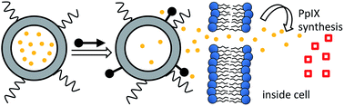
5-Aminolevulinic acid (5-ALA), a prodrug of protoporphyrin IX (PpIX), is used for photodynamic therapy of several medical conditions, and as an adjunct for fluorescence guided surgery. The clinical problem of patient photosensitivity after systemic administration could likely be ameliorated if the 5-ALA was delivered more selectivity to the treatment site. Liposomal formulations are inherently attractive as targeted delivery vehicles but it is hard to regulate the spatiotemporal release of aqueous contents from a liposome. Here, we demonstrate chemically triggered leakage of 5-ALA from stealth liposomes in the presence of cell culture. The chemical trigger is a zinc(II)-dipicolylamine (ZnBDPA) coordination complex that selectively targets liposome membranes containing a small amount of anionic phosphatidylserine. Systematic screening of several ZnBDPA complexes uncovered a compound with excellent performance in biological media. Cell culture studies showed triggered release of 5-ALA from stealth liposomes followed by uptake into neighboring mammalian cells and intracellular biosynthesis to form fluorescent PpIX.
Introduction
5-Aminolevulinic acid (5-ALA)‡ is a naturally occurring amino acid that is converted by the intracellular heme biosynthetic pathway into red fluorescent protoporphyrin IX (PpIX, Fig. 1).1 The process occurs to varying extents in virtually all tissue types. Addition of exogenous 5-ALA leads to selective accumulation of PpIX in cells undergoing high metabolic turnover, including cancer cells.2,3 5-ALA is finding increasing clinical use as an adjunct in fluorescence-guided surgery, where it is especially effective for guiding resection of high grade glioma.4 Low intensity, blue light illumination of the surgical field produces a two-color fluorescence image that helps surgeons delineate tumor margins. 5-ALA is also clinically important as a prodrug for photodynamic therapy (PDT) of several medical conditions.1,5,6 In the USA, it is approved for treatment of actinic keratosis and esophageal dysplasia.7 In these PDT applications, the PpIX is exploited as a photosensitizer, whereby red light irradiation of the diseased tissue that has been treated with 5-ALA produces a localized burst of cytotoxic reactive oxygen species.8,9
 |
| Fig. 1 Chemical structures. | |
In the oncology clinic, 5-ALA is administered either topically or systemically, and in both cases only a small fraction of the dose enters cells, primarily via endogenous amino acid transporters. After systemic treatment, the patient remains photosensitive until all of the 5-ALA is cleared from the body, which can take up two days.4,10 In principle, this clinical drawback could be ameliorated if the 5-ALA was delivered selectively to tumors. Recent research efforts have explored covalent modification11,12 or colloidal encapsulation8,13,14 as new methods to enhance cellular uptake of 5-ALA after topical administration. But to the best of our knowledge, there are no reported studies of living subjects treated systemically with nanoparticles containing 5-ALA. This is somewhat surprising since stealth liposomes are well-known as drug delivery vehicles that accumulate within solid tumors due to the enhanced permeation retention (EPR) effect. However, the classic dilemma with stealth liposomes is that the polyethylene glycol (PEG) corona that surrounds the stealth liposomes and ensures avoidance of the reticuloendothelial system, also makes it hard for drugs to leak from the liposomes. Thus, the technical challenge with stealth liposomes is to induce drug release after the liposomes have reached the tumor tissue.15 Various methods have been investigated to trigger liposome leakage including changes in temperature, pH, light, ultrasound, and covalent bond cleavage.16–18 Our lab is interested in developing an alternative release strategy using a non-covalent chemical trigger.19 The general concept is envisioned as a two-step dosing procedure that first administers drug-filled stealth liposomes, followed by a waiting period for tumor accumulation, and then a dose of non-toxic chemical trigger to rapidly release the drug from the liposomes at the tumor (Scheme 1).20 An attractive feature with this strategy is that knowledge of the tumor location(s) is not a necessary requirement. For effective performance, the chemical trigger must have high and selective affinity for the stealth liposomes. This requires the chemical trigger to be sterically small enough to avoid the PEG chains protruding from the stealth liposome surface, yet able to recognize a suitable structural target that is embedded within the liposome membrane. These design criteria have led us to pursue a triggering process that employs a zinc(II)-bis(dipicolylamine) (ZnBDPA) coordination complex as the chemical trigger and phosphatidylserine (PS) as the liposome membrane surface target. There is a large body of published biological imaging work showing that ZnBDPA complexes can selectively associate with anionic PS-containing membranes and distinguish them from the uncharged membrane surfaces of healthy mammalian cells.21–23 Furthermore, studies using living animals models have shown that the anionic membrane recognition process works effectively in vivo.
 |
| Scheme 1 Cartoon depiction of extracellular triggered release of 5-ALA (yellow circles) from stealth liposomes (grey donut with protruding PEG chains) upon exposure to chemical trigger (black arrow) with subsequent cellular uptake via endogenous amino acid transporters and biochemical conversion to PpIX (red squares). | |
Building on this knowledge, we recently demonstrated the first part of this chemically triggered release concept by showing that ZnBDPA coordination complex 1 can induce leakage of aqueous contents from a stealth liposome system composed of 67
:
28
:
8
:
5 DPPC
:
cholesterol
:
DPPE-PEG2000
:
POPS (Fig. 2).19 The small fraction of DPPE-PEG2000 provides the liposomes with steric protection24 and together with the POPS gives the liposome surface a net anionic charge. Mechanistic studies indicate that selective association of cationic chemical trigger 1 with the anionic PS embedded in the liposome membrane causes lateral phase separation of ordered and disordered phases.25–27 This leads to mismatched membrane thicknesses at the phase interface and defects that allow leakage of aqueous contents.28 Here, we advance this controlled release concept by reporting the discovery of a next-generation chemical trigger that operates much more effectively than the old trigger 1 in physiological conditions. Using this new chemical trigger, we demonstrate greatly enhanced release of 5-ALA prodrug from liposomes in the presence of healthy mammalian cells, followed by cell entry of the 5-ALA and subsequent biosynthesis into photoactive PpIX (Scheme 1).
 |
| Fig. 2 (Top) Chemical structures of relevant phospholipids; (Bottom) chemical structures of ZnBDPA triggers 1–5. | |
Results and discussion
Chemical synthesis
The structures of chemical triggers 2, 3, 4, and 5 are provided in Fig. 2 and the synthetic pathways are shown in Fig. 3. Chemical triggers 2 and 3 were prepared by treating compound apo-1 with the appropriate bis(succinate ester) to give apo-2 and apo-3, followed by complexation with Zn(NO3)2 in MeOH. Compound 6 was prepared according to literature precedent and was treated with butylisocyanate to prepare 7 in modest yield.21 Treatment of 7 with hydrazine monohydrate effectively removed the phthalimide protecting group and produced primary amine apo-4 that was complexed with Zn(NO3)2 to yield chemical trigger 4 (Fig. 2). Similar treatment of 6 with 4-fluorophenethyl isocyanate led to formation of 8 in excellent yield. Subsequent treatment of 8 with hydrazine monohydrate effectively removed the phthalimide protecting group and produced primary amine apo-5 that was complexed with Zn(NO3)2 to yield chemical trigger 5.
 |
| Fig. 3 Synthesis of chemical triggers 2, 3, 4, and 5. | |
Liposome leakage studies
Our previous study monitored the leakage of fluorescent carboxyfluorescein (CF) from liposomes in TES buffer and after some experimentation we settled on the preferred liposome membrane composition of 67
:
28
:
8
:
5 DPPC
:
cholesterol
:
DPPE-PEG2000
:
POPS.§ With a goal of successful operation in physiological samples, we started the current study by measuring the effect of buffer system on triggered CF leakage, and found that the amount of CF leakage caused by first generation trigger 1 (10 μM) dropped from 84% in TES to 34% in phosphate buffered saline (PBS). We attribute this decrease to competitive binding of phosphate anions to the ZnBDPA units in 1, thus reducing the affinity of 1 for the anionic liposome surface.29 This result led us to determine if alternative ZnBDPA structures, with higher affinity for PS-rich membranes, would perform better as chemical triggers in physiologically relevant media. We speculated that dimeric ZnBDPA structures might facilitate leakage by strongly sequestering the PS and promoting lateral phase separation.22,30 Thus, we tested the dimeric complexes 2 and 3 with covalent linkers of different lengths (Fig. 2). But these dimeric structures only produced marginally better CF leakage in PBS of 49% compared to the 34% seen with 1 (compare entry 1 with entries 4 and 6, Table 1). We also tested ZnBDPA complexes 4 and 5, which are modified structures with a ureido group covalently attached to each of the BDPA units (Fig. 2). We recently showed that ureido modified ZnBDPA structures have selective and enhanced affinity for PS-rich membranes and furthermore they are able to permeate through the membranes.21 Thus, we expected structures 4 and 5 to perform well as selective chemical triggers of leakage from PS-rich liposomes. A modest CF leakage enhancement in PBS was seen with modified ZnBDPA structure 4 (43%, entry 8, Table 1), but we were pleased to observe a much larger amount of leakage (71%, entry 10, Table 1) using modified ZnBDPA 5. This led to a performance comparison of original chemical trigger 1 and next generation version 5 in growth media (Fig. 4). Under these more complex and competitive conditions, there was very little CF leakage induced by 1 (2%, entry 3, Table 1) but still significant leakage induced by 5 (36%, entry 12, Table 1).
Table 1 Percent release of CF from 67
:
28
:
8:5 DPPC
:
cholesterol
:
DPPE-PEG2000
:
POPS liposomes upon exposure to a chemical triggera
Entry |
Bufferb |
Trigger |
[Trigger], μM |
%Releasec |
Percent release at 120 seconds after addition of chemical trigger in buffer; [total lipid] = 10 μM; 37 °C. F-12K media containing 10% FBS and 2% Strep. Pen. Uncertainty <10% of the value. |
1 |
PBS |
1 |
10.0 |
34 |
2 |
PBS |
1 |
1.0 |
3 |
3 |
F12-K media |
1 |
10.0 |
2 |
4 |
PBS |
2 |
10.0 |
49 |
5 |
PBS |
2 |
1.0 |
5 |
6 |
PBS |
3 |
10.0 |
49 |
7 |
PBS |
3 |
1.0 |
7 |
8 |
PBS |
4 |
10.0 |
43 |
9 |
PBS |
4 |
1.0 |
28 |
10 |
PBS |
5 |
10.0 |
71 |
11 |
PBS |
5 |
1.0 |
38 |
12 |
F-12K media |
5 |
10.0 |
36 |
13 |
F-12K media |
5 |
1.0 |
3 |
 |
| Fig. 4 Percent CF release from liposomes (67 : 28 : 8:5 DPPC : chol : DPPE-PEG2000 : POPS, 10 μM total lipid containing 50 mM CF) treated with either chemical trigger 1 (10 μM, A) or 5 (10 μM, B) at 60 seconds followed by liposome lysis with Triton-X-100 (20% v/v, 20 μL) at 180 seconds in either PBS buffer (blue crosses) or F12-K growth media (red squares). | |
The next step was to confirm that next-generation chemical trigger 5 was able to release 5-ALA from stealth liposomes. Liposomes encapsulating 5-ALA were prepared by the thin film hydration method. Note that a very small amount of fluorescent DiIC18, a lipophilic membrane tracer, was added to the liposome composition to facilitate purification and characterization of the final liposomal solution. The concentration of 5-ALA was measured using a simple colorimetric assay reported by Tomokuni and Ogata with only minor modification.31 Final preparations of liposomes encapsulating 5-ALA were typically composed of 2.7 mM total lipid and 5.1 mM 5-ALA; these results correspond to 2% encapsulation efficiency (which is consistent with literature precedent) and 30% liposome loading by weight. The average liposome size was measured to be 200 ± 3 nm using dynamic light scattering and was unchanged after storage at room temperature for several days (Fig. S1†). To measure release of 5-ALA a dialysis assay was developed using a dialysis device with source and receiver compartments that were separated by a porous membrane with 8000 molecular weight cutoff. Initially, liposomes filled with 5-ALA were added to the source compartment and the amount of 5-ALA in both the source and receiver compartments were measured over time. In the absence of chemical trigger the liposomes remained intact in the source compartment, releasing <10% of the encapsulated 5-ALA (Fig. S2†). Exposure to Triton-X-100 led to liposome lysis and complete equilibration of the released 5-ALA between source and receiver compartments (100% release) within 24 hours. In comparison, treatment of the liposomal 5-ALA with chemical trigger 5 led to 80% release of the encapsulated 5-ALA after 24 hours.
Cell uptake studies
The next goal was to demonstrate that ZnBDPA complex 5 could trigger 5-ALA release from liposomes in the presence of healthy mammalian cells, with subsequent permeation of the released 5-ALA into the cells, followed by intracellular biosynthesis into red fluorescent PpIX. For experimental convenience we conducted these experiments using CHO-K1 (Chinese hamster ovary) cells which are easy to cultivate and known to have moderate intracellular PpIX biosynthesis activity.32 An MTT cell viability assay of these cells showed that there was negligible cytotoxicity (greater than 80% cell viability) when the concentration of trigger 5 was kept under 100 μM (Fig. S3†). The amount of biosynthesized PpIX was determined using a standard fluorescence assay that measured the increase in red PpIX fluorescence produced by the cell culture. The bar graph in Fig. 5 shows the amount of PpIX produced at six hours after various treatments. In each case, the concentration of 5-ALA added to the cell culture well was 100 μM. Cell treatment with free 5-ALA increased the relative concentration of PpIX by a factor of three, whereas cell treatment with liposomal 5-ALA hardly changed the relative concentration of PpIX, which was expected since the encapsulated 5-ALA is prevented from entering the cells. In contrast, a sequential treatment of cells with liposomal 5-ALA followed by chemical trigger 5 (6.6 μM) led to the expected three-fold increase in PpIX production.
 |
| Fig. 5 PpIX production by CHO-K1 cells at six hours after addition of media alone, free 5-ALA (100 μM), liposomal-5-ALA (Lip-5-ALA, 100 μM 5-ALA, 66 μM total lipid), or liposomal-5-ALA (100 μM 5-ALA, 66 μM total lipid) for five minutes followed by chemical trigger 5 (6.6 μM). Emission intensities are normalized to cells incubated with media alone, λex = 406 nm, λem = 604 nm. | |
A visual demonstration of the trigger release effect is shown in Fig. 6 which compares cells that have been treated with either liposomal 5-ALA or a sequential treatment of liposomal 5-ALA followed by chemical trigger 5. The micrographs show clearly that the latter treatment produces a much higher red fluorescence intensity due to conversion of the released 5-ALA to PpIX. The distribution of PpIX is not homogenous throughout the field of cells, which is consistent with localized concentrations gradients of 5-ALA caused by uneven rates of triggered liposome release.
 |
| Fig. 6 Fluorescence micrographs (Brightfield = top; Cy 3 (PpIX) = middle; merge = bottom) (60×) of CHO-K1 cells at six hours after treatment with: (Left column) liposomal 5-ALA (100 μM total 5-ALA, 66 μM total lipid), (Right column) liposomal-5-ALA (100 μM total 5-ALA, 66 μM total lipid) and 6.6 μM chemical trigger 5 added five minutes later. Scale bar = 25 μm. | |
Conclusion
A next-generation ZnBDPA chemical trigger, 5, is shown to promote rapid release of aqueous contents from stealth liposomes containing a small fraction of phosphatidylserine. The chemical trigger selectively targets the anionic liposome membrane and avoids the surrounding corona of PEG chains. Cell culture studies demonstrated triggered release of 5-ALA from stealth liposomes, followed by 5-ALA uptake into neighboring mammalian cells and intracellular biosynthesis to form fluorescent PpIX. The next step in the project is to conduct in vivo studies that test the two-step dosing procedure described in the introduction section with the expectation that the initial dose of stealth liposomal 5-ALA will selectively accumulate within solid tumors.
Experimental
Materials
All lipids were purchased from Avanti Polar lipids (Alabaster, AL, USA). Dialysis membranes were purchased from Spectrum Labs (Rancho Dominquez, CA, USA). PD-10 pre-packed Sephadex™G-25M gel filtration columns were purchased from GE Healthcare (Knox, IN, USA). Bis-dPEG®2-NHS ester and Bis-dPEG®4-NHS ester were purchased from Quanta Biodesign (Plain City, OH, USA). All other reagents and chemicals were purchased from Sigma Aldrich (St. Louis, MO, USA) and used without further purification. Chinese hamster ovary (CHO-K1) cells were purchased from American Type Culture Collection (Manassas, VA, USA). RPMI and F-12K media were also purchased from American Type Culture Collection (Manassas, VA, USA). Fetal bovine serum was purchased from Atlanta Biologicals Inc., (Flowery Branch, GA, USA). Corning cellgro® penicillin–streptomycin solution was purchased from Corning Inc. (Corning, NY, USA).
Synthetic chemistry
Apo-2. To a solution of apo-1 (ref. 21) (29 mg, 49 μmol) in CH2Cl2 (1 mL) was added Bis-dPEG®2-NHS ester (8.9 mg, 22 μmol). The reaction mixture was allowed to stir overnight at room temperature. Solvent was removed under reduced pressure and the residue was purified by silica gel column chromatography with 0–10% MeOH in CHCl3 as the eluent to yield apo-2 (19.6 mg, 66% yield) as an opaque solid. 1H NMR (500 MHz, CDCl3) δ 1.66 (p, J = 8 Hz, 4H), 1.79 (p, J = 9 Hz, 4H), 2.43 (t, J = 6 Hz, 4H), 3.295 (q, J = 6 Hz, 4H), 3.58 (s, 4H), 3.64 (s, 8H), 3.71 (t, J = 6 Hz, 4H), 3.79 (s, 16H), 3.94 (t, J = 7 Hz, 4H) 6.51 (t, J = 6 Hz, 2H), 6.82 (s, 4H), 7.07 (s, 2H), 7.10–7.13 (m, 8H), 7.57–7.63 (m, 16H), 8.492 (dq, J = 5 Hz, J = 1 Hz, 8H) ppm; 13C NMR (500 MHz, CDCl3) δ 26.4, 26.7, 37.0, 39.1, 58.5, 60.0, 67.3, 70.1, 113.5, 121.5, 121.9, 122.7, 136.4, 140.5, 148.9, 159.0, 159.7, 171.3 ppm; HRMS (ESI, MeCN): m/z = 1345.7375 ([M + H]+).
Apo-3. To a solution of apo-1 (48 mg, 82 μmol) in CH2Cl2 (1 mL) was added Bis-dPEG®4-NHS ester (18.3 mg, 37 μmol). The reaction mixture was allowed to stir overnight at room temperature. Solvent was removed under reduced pressure and the residue was purified by silica gel column chromatography with 0–10% MeOH in CHCl3 as the eluent to yield apo-3 (31.7 mg, 59% yield) as a brown sticky oil. 1H NMR (500 MHz, CDCl3) δ 1.68 (p, J = 8 Hz, 4H), 1.80 (p, J = 6 Hz, 4H), 2.45 (t, J = 6 Hz, 4H), 3.303 (q, J = 7 Hz, 4H), 3.58–3.60 (m, 12H), 3.65 (s, 8H), 3.71 (t, J = 6 Hz, 4H), 3.80 (s, 16H), 3.95 (t, J = 6 Hz, 4H), 6.74 (t, J = 5 Hz, 2H), 6.83 (s, 4H), 7.08 (s, 2H), 7.11–7.14 (m, 8H), 7.57–7.64 (m, 16H), 8.49–8.50 (m, 8H) ppm; 13C NMR (500 MHz, CDCl3) δ 26.4, 26.7, 37.0, 39.0, 58.5, 59.9, 67.3, 70.1, 70.3, 70.5, 113.5, 121.4, 122.0, 122.6, 136.5, 140.4, 148.9, 159.0, 159.6, 171.5 ppm; HRMS (ESI, MeCN): m/z = 1433.7949 ([M + H]+).
Compound 7. To a solution of 6 (14 mg, 18 μmol) in CHCl3 (100 μL) was added butylisocyanate (20 μL, 180 nmol). The reaction was allowed to stir at room temperature for 12 hours. Solvent was removed and the crude material was purified using alumina gel column chromatography with 0–10% MeOH
:
CHCl3 as the eluent to yield the desired product as a clear oil (8.0 mg, 47% yield). 1H NMR (600 MHz, CDCl3) δ 0.91 (t, J = 7 Hz, 6H), 1.39 (s, J = 7 Hz, 4H), 1.54 (p, J = 7 Hz, 4H), 1.80–1.90 (m, 4H), 3.34 (q, J = 6 Hz, 4H), 3.63 (s, 4H), 3.66 (s, 4H), 3.76 (t, J = 6 Hz, 2H), 3.78 (s, 4H), 3.96 (t, J = 6 Hz, 2H), 6.64 (s, 2H), 6.83 (s, 2H), 6.98 (s, 1H), 7.09 (d, J = 7 Hz, 2H), 7.12–7.13 (m, 2H), 7.49–7.54 (m, 4H), 7.62 (t, J = 7 Hz, 2H), 7.69–7.70 (m, 2H), 7.82–7.83 (m, 2H), 8.37 (s, 2H), 8.50 (d, J = 7 Hz, 2H), 9.40 (s, 2H) ppm; 13C NMR (600 MHz, CDCl3) δ 13.8, 20.2, 25.4, 26.7, 32.0, 37.6, 39.5, 58.7, 59.8, 60.1, 67.1, 110.0, 113.4, 115.2, 121.2, 122.0, 122.6, 123.2, 132.1, 134.0, 136.5, 138.6, 140.5, 149.0, 152.3, 156.1, 156.3, 159.2, 159.6, 168.4 ppm; HRMS (ESI, MeCN): m/z = 946.5089 ([M + H]+).
Apo-4. To a solution of 7 (83 mg, 88 μmol) in CH2Cl2 (400 μL) and EtOH (1.5 mL) was added hydrazine monohydrate (140 μL, 2.89 mmol). The reaction mixture was allowed to stir at room temperature overnight during which a large amount of white precipitate formed. The reaction mixture was filtered, concentrated, dissolved in dichloromethane, filtered, and concentrated again to yield the desired product as an off-white solid (37 mg, 52% yield). 1H NMR (600 MHz, CDCl3) δ 0.86 (t, J = 7 Hz, 6H), 1.35 (s, J = 7 Hz, 4H), 1.50 (p, J = 7 Hz, 4H), 1.73–1.80 (m, 4H), 2.90 (t, J = 6 Hz, 2H), 3.31 (q, J = 7 Hz, 4H), 3.60 (s, 4H), 3.63 (s, 4H), 3.80 (s, 4H), 3.85 (t, J = 7 Hz, 2H), 4.44 (br s, NH, 2H), 6.74 (s, 2H), 6.79 (d, J = 7 Hz, 2H), 6.89 (s, 1H), 6.99 (d, J = 7 Hz, 2H), 7.11–7.13 (m, 2H), 7.45 (t, J = 7 Hz, 2H), 7.50 (d, J = 7 Hz, 2H), 7.60 (d of t, J = 1, 7 Hz, 2H), 8.50 (d, J = 7 Hz, 2H), 8.98 (s, 2H), 8.56 (s, 2H) ppm; 13C NMR (600 MHz, CDCl3) δ 13.8, 20.2, 26.5, 27.5, 32.0, 39.5, 40.7, 58.6, 59.9, 60.5, 67.2, 110.3, 113.5, 115.5, 121.3, 122.1, 122.7, 136.5, 138.5, 140.2, 148.9, 153.0, 156.0, 156.4, 159.1, 159.4 ppm; HRMS (ESI, MeCN): m/z = 816.5025 ([M + H]+).
Compound 8. To a solution of 6 (200 mg, 270 μmol) in CHCl3 (500 μL) was added 4-fluorophenethyl isocyanate (200 μL, 1.37 mmol). The reaction was allowed to stir at room temperature for 12 hours. Solvent was removed and the crude material was purified using silica gel column chromatography with 0–10% MeOH
:
CHCl3 as the eluent to yield the desired product as a yellow oil (231 mg, 80% yield). 1H NMR (500 MHz, CDCl3) δ 1.78–1.89 (m, 4H), 2.84 (t, J = 7 Hz, 4H), 3.42 (s, 4H), 3.57 (s, 4H), 3.62 (q, J = 7 Hz, 4H), 3.73 (s, 4H), 3.74 (t, J = 7 Hz, 2H), 3.96 (t, J = 7 Hz, 2H), 6.64 (d, J = 8 Hz, 2H), 6.84 (s, 2H), 6.89–6.94 (m, 4H), 7.01 (s, 1H), 7.09–7.16 (m, 8H), 748 (t, J = 8 Hz, 2H), 7.52 (d, J = 8 Hz, 2H), 7.60 (td, J = 2, 8 Hz, 2H), 7.68–7.69 (m, 2H), 7.81–7.82 (m, 2H), 8.49–8.51 (m, 2H), 8.75 (br s, NH, 2H), 9.52 (br s, NH, 2H) ppm; 13C NMR (600 MHz, CDCl3) δ 25.4, 26.7, 35.4, 37.6, 40.9, 58.6, 59.5, 60.1, 67.1, 110.0, 113.3, 115.0, 115.2, 115.4, 122.0, 122.5, 123.2, 130.1, 132.1, 134.0, 135.1, 136.5, 140.6, 149.0, 152.7, 155.4, 156.2, 159.2, 159.6, 160.6, 162.3, 168.4 ppm; HRMS (ESI, MeCN): m/z = 1078.4911 ([M + H]+).
Apo-5. To a solution of 8 (194 mg, 180 μmol) in CH2Cl2 (0.82 mL) and EtOH (3.0 mL) was added hydrazine monohydrate (80 μL, 1.65 mmol). The reaction mixture was allowed to stir at room temperature overnight during which a large amount of white precipitate formed. The reaction mixture was filtered, concentrated, dissolved in dichloromethane, filtered, and concentrated again to yield the desired product as an off-white solid (72.6 mg, 43% yield). 1H NMR (600 MHz, CDCl3) δ 1.63 (p, J = 7 Hz, 2H), 1.78 (p, J = 7 Hz, 2H), 2.27 (s, 2H), 2.76 (t, J = 8 Hz, 2H), 2.83 (t, J = 8 Hz, 4H), 3.43 (s, 4H), 3.57 (s, 4H), 3.61 (q, J = 6 Hz, 4H), 3.74 (s, 4H), 3.91 (t, J = 7 Hz, 2H), 6.68 (d, J = 8 Hz, 2H), 6.83 (s, 2H), 6.90 (t, J = 8 Hz, 4H), 6.98 (s, 1H), 7.07 (d, J = 8 Hz, 2H), 7.10–7.15 (m, 6H), 7.46 (t, J = 8 Hz, 2H), 7.51 (d, J = 7 Hz, 2H), 7.59 (d of t, J = 2, 8 Hz, 2H), 8.48–8.50 (m, 2H), 8.74 (br s, NH, 2H), 9.52 (br s, NH, 2H) ppm; 13C NMR (600 MHz, CDCl3) δ 26.6, 29.7, 35.4, 40.9, 41.6, 58.6, 59.5, 60.2, 67.6, 109.8, 113.4, 115.2, 115.4, 121.1, 122.1, 122.6, 130.2, 130.2, 135.1, 136.5, 138.6, 140.4, 149.0, 152.5, 155.8, 156.5, 159.2, 159.5, 160.6, 162.3 ppm; HRMS (ESI, MeCN): m/z = 948.4809 ([M + H]+).
Zinc complexation. Stock solutions of Zn(NO3)2·6H2O (25 mM) and ZnBDPA scaffolds were prepared in MeOH and mixed such that the molar ratio of [Zn2+]
:
[DPA scaffold] was 1
:
1. The solutions were allowed to shake for 1 h before solvent was removed by rotary evaporation. Residual solvent was removed under vacuum over a period of at least 1 h.
Liposome preparation
All phospholipids were purchased from Avanti Polar Lipids (Alabaster, AL) and stored at −20 °C until use. Liposomes were prepared using the thin film hydration method, followed by membrane extrusion for size control. Appropriately sized aliquots of lipid solutions were added to a clean, dry test tube. Solvent was removed by evaporation using a gentle stream of N2 gas. Residual solvent was removed under vacuum over a period of at least 1 h. Lipids were rehydrated with the desired buffer. A glass ring was added to the solution to ensure complete removal of all lipid from the test tube wall and the solution was vortexed. The suspension was extruded 21 times through a 19 mm polycarbonate membrane with 200 nm diameter pores. Liposomes were used on the day of preparation.
Carboxyfluorescein release from liposomes
Liposomes were prepared using the thin film hydration method upon hydration with TES buffer (5 mM TES, 145 mM NaCl, pH 7.4) containing carboxyfluorescein (CF, 50 mM).33 Unencapsulated CF was removed by either overnight dialysis (12–14
000 MWCO tubing) against TES buffer (5 mM TES, 145 mM NaCl, pH 7.4) or gel filtration through PD-10 columns packed with Sephadex™G-25M. From the stock solution of CF liposomes, samples (10 μM total phospholipid) were assayed for release of CF (λex = 492 nm, λem = 517 nm) upon addition of chemical trigger (10 μM) at 60 seconds and Triton X-100 (20% v/v, 20 μL) at 180 seconds in different media at 37 °C. The ‘% CF release’ was calculated from eqn (1) where Fi and Ff are the initial and final fluorescent intensities and Ft is the fluorescent intensity at time t. Uncertainties are reported as the standard deviation of the mean for CF release experiments reproduced in at least triplicate. |
 | (1) |
5-ALA release from liposomes
Liposomes composed of DPPC
:
cholesterol
:
DPPE-PEG2000
:
POPS
:
DiIC18 in the mole ratio of 67
:
28
:
8
:
5
:
1 were prepared using the thin film hydration method upon hydration with HEPES buffer (10 mM HEPES, 137 mM NaCl, 3.2 mM KCl, pH 7.4) containing 5-ALA (1.0 M 5-ALA). It is important to note that a small amount of fluorescent DiIC18, a lipophilic membrane tracer, was added to the liposome composition to facilitate purification and characterization of the final liposomal solution. Unencapsulated 5-ALA was removed by gel filtration through PD-10 columns packed with Sephadex™G-25M. Liposome dilution from the gel filtration process was measured by comparing the emission intensity of DiIC18 (λex = 490 nm, λem = 566 nm) to a standardized curve. The concentration of 5-ALA was measured using a simple colorimetric assay reported by Tomokuni and Ogata with only minor modifications.31 Briefly, samples containing 5-ALA (100 μL) were treated with Triton-X-100 (20% v/v, 20 μL) and diluted to a final volume of 500 μL with HEPES buffer (10 mM HEPES, 137 mM NaCl, 3.2 mM KCl, pH 7.4). Samples were then further diluted upon addition of acetate buffer (500 μL; 2.0 M sodium acetate buffer at pH 4.6) and ethyl acetoacetate (100 μL) prior to heating at 100 °C for 10 minutes. Samples were cooled to room temperature, treated with ethyl acetate (1.5 mL) and were shaken by hand approximately 50 times. The organic layer (1.0 mL) was removed and treated with modified Ehrlich's reagent (1.0 mL) for 10 minutes prior to spectroscopic analysis at 555 nm. Liposomal encapsulation of 5-ALA was determined by comparing the 5-ALA concentration of liposome samples before and after gel filtration. Final preparations of liposomes encapsulating 5-ALA were typically composed of 2.7 mM total lipid and 5.1 mM 5-ALA; these results correspond to 2% encapsulation efficiency (which is consistent with literature precedent) and 30% liposome loading by weight. The average liposome size was measured to be 200 ± 3 using dynamic light scattering (Fig. S1†). Dialysis experiments show <10% leakage over 24 hours and the liposomes appear to be stable for days at room temperature.
Preparation of modified Ehrlich's reagent
To a solution of 30 mL glacial acetic acid was added p-dimethylaminobenzaldehyde (1.0 g, 6.7 mmol), 60% perchloric acid (5 mL), and distilled water (5 mL). The solution was mixed and diluted to a final volume of 50 mL using glacial acetic acid.
Dialysis experiments
Release of 5-ALA from liposomes upon exposure to external stimuli was measured using dialysis experiments. Liposomes encapsulating 5-ALA (750 μM total lipid; 67
:
28
:
8
:
5
:
1 DPPC
:
cholesterol
:
DPPE-PEG2000
:
POPS
:
DiIC18) in HEPES buffer (10 mM HEPES, 137 mM NaCl, 3.2 mM KCl, pH 7.4) were added to the source compartment of a dialysis apparatus while either chemical trigger 5 (75 μM) or Triton-X-100 (20% v/v, 50 μL) were added to the receiver compartment. The two dialysis compartments were separated by a 8000 molecular weight cutoff membrane that prevents liposomes from equilibrating between the two compartments. The concentrations of 5-ALA were measured as described above.
Cell culture
Chinese hamster ovary (CHO-K1) cells were purchased from American Type Culture Collection, seeded into 96-microwell plates, and grown to confluency of 8 × 10⁴ cells per well in F-12K media supplemented with 10% fetal bovine serum, and 1% penicillin–streptomycin solution at 37 °C and 5% CO2, respectively.
Determination of cellular PpIX production
Cells were grown to confluency of 8 × 10⁴ cells per well, then different microwells were treated with cell media alone, 5-ALA, liposomes encapsulating 5-ALA, liposomal-5-ALA for 5 min followed by chemical trigger 5. After treatment the cells were incubated for 6 hours at 37 °C and 5% CO2 in serum-free F-12K media. The amount of biosynthesized PpIX was measured by replacing the cell media in each microwell with 200 μL of 5% HCl, and incubating for an additional 30 min at 37 °C. Each sample was subjected to a 5-fold dilution in 5% HCl prior to collecting fluorescence emission spectra (λex = 406 nm, λem = 604 nm) on a Fluoromax-4 spectrofluorometer with FluorEssence software (Horiba Scientific, Edison, NJ, USA). The experiment was performed in triplicate. Data and error bars correspond to the mean ± the standard error of the mean (SEM) for each treatment. Data manipulation was performed using Microsoft Excel and graphs generated using Graphpad Prism 5 (Graphpad Software Inc., San Diego, CA).
Cell microscopy
CHO-K1 cells were seeded in an 8-well chambered plate and grown to confluency as described above. Wells were either treated with liposomal-5-ALA (100 μM total 5-ALA, 66 μM total lipid) or liposomal-5-ALA (100 μM total 5-ALA, 66 μM total lipid) and 6.6 μM chemical trigger 5, five minutes post 5-ALA treatment for a total of 200 μL in each well, followed by incubation for 6 hours at 37 °C and 5% CO2 in serum-free F-12K media. Media in each well was then removed; cells were washed 1× with PBS and resuspended in PBS buffer. Live cell imaging was performed using a Nikon TE-2000U epifluorescence microscope equipped with a Cy 3 filter set (λex = 535/50, λem = 610/75) (Nikon Instruments Inc., Melville, NY, USA). Brightfield images were captured and corresponding fluorescence images were normalized to the highest intensity (60× magnification, 46 ms exposure time).
MTT cell viability assay
Cell viability was measured using the 3-(4,5-dimethylthiazol-2-yl)-2,5-diphenyltetrazolium bromide (MTT) cell viability assay. Cells were grown to confluency of 8 × 10⁴ cells per well and the Vybrant MTT cell proliferation Assay Kit (Invitrogen, Eugene, USA) was used according to the manufacturer's protocol and validated using etoposide (50 μM) as a positive control for high toxicity. Cells were treated with chemical trigger 5 (0–100 μM) and incubated for 6 hours at 37 °C. The medium was removed and replaced with 100 μL of F-12K media containing [3-(4,5-dimethylthiazol-2-yl)-2,5-diphenyltetrazolium bromide] (MTT, 1.2 mM). An SDS–HCl detergent solution was added and incubated at 37 °C and 5% CO2 for an additional 4 hours. The absorbance of each well was read at 570 nm and normalized to wells containing no cells or added 5 (measured in quadruplicate).
Statistical analysis
Results are depicted as mean ± standard error of the mean (SEM). Statistical analysis was performed using a Student's t test.
Acknowledgements
This work was supported by the NIH (USA) and the University of Notre Dame.
References
- B. Nokes, M. Apel, C. Jones, G. Brown and J. E. Lang, J. Surg. Res., 2013, 181, 262–271 CrossRef CAS PubMed.
- S. L. Gibbs, B. Chen, J. A. O'Hara, P. J. Hoopes, T. Hasan and B. W. Pogue, Photochem. Photobiol., 2006, 82, 1334–1341 CrossRef CAS PubMed.
- R. C. Krieg, S. Fickweiler, O. S. Wolfbeis and R. Knuechel, Photochem. Photobiol., 2000, 72, 226–233 CrossRef CAS.
- S. Marbacher, E. Klinger, L. Schwyzer, I. Fischer, E. Nevzati, M. Diepers, U. Roelcke, A. R. Fathi, D. Coluccia and J. Fandino, Neurosurg. Focus, 2014, 36, E10 CrossRef PubMed.
- D. Bechet, S. R. Mordon, F. Guillemin and M. A. Barberi-Heyob, Cancer Treat. Rev., 2014, 40, 229–241 CrossRef PubMed.
- Y. Zhang and J. F. Lovell, Theranostics, 2012, 2, 905–915 CrossRef CAS PubMed.
- Q. Peng, K. Berg, J. Moan, M. Kongshaug and J. M. Nesland, Photochem. Photobiol., 1997, 65, 235–251 CrossRef CAS PubMed.
- C. W. Chung, K. D. Chung, Y. I. Jeong and D. H. Kang, Int. J. Nanomed., 2013, 8, 809–819 CrossRef PubMed.
- E. Buytaert, M. Dewaele and P. Agostinis, Biochim. Biophys. Acta, Rev. Cancer, 2007, 1776, 86–107 CrossRef CAS PubMed.
- N. Dragicevic-Curic and A. Fahr, Expert Opin. Drug Delivery, 2012, 9, 1015–1032 CrossRef CAS PubMed.
- J. M. Gaullier, K. Berg, Q. Peng, H. Anholt, P. K. Selbo, L. W. Ma and J. Moan, Cancer Res., 1997, 57, 1481–1486 CAS.
- A. Casas, C. Perotti, M. Saccoliti, P. Sacca, H. Fukuda and A. M. D. Batlle, Br. J. Cancer, 2002, 86, 837–842 CrossRef CAS PubMed.
- G. Di Venosa, L. Hermida, H. Fukuda, M. V. Defain, L. Rodriguez, L. Mamone, A. MacRobert, A. Casas and A. Batlle, J. Photochem. Photobiol., B, 2009, 96, 152–158 CrossRef CAS PubMed.
- A. Casas and A. Batlle, Curr. Med. Chem., 2006, 13, 1157–1168 CrossRef CAS.
- Y. Malam, M. Loizidou and A. M. Seifalian, Trends Pharmacol. Sci., 2009, 30, 592–599 CrossRef CAS PubMed.
- D. Mellal and A. Zumbuehl, J. Mater. Chem. B, 2014, 2, 247–252 RSC.
- T. M. Allen and P. R. Cullis, Adv. Drug Delivery Rev., 2013, 65, 36–48 CrossRef CAS PubMed.
- S. Bibi, E. Lattmann, A. R. Mohammed and Y. Perrie, J. Microencapsulation, 2012, 29, 262–276 CrossRef CAS PubMed.
- A. J. Plaunt, M. B. Courbanou, K. D. Cuison, K. M. Harmatys and B. D. Smith, Chem. Commun., 2012, 48, 8123–8125 RSC.
- A. S. L. Derycke and P. A. M. de Witte, Adv. Drug Delivery Rev., 2004, 56, 17–30 CrossRef CAS PubMed.
- A. J. Plaunt, K. M. Harmatys, W. R. Wolter, M. A. Suckow and B. D. Smith, Bioconjugate Chem., 2014, 25, 724–737 CrossRef CAS PubMed.
- B. A. Smith, K. M. Harmatys, S. Xiao, E. L. Cole, A. J. Plaunt, W. Wolter, M. A. Suckow and B. D. Smith, Mol. Pharm., 2013, 10, 3296–3303 CrossRef CAS PubMed.
- B. A. Smith, W. J. Akers, W. M. Leevy, A. J. Lampkins, S. Xiao, W. Wolter, M. A. Suckow, S. Achilefu and B. D. Smith, J. Am. Chem. Soc., 2010, 132, 67–69 CrossRef CAS PubMed.
- C. Allen, N. Dos Santos, R. Gallagher, G. N. C. Chiu, Y. Shu, W. M. Li, S. A. Johnstone, A. S. Janoff, L. D. Mayer, M. S. Webb and M. B. Bally, Biosci. Rep., 2002, 22, 225–250 CrossRef CAS.
- N. Shimokawa, M. Hishida, H. Seto and K. Yoshikawa, Chem. Phys. Lett., 2010, 496, 59–63 CrossRef CAS PubMed.
- D. Y. Sasaki, Cell Biochem. Biophys., 2003, 39, 145–161 CrossRef CAS.
- S. Ohnishi and T. Ito, Biochemistry, 1974, 13, 881–887 CrossRef CAS.
- S. G. Clerc and T. E. Thompson, Biophys. J., 1995, 68, 2333–2341 CrossRef CAS.
- R. G. Hanshaw, S. M. Hilkert, H. Jiang and B. D. Smith, Tetrahedron Lett., 2004, 45, 8721–8724 CrossRef CAS PubMed.
- S. Z. Xiao, L. Abu-Esba, S. Turkyilmaz, A. G. White and B. D. Smith, Theranostics, 2013, 3, 658–666 CrossRef PubMed.
- K. Tomokuni and M. Ogata, Clin. Chem., 1972, 18, 1534–1536 CAS.
- M. Wolun-Cholewa and W. Warchol, Folia Histochem. Cytobiol., 2004, 42, 131–135 CAS.
- K. M. DiVittorio, W. M. Leevy, E. J. O'Neil, J. R. Johnson, S. Vakulenko, J. D. Morris, K. D. Rosek, N. Serazin, S. Hilkert, S. Hurley, M. Marquez and B. D. Smith, ChemBioChem, 2008, 9, 286–293 CrossRef CAS PubMed.
Footnotes |
† Electronic supplementary information (ESI) available. See DOI: 10.1039/c4ra10340h |
‡ Abbreviations: 1,2-dipalmitoyl-sn-glycero-3-phosphocholine (DPPC), 1,2-dipalmitoyl-sn-glycero-3-phosphoethanolamine-N-[methoxy(polyethylene glycol)-2000] ammonium salt (DPPE-PEG2000), 1,2-distearoyl-sn-glycero-3-phosphoethanolamine-N-[methoxy(polyethylene glycol)-2000] ammonium salt (DPPE-PEG2000), 1-palmitoyl-2-oleoyl-sn-glycero-3-phospho-L-serine sodium salt (POPS), 5-Aminolevulinic acid (5-ALA), Carboxyfluorescein (CF), Hydrogenated Soy Phosphatidylcholine, (HSPC), Phosphate Buffered Saline (PBS), Phosphatidylserine (PS), Photodynamic Therapy (PDT), Polyethylene Glycol (PEG), Protoporphyrin IX (PpIX), Zinc(II)-bisdipicolylamine (ZnBDPA). |
§ Comparative studies using liposomes with a membrane containing the thicker HSPC as a substitute for DPPC (i.e., liposomes composed of 57 : 38 : 5 : 5 HSPC : cholesterol : DSPE-PEG2000 : POPS) showed that the CF leakage caused by chemical trigger 1 was greatly diminished. |
|
This journal is © The Royal Society of Chemistry 2014 |
Click here to see how this site uses Cookies. View our privacy policy here.