Design and development of papain–urea loaded PVA nanofibers for wound debridement

Ekambaram Shoba, Rachita Lakra, Manikantan Syamala Kiran and Purna Sai Korrapati*
Biomaterials Department, CSIR - Central Leather Research Institute, Chennai, India-600 020. E-mail: purnasaik.clri@gmail.com; Tel: +91-44-24437263/+91-44-24453491

Received 11th September 2014 , Accepted 5th November 2014

First published on 6th November 2014


Abstract

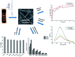
Devitalized tissues present in a wound bed serve as a reservoir for bacterial growth and contain elevated levels of inflammatory mediators that promote chronic inflammation and impair cellular migration necessary for wound repair. Effective wound cleansing and debridement are essential for granulation and re-epithelization. Among various debridement methods, enzymatic debridement is a highly selective method that uses naturally occurring proteolytic enzymes. Papain combined with urea has been widely used to remove necrotic/devitalized tissues. Our approach is to encapsulate papain and urea in PVA nanofibers to bring out sustained release to enable breakdown of fibrinous material in necrotic tissue and enhance wound healing. Physico-chemical characterization of nanofibers depicted the enzyme interaction with the polymer and also confirmed that the enzyme was evenly distributed in the nanofibers in an amorphous state. Fluorescence spectroscopy confirmed that the structural integrity of the enzyme was maintained after encapsulation. The results of antibacterial activity along with cell compatibility assays confirm the structural and functional integrity of the enzyme preparation along with the biocompatibility of the electrospun nanofiber and thereby provide more suitability as a dressing for wound debridement.
Introduction

Papain is an endolytic cysteine protease (EC: 3.4.22.2) widely used in food, pharmaceutical, leather, cosmetic and textile industries.1–3 It is a highly functional proteolytic enzyme with bactericidal, bacteriostatic, and anti-inflammatory properties hence has been proven to be effective in healing process of wounds, lesions, and ulcers.4 Papain breaks down fibrinous material in necrotic tissue and requires the presence of sulfhydryl groups, such as cysteine, for its activity. Urea is usually combined with papain in order to expose the activators of papain in the necrotic tissue. Urea also denatures proteins, making them more susceptible to proteolysis by papain. The combination of papain and urea is approximately twice as effective in degrading proteins compared with papain alone.5 Papain gets deactivated quickly in their free state in solution therefore; it must be immobilized in a matrix system for its effective use. Enzyme immobilization and their application have attracted continuous attention in the field of biomedicine, biosensors fabrication, bioremediation, fine chemistry and protein digestion in protein analysis.6 Immobilized enzyme performance at various fields largely depends on the structure of the supporting material.

Nanostructured supports are believed to retain the catalytic activity as well as ensure the immobilization efficiency of enzyme to a greater extent.7 Various nanostructured supports such as nanoparticles, nanospheres, nanofibers etc. are used for enzyme immobilization. Uniform distribution, recycling and losses during encapsulation are the predominant drawbacks of nanoparticles and nanospheres. However, nanofibers overcome these drawbacks and also provide several attractive aspects like high porosity, interconnectivity, high surface to volume ratio and easy handling.8 The electrospun nanofibers provide an ideal wound dressing material due to its useful properties, including oxygen permeable high porosity, variable pore size distribution and most importantly, morphological similarity to the natural extracellular matrix (ECM) in the skin, all of which promote cell adhesion, migration, and proliferation.9 Enzyme immobilization can be achieved by either binding onto the surface of the support or encapsulating in the supporting matrix. Currently nanofibers based supporting matrixes are gaining much attention because of the versatility to fabricate the polymer fiber with larger length, diameter aspect ratio.10 Encapsulation of enzyme with supporting matrix brings high enzyme loading efficacy and allow possibility of mixing more than one type of enzyme into nanofibers.11 Immobilization of papain onto various supports such as mesoporous silica,12 macroporous bead carrier of copolymer,13 polyacrolein microspheres, anion-exchange resins,14,15 epoxy polymer,16 superparamagnetic nanoparticles17 and polymeric nanofibers has been previously reported. PVA was selected as a matrix owing to its biocompatibility and good thermo-chemical behaviour.18,19 The present study essentially deals with encapsulation of papain and urea into PVA nanofiber so as to achieve the combined beneficial effects of papain and urea for wound debridement. In vitro release, encapsulation efficiency, structural integrity of enzyme in nanofiber was analyzed. The functional integrity was confirmed through antibacterial activity, cell compatibility assays suggesting the possibilities of exploring the potential of nanofiber as biocompatible material. The study therefore, opens up venues for multiple methods of devitalizing the wounds and thereby enhances healing process.

Materials and methods

Materials

Poly(vinyl alcohol) (PVA; 99% hydrolysis (Cat. no. 363146), MW ∼ 85[thin space (1/6-em)]000–125[thin space (1/6-em)]000) and cysteine hydrochloride was purchased from Sigma-Aldrich (St. Louis, USA). Papain (source Carica papaya) was obtained from Sigma-Aldrich. Casein, trisodium citrate dihydrate, ethylenediaminetetraacetic acid (EDTA), L-cysteine hydrochloride, sodium carbonate (Na2CO3), trichloroacetic acid (CCl3·COOH), ethanol, Folin Ciocalteu's reagent and urea were purchased from SD fine chem. Ltd, Mumbai, India. All other chemicals and solvents were of analytical grade and used without further purification. Deionized water was used throughout the study.

Preparation of papain–urea loaded PVA nanofiber

Papain–urea/PVA was prepared at various ratios to understand the highest encapsulation efficiency and release of papain and urea from PVA nanofiber. 10% (w/v) of PVA solution were prepared by dissolving in deionized water at 80 °C with constant stirring for 4–6 hours. Papain (in range of 5–50 wt% to polymer) activated with urea in a ratio of 2[thin space (1/6-em)]:[thin space (1/6-em)]1 were mixed with 10% PVA solution and stirred at room temperature to obtain a homogenous mixture. The freshly prepared P–U/PVA (papain–urea/PVA) blend was loaded into 5 mL syringe equipped with an 18-gauge blunt stainless steel needle. The feeding rate was controlled by a syringe pump (physics instrument & co., India) and was fixed at 0.3 mL h−1. The electric potential was applied to the metallic needle (0.5 mm inner hole diameter) and the tip to collector distance was set to 10–12 cm. As the jet breaks up into fibers from the Taylor cone, the liquid was evaporated and gave rise to relatively dry fibers which were subsequently spun on the aluminium foil until multi-layered fiber mat was obtained. The electrospinning process was carried out at 24 ± 1 °C and relative humidity of 68 ± 3 °C in an enclosed Plexiglas box.

Characterization of papain and urea loaded PVA nanofibers

Fourier transform infrared spectrum of the prepared samples was measured using FTIR spectrometer (Perkin-Elmer). The electrospun nanofibers were cut into small pieces and mixed with KBr to make sample pellets. Measurements were taken in a range between 4000–400 cm−1 with a resolution of 2 cm−1. The surface morphology and the diameter were analysed by scanning electron microscopy (Hitachi, Japan) using an acceleration voltage of 10 kV. Prior to the experiment the samples were gold sputter coated under argon atmosphere to render them electrically conductive. The average diameter of the electrospun fibrous mats was measured using the inbuilt software attachment of SEM. To visualize the presence and distribution of the proteins into electrospun PVA nanofiber scaffolds, a model protein BSA conjugated with FITC was added to the polymer solution. A thin layer of electrospun fibers was collected on a glass slide and observed under fluorescence microscope (Leica microsystems, Germany). The thermal characteristics were determined using a differential scanning calorimeter (Q-200 differential scanning calorimeter TA, instruments Co., USA). Samples were crimped in a standard aluminium pan and heated from 40° C to 250° C at a heating rate 10° C per minute under constant purging of nitrogen at 20 mL min−1.

Measurement of bio catalytic activity of immobilized and native papain

The catalytic activity of papain and papain released from PVA NF was determined by reaction with 2% casein at 37° C in 0.01 M PBS in accordance to Sahoo et al.20 Catalytic activity was carried by activation of papain according to the following procedure. 20 mg of encapsulated papain in 50 mmol L−1 phosphate buffer was mixed with 50 mmol L−1 L-cysteine hydrochloride and 3 mmol L−1 ethylenediaminetetraacetic acid (EDTA) and incubated for 30 min at 30° C. The activated papain (0.5 mL) was added to test tube containing 0.5 mL of 2% casein. The reaction was carried out at a 37° C for 15 min and stopped by the addition of 2 mL of 30% w/v trichloroacetic acid solution (TCA) and centrifuged. To 0.5 mL of the supernatant 1 mL of 0.5 mol L−1 Na2CO3 was added and kept for 10 min. After that, 1 mL of Folin Ciocalteu's reagent was added. The tubes were incubated 30 min in dark condition for color development. The color solution was measured against the reagent blank at 660 nm using spectrophotometer (Perkin-Elmer, USA). Papain activity was expressed in terms of amount of enzyme required to release 1 μg of tyrosine per minute per milliliter. In case of native papain, the activity was measured following the above procedures and instead of NF released papain 0.5 mL of 1 mg mL−1 native papain solution in 10 mM PBS (pH 7.4) was used.

Estimation of encapsulation efficiency

Ten milligram of papain loaded nanofibers were weighed accurately and dissolved in 1 mL of phosphate buffer (pH 7.4). The resulting solution was centrifuged at 2500 rpm for 10 minutes (Remi Instruments Ltd, Mumbai, India), and the supernatant was assayed (n = 3) for enzyme content by modified casein digestion method. Results were expressed as a mean ± standard deviation of three experiments. Encapsulation efficiency was determined using the following equations
image file: c4ra10239h-t1.tif

image file: c4ra10239h-t2.tif

In vitro enzyme release

The release characteristics of papain from the papain loaded PVA nanofiber mats were determined using PBS at pH of 7.4. A weighed quantity of enzyme loaded composite nanofibers (approximately 10 mg) was immersed in a 10 mL of phosphate buffer (pH 7.4). The samples were incubated at 37 ± 5° C with continuous shaking at 100 rpm. At predetermined time intervals (every 60 min) aliquots of 1 mL were withdrawn and replaced by an equal volume of respective medium to maintain constant volume. After filtrations through Whatmann membrane (no. 1), the amount of enzyme released from the composite nanofibers were calculated by analyzing the samples using an UV spectrophotometer at a wavelength of 280 nm. Enzyme released at specified time periods were plotted as percentage released versus time. All measurements were conducted in triplicate and results are reported as average values.

Fluorescence spectroscopy

A fluorescence spectrometer (Hitachi F-4500) was used to detect changes in the tertiary structure of the papain-loaded in nanofibers. The emission spectrum (300–500 nm) of each sample was collected at an excitation wavelength of 280 nm. When excited at 280 nm, the emission spectrum is a result of contributions from both tryptophan and tyrosine residues in the protein. Each protein spectrum was corrected by subtracting the spectrum of the appropriate blank solution (phosphate buffer pH 7.4).

Antibacterial assessment

Antibacterial activity of the electrospun nanocomposite fibrous mats was studied using kinetic testing method with E. coli Gram negative bacteria. Approximately 10 mg of sample nanofibers were added to 50 mL of bacterial nutrient solution with a bacterial concentration of ∼109 colony forming units per mL. A bacterial nutrient solution with the same concentration of bacteria was used as the control. The optical density of the bacterial broth at 600 nm was measured by ultraviolet-visible spectrophotometry.

In vitro biocompatibility assays

The cytotoxic effect of nanofiber was assessed by MTT 3-[4,5-dimethlythiazol-2-yl]-2,5-diphenly tetrazolium bromide (MTT, sigma, USA) assay.21 The sample nanofiber mats were placed in 12 well plates and sterilized by UV radiation for 1 h. 8 × 10³ cells per well (HaCaT) were seeded onto it and allowed to adhere and proliferate overnight in a CO2 incubator at 37 °C. After 24 h incubation with nanofibers at 37 °C, the cells were treated with MTT (0.5 mg mL−1) for 3 h. The formazan complex formed was solubilized with DMSO (dimethyl sulphoxide) and the absorption was measured at 570 nm with a reference wavelength of 630 nm, in a micro plate reader (Biorad). The percentage of viability was calculated using the optical density of the control and treated cells.

The haemolytic assay was done for nanofiber samples as described by Sai et al.22 RBCs were isolated from heparinized blood by centrifugation. The cells were washed three times with 5 mM HEPES buffer containing 150 mM sodium chloride. Aliquots of 1 mL suspension containing 107 cells in the microfuge tubes were incubated with different nanofiber samples in triplicate at 37° C for 30 min with gentle mixing. The tubes were then centrifuged and absorbance (Abs) of the supernatant was measured at 540 nm. The lysis obtained with water was considered as 100%. Percent hemolysis was calculated using the following equation:

image file: c4ra10239h-t3.tif

The anticoagulant properties of the nanofiber samples were determined by a kinetic clotting time method described in the previous studies.23 The sample nanofibrous mats were cut into small pieces in a dimension of 20 × 20 mm2 in triplicate and were placed into individual well of 12-well tissue culture plate. Coverslips without nanofibers were used as control. Then, fresh human blood (20 μl) was dropped onto the surface of the nanofibrous samples and the cover slips, respectively, followed by adding 10 μl of CaCl2 solution (0.2 mol L−1) to each blood drop and incubating at 37° C for a predetermined period of (5, 10, 20, 40, and 60 min). After that, 5 mL water was added into each well carefully and incubated at 37° C for 5 min. The concentration of free hemoglobin in water was measured at 540 nm using spectrophotometer.

Statistical analysis

Enzyme content and all the in vitro studies were repeated three times and the results were expressed as mean and standard deviation from the three replicates.

Results and discussion

Morphology and structure of P–U/PVA nanofibers

Papain–urea loaded PVA nanofiber has been developed and various in vitro characterizations were successfully performed. Most enzymes are not directly electrospinnable because of low viscosity (even at very high concentration). To facilitate the electropinning of enzymes, the most convenient method is to make solution with electrospinnable polymer. The polymer concentration and electrical conductivity are important factors that influence the fiber diameter and distribution in electrospinning. In this regard various concentration of PVA from 7 to 10% and the potential was altered from 15 to 25 kV to develop fine nanofibers. Fig. 1(a and b) shows the SEM image of 10% PVA containing 30 wt% papain and 15 wt% urea mixture resulted in randomly oriented continuous nanofiber (average diameter of 200–400 nm) with porous interconnected network structure. PVA at 10% was identified as the optimum concentration for electrospinning since spinning at lower concentrations resulted in fibers with larger diameters along with spray formation. Desired size range of nanofibers was obtained at a high potential of 25 kV. Therefore, a combination of 10% of PVA at a potential of 25 kV was selected as the ideal condition for obtaining the composite fibers at the desired nano size range. Beaded nanofiber (Fig. 1(c and d)) was formed when the papain concentration was increased to more than 30 wt% to polymer. This is due to the increased interaction of PVA and papain which resulted in the reduced stability of PVA solution.24 High encapsulation and catalytic efficiency was optimized at 30 wt% papain and 15 wt% urea, which resulted in smooth and bead free distribution of nanofibers. Based on the fiber quality, the ratio of papain and urea was optimized as 2[thin space (1/6-em)]:[thin space (1/6-em)]1 and the same was fixed for subsequent studies. When the ratio of urea was enhanced, the fiber yield was not good owing to foaming of electrospinning solution. The encapsulation and distribution of enzyme/protein into PVA nanofibers was further confirmed by encapsulating BSA–FITC into the nanofibers as shown in Fig. S1.
image file: c4ra10239h-f1.tif
Fig. 1 SEM image of papain loaded PVA nanofiber mats with different concentration of papain: (a and b) 30 wt% (c and d) 50 wt% [bar = 5 μm in (a and c), bar = 1 μm in (b and d)].

Characterizations

FTIR spectrum (Fig. 2) shows the spectra of PVA carboxyl vibration at around 1665 cm−1. The peak 3429 cm−1 is due to a polyhydroxyl(–OH) group and 1064.4 cm−1 is due to C–O stretch of cyclic ether. The specific absorption peaks at 1413 cm−1 and 1319 cm−1 corresponding to the C–C and –CH2 stretch band.25 The spectra of native papain showed a peak at 3385 cm−1 due to the N–H stretch of a secondary N-substituted amide, 868 cm−1 and 850 cm−1 due to aromatic residue of tryptophan or tyrosine, with C[double bond, length as m-dash]O stretch of a carboxylate anion and an amide group at 1645 cm−1, and strong peaks between 1150–1050 cm−1 and 705–570 cm−1 due to C–S stretch of sulphide and disulfides.26 The N–H stretching frequency of pure urea appeared in a broad peak at 3500 cm−1, carbonyl stretching frequency and N–C–N stretching frequency occurred at 1680 cm−1 and 1460 cm−1 respectively.27 The spectra of the P/PVA and P–U/PVA nanofiber showed peaks at 3422 cm−1 and 3416 cm−1 for the substituted secondary amide which is wider than that of PVA fibers due to the superposition of stretching vibration of O–H and N–H bonds.28
image file: c4ra10239h-f2.tif
Fig. 2 FTIR spectra of papain and urea loaded PVA nanofiber preparations (a) PVA NF, (b) papain powder, (c) P/PVA NF, (d) P–U/PVA NF and (e) urea powder.

The physical state of the components in the nanofiber is depicted in Fig. 3. Pure papain showed endothermic peaks at 183.05 °C, 211.02 °C, while urea exhibited at 136.87 °C and 226.78 °C. The melting point of only PVA NF was observed at approximately 223.67 °C. P/PVA and P–U/PVA NF showed a single peak at 211.24 °C and 215.24 °C which is similar to thermogram of the polymer but the polymeric peak shifted to lower temperature due to the presence of papain and urea. The reduction of endothermic peak in the nanofiber suggested that papain and urea was embedded in PVA and existed in an amorphous state indicating a thermodynamic compatibility.


image file: c4ra10239h-f3.tif
Fig. 3 DSC thermogram of papain and urea loaded PVA nanofiber preparations (a) PVA NF, (b) P/PVA NF, (c) P–U/PVA NF, (d) papain powder and (e) urea powder.

Biocatalytic activity of immobilized and native papain

Enzyme delivery through nanofibers is considered to be efficiently achieved only if its activity is retained. To ensure that the process of electrospinning and the strong polar interaction with PVA do not hinder the functional integrity of the enzyme, the biocatalytic activity was determined. The graphical representation of the activity (Fig. 4) clearly indicates that there is dose dependent increase in the activity with reference to the enzyme loading. At low loading percentages of enzyme corresponding to 5% and 10%, the activity was minimum which may be attributed to the low concentration of the active enzyme available on release from the fibers. The optimum activity was observed with a loading efficiency of 30 wt% of enzyme. However, at high concentration, the activity again diminished probably owing to the steric hindrance effect.29 The study confirms that the biological activity of papain was therefore not affected during electrospinning and it is a viable approach to entrap enzyme protein and possibly other peptides/proteins into ultrafine fibers.
image file: c4ra10239h-f4.tif
Fig. 4 Biocatalytic activities of native papain and papain immobilized in PVA NF with 5–50 wt% papain contents.

Papain loading efficiency

The papain loading in the PVA NF mats are shown in Table 1. The encapsulation efficiency of papain in the nanofiber mat was determined as 50% with respect to total amount of papain used in electrospinning which shows the good incorporation of papain in the nanofibers. Papain is a water soluble enzyme; formed homogenous mixture with the hydrophilic polymer without compromising on its activity and also did not denature under high electric voltage condition during the process of electrospinning. Lower encapsulation efficiency was obtained when the concentration of papain and urea were increased more than 30 wt% of papain and 15 wt% of urea.
Table 1 Loading and encapsulation efficiency of papain in PVA nanofiber
Conc. of papaina (%wt) to polymer Theoretical loading (% w/w) Actual loading (% w/w) Encapsulation efficiency (% w/w)
a Papain was activated with urea in a ratio of 2[thin space (1/6-em)]:[thin space (1/6-em)]1.
5 4.7 1.86 ± 0.26 39.63 ± 5.6
10 9.09 3.58 ± 0.61 39.50 ± 6.73
20 16.6 7.56 ± 0.87 45.53 ± 5.3
30 23 11.56 ± 0.89 50.33 ± 3.70
50 33 10.68 ± 0.91 32.30 ± 2.76


In vitro release kinetics

The release rate of papain was examined by immersing papain loaded electrospun fiber mats into PBS solutions with pH of 7.4. Fig. 5 shows the papain release profiles from P/PVA and P–U/PVA nanofiber mats. The percentage of cumulative papain release reaches approximately 40% within 30 min. A burst release was observed initially due to diffusion owing to high surface and porosity of the hydrophilic polymer which allowed more papain to diffuse from the nanofibers to the surrounding medium.30 After the burst release, a sustained release was observed, up to 24 hours.
image file: c4ra10239h-f5.tif
Fig. 5 Papain release profile from P/PVA and P–U/PVA nanofiber mats.

Fluorescence spectroscopy

The fluorescence spectra of papain and papain released from the PVA nanofibers were compared in Fig. 6. The fluorescence studies of native papain and papain released from nanofiber didn't show clear changes in the microenvironment of aromatic chromophore. The λmax of the non-entrapped papain spectrum was 345 nm. The λmax value in the range of 300–350 nm corresponds to intense emission of tryptophan.25 Both native papain and papain released from nanofiber showed λmax in this range. Thus PVA nanofiber maintained the tertiary structure of papain under encapsulated condition.
image file: c4ra10239h-f6.tif
Fig. 6 Intrinsic fluorescence spectra of native papain and papain released from optimized formulation of P/PVA and P–U/PVA nanofibers.

Antibacterial activity measurement

Papain is known to exhibit antimicrobial activity thereby to understand the functional integrity of the enzyme in the nanofiber preparation bactericidal testing was carried out. Fig. 7 showed antibacterial activity of free papain, PVA NF, P/PVA NF, U/PVA NF, and P–U/PVA NF against Gram negative bacteria E. coli, commonly found on contaminated tissues in acute and chronic lesions of wounds. The control PVA NF itself showed a 50% of bactericidal activity and incorporation of papain and urea in the PVA NF (i.e. P–U/PVA NF) exhibited a continuous and stable antibacterial activity than the free papain.
image file: c4ra10239h-f7.tif
Fig. 7 Comparison of antibacterial activity of papain, PVA, urea and their combinations in nanofiber.

In vitro biocompatibility assay

Cytocompatibility assay. The cytocompatibility was carried out to investigate the biocompatibility of nanofiber membranes. The cell viability was evaluated using ISO10993-5 standard test method of indirect MTT assay. The viability of HaCaT cells that were cultured directly on the nanofiber membrane is shown in Fig. 8. Literature also indicated that urea at a concentration of 0.1 mg mL−1 is not toxic.31 After 24 hours of incubation the cell viability remained more than 80% indicating that the nanofiber membranes were favourable to the adherence and proliferation of cells.
image file: c4ra10239h-f8.tif
Fig. 8 MTT assay of HaCaT cell proliferation after the cell was treated with electrospun P/PVA, P–U/PVA, PVA nanofibers and free papain and urea powder.
Hemolytic assay. For application in therapeutics as wound dressing, one main concern of the fibrous materials is their potency of hemolysis when contacting blood.32 Thus biocompatibility, especially blood compatibility, is the most important property with regard to biomedical materials. Table 2 shows the hemolytic activity of control and nanofiber samples.
Table 2 Hemolysis percentage of water, buffer and papain loaded nanofiber samples
Sample Hemolysis%
Positive control 100
Negative control 0.03
PVA NF 8.18
P/PVA N 9.09
U/PVA NF 7.27
P–U/PVA NF 6.36


Less than 10% of hemolysis was observed with PVA, P/PVA, U/PVA, P–U/PVA nanofibers preparations. The hemolysis with water is considered as 100%. The nanofiber being non-hemolytic thereby is suitable for biological application.

Anticoagulation of electrospun composite PVA nanofiber. The blood clotting behaviour of PVA NF, P/PVA NF, U/PVA NF, P–U/PVA NF were shown in Fig. 9. The main aim of in vitro dynamic clotting time test was to measure the extent of blood clotting time influenced by the materials. The figure shows the optical density (OD) values of hemoglobin at 540 nm in a predetermined time intervals. A higher OD values was due to higher hemoglobin concentration suggesting less clotting behaviour of the nanofiber membranes. There is no significant difference in the OD values between the pure PVA and P/PVA, P–U/PVA nanofibers due to the fact that papain and urea are well incorporated within the nanofiber. The surface property of PVA nanofiber loaded with papain and urea does not have much change when compared with that of the PVA nanofibers without loading.33 The longer the clotting time was the better anticoagulation the material possessed. The blood incubated with sample nanofiber membranes had a significantly higher absorbance than glass at each time point measured. Thus, the results indicate that all the PVA fibrous mat with or without loading of papain and urea possess good anticoagulant property.
image file: c4ra10239h-f9.tif
Fig. 9 Anticoagulation assay of papain, urea and papain urea loaded PVA nanofibers at different time intervals.

Conclusions

The study enables simultaneous and multiple protein/drug delivery through nanofibers with retention of structural and functional integrity. The current study therefore paves way for designing and development of biomaterial dressing for wound debridement.

Note added after first publication

This article replaces the version published on 6th November 2014, which contained errors in the Introduction section.

Acknowledgements

The authors gratefully acknowledge the financial support from CSIR (Govt. of India) for the project Advanced Drug Delivery (ADD – CSC 0302). We thank Dr A. B. Mandal, Director, CSIR – Central Leather Research Institute for providing the facilities to carry out this work.

References

  1. K. Sangeetha and T. Emilia Abraham, J. Mol. Catal. B: Enzym., 2006, 38, 171 CrossRef CAS PubMed.
  2. J. Jegan Roy, S. Sumi, K. Sangeetha and T. Emilia Abraham, J. Chem. Technol. Biotechnol., 2005, 80, 184 CrossRef.
  3. J. Dreuth, J. Jansonius, R. Koekock, H. Swen and B. Wolters, Nature, 1968, 218, 929 CrossRef.
  4. K. M. Gregorio Jauregui, J. C. Cano Cabrera, E. P. Segura Ceniceros, J. L. Martinez Hernandez and A. Ilyina, Biotechnol. Bioprocess Eng., 2009, 14, 450 CrossRef PubMed.
  5. P. Silverstein, F. J. Ruzicka, G. M. Helmkamp, R. A. Lincoln and A. D. Mason, Surgery, 1973, 73, 15 CAS.
  6. Z. Wang, L. Wan, Z. Liu, X. Huang and Z. Xu, J. Mol. Catal. B: Enzym., 2009, 56, 189 CrossRef CAS PubMed.
  7. J. Kim, J. W. Grate and P. Wang, Chem. Eng. Sci., 2006, 61, 1017 CrossRef CAS PubMed.
  8. R. Jayakumar, M. Prabaharan, P. T. S. Kumar, S. V. Nair and H. Tamura, Biotechnol. Adv., 2011, 29, 322 CrossRef CAS PubMed.
  9. C. P. Barnes, S. A. Sell, E. D. Boland, D. G. Simpson and G. L. Bowlin, Adv. Drug Delivery Rev., 2007, 59, 1413 CrossRef CAS PubMed.
  10. N. Bhardwaj and S. C. Kunder, Biotechnol. Adv., 2010, 28, 325 CrossRef CAS PubMed.
  11. Y. Wang and L. Y. Hsieh, J. Membr. Sci., 2008, 309, 73 CrossRef CAS PubMed.
  12. S. Solis, J. Paniagua, J. C. Martinez and M. J. Asomoza, J. Sol–Gel Sci. Technol., 2006, 37, 125 CrossRef CAS PubMed.
  13. Y. F. Li, J. R. Li, L. D. Fu and Y. Z. Li, Chin. J. Polym. Sci., 2000, 18, 25 CAS.
  14. T. Hayashi and Y. Ikada, Biotechnol. Bioeng., 1990, 35, 518 CrossRef CAS PubMed.
  15. R. Y. Y. Chiou and L. R. Beuchat, J. Food Biochem., 1987, 11, 163 CrossRef CAS PubMed.
  16. H. Eckstein, H. J. Renner and H. Brun, Biomed. Biochim. Acta, 1991, 50, S114 CAS.
  17. Y. Y. Liang and L. M. Zhang, Biomacromolecules, 2007, 8, 1480 CrossRef CAS PubMed.
  18. B. Ding, H. Y. Kim, S. C. Lee, C. L. Shao, D. R. Lee, S. J. Park, G. B. Kwag and K. J. Choi, Fibers Polym., 2002, 3, 73 CrossRef CAS.
  19. Y. C. Liu, X. H. Zhang, H. Y. Liu, T. Y. Yu and J. Deng, J. Biotechnol., 1996, 46, 131 CrossRef CAS.
  20. B. Sahoo, S. K. Sahu, D. Bhattacharya, D. Dhara and P. Pramanik, Colloids Surf., B, 2013, 101, 280 CrossRef CAS PubMed.
  21. T. Mosmann, J. Immunol. Methods, 1983, 65, 55 CrossRef CAS.
  22. K. P. Sai, M. V. Jagannadham, M. Vairamani, N. P. Raju, A. S. Devi and R. Nagaraj, J. Biol. Chem., 2001, 276, 2701 CrossRef CAS PubMed.
  23. Y. Imai and Y. Nose, J. Biomed. Mater. Res., 1972, 6, 165 CrossRef CAS PubMed.
  24. L. Wu, X. Yuan and J. Sheng, J. Membr. Sci., 2005, 250, 167 CrossRef CAS PubMed.
  25. N. Gobi and K. Tarun, Indian J. Fibre Text. Res., 2011, 37, 127 Search PubMed.
  26. M. Sharma, V. Sharma, A. K. Panda and D. K. Majumdar, Int. J. Nanomed., 2011, 6, 2097 CAS.
  27. N. Kottegoda, I. Munaweera, N. Madusanka and V. Karunaratne, Curr. Sci., 2011, 101, 73 CAS.
  28. A. H. Kuptsov and G. N. Zhizhin, Handbook of Fourier Transform Raman and Infrared Spectra of Polymers, Elsevier Science B.V, Amsterdam, 1998 Search PubMed.
  29. X. Mao, G. Guo, J. Huang, Z. Du, Z. Huang, L. Ma, P. Li and L. Gu, J. Chem. Technol. Biotechnol., 2006, 81, 189 CrossRef CAS.
  30. Z. X. Meng, X. X. Xu, W. Zheng, H. M. Zhou, L. Li, Y. F. Zheng and X. Lou, Colloids Surf., B, 2011, 84, 97 CrossRef CAS PubMed.
  31. A. D. Glinos, G. N. Bardi, K. C. Dermitzaki, S. A. Perez and M. J. Talieri, J. Natl. Cancer Inst., 1983, 75, 1211 Search PubMed.
  32. R. K. Dey and A. R. Ray, Biomaterials, 2003, 24, 2985 CrossRef CAS.
  33. Y. Zhao, S. Wang, Q. Guo, M. Shen and X. Shi, J. Appl. Polym. Sci., 2012, 127, 4825 CrossRef.

Footnote

Electronic supplementary information (ESI) available. See DOI: 10.1039/c4ra10239h

This journal is © The Royal Society of Chemistry 2014
Click here to see how this site uses Cookies. View our privacy policy here.