DOI:
10.1039/C4RA10204E
(Paper)
RSC Adv., 2015,
5, 1873-1882
Physicochemical properties of inclusion complexes of sanguinarine with natural cyclodextrins: spectroscopy, calorimetry and NMR studies†
Received
11th September 2014
, Accepted 27th November 2014
First published on 28th November 2014
Abstract
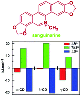
The supramolecular interactions for the formation of inclusion complexes of sanguinarine with α-, β-, and γ-cyclodextrins (CDs) were studied by UV-vis absorbance, fluorescence, circular dichroism and proton nuclear magnetic resonance spectroscopy, and isothermal titration calorimetry. The results revealed that sanguinarine binds with the three natural CDs in 1
:
1 stoichiometry. The binding affinity followed the order β > α > γ-CDs; the affinity to β-CD was the highest compared to the other two CDs apparently due to the perfect cavity size for the inclusion of sanguinarine into the β-CD cavity. The association of sanguinarine with α-, and β-CDs is synergistically driven by a greater entropy contribution to the Gibbs energy of the association, whereas it was favoured by both enthalpy and entropy contributions for the γ-CD. The NMR study indicated that encapsulation of sanguinarine in α-, and β-CDs, involves partial inclusion and to γ-CD it is non-specific and complete inclusion i.e., both ends of sanguinarine, can be included into the CD cavity.
Introduction
Sanguinarine (SGR) (Fig. 1) is a natural plant alkaloid abundantly distributed in the plants like Sanguinaria canadensis and Chelidonium majus.1–5 Sanguinarine has been the focus of extensive research for its potential drug value. In recent years considerable research on its antimicrobial, antifungal, antioxidant and anti-inflammatory effects,6–10 and ability for selective/preferential elimination of cancer cells has been undertaken.11–14 Sanguinarine is a putative anticancer agent that can modify the genetic material and inflict DNA damage.15,16 A critical limitation of its medicinal application, however, stems from its poor aqueous solubility (<0.3 mg mL−1 or 0.9 mM at 298.15 K), and high sensitivity to external agents such as air, light and nucleophiles, which severely restricts its utility. Several approaches to improve the solubility of the alkaloid, including formation of ‘host–guest’ complexes, have been attempted. Supramolecular complex formations of guest molecules have been shown to aid to circumvent the low stability and solubility problems enabling more efficient bioavailability and enhanced physical and chemical properties to the guest molecules. Moreover, supramolecular complexation has been shown to confer protection to alkaloids against nucleophilic attack, thermal and radiation-induced degradation etc.17 In this regard, supramolecular complexes of alkaloids with cucurbit[n]urils,18,19 cyclodextrins (CDs) and their derivatives are studied in extensive details.20–22 Among these approaches inclusion complexes with natural cyclodextrins offer a number of advantages over the others. Firstly, complexation with natural cyclodextrins does not alter the chemical structure of the alkaloid and does not generate any toxic waste. Secondly, it does not introduce synthetic polymers as some other microencapsulation does, which may hinder its applications.
 |
| Fig. 1 Molecular structure of sanguinarine. | |
Cyclodextrins (CDs) are a family of molecules comprising of several glucopyranose units bound together to form a ring, linked by α-1,4 glycosidic bonds.23 CDs have been named depending on the number of glucose units. The natural CDs namely α-, β-, and γ-CDs containing six, seven, and eight glucopyranose units, respectively, are most common among all the CDs and used mainly as entrapping vehicles. CDs are rigid, cone shaped with a semipolar interior cavity24,25 and have the unique ability to form stable inclusion complexes with a variety of molecules in which guest molecules are included in the hydrophobic cavities of the CDs through ‘host–guest’ complexation in aqueous solution.26,27 This ability of inclusion by CDs leads them to be of extensive use in the pharmaceutical applications. Among the three natural CDs, β-CD is the most widely used one for this purpose as its cavity size is suitable for common drug molecules with molar masses in the range 200 to 800 g mol−1.28
Several papers have been published studying the physicochemical properties of sanguinarine inclusion with cucurbits and the derivatives of CDs17,29–31 but to the best of our knowledge there are no reports on the comparative association and thermodynamics of inclusion with natural CDs. Therefore, in this work, inclusion complexes of SGR with α-, β-, and γ-CDs are studied by various techniques such as UV-vis absorption, fluorescence, circular dichroism and NMR spectroscopy, and isothermal titration calorimetry. Among these techniques calorimetry is an extremely powerful and highly sensitive one for the characterization of thermodynamic parameters and interaction mechanisms of drugs with CDs.32 It is used for the determination of thermodynamic parameters of host–guest complexation, particularly that of weak interactions with great success. The comparative data with these natural CDs may give information for use as improved drug carrier and promote its application in pharmaceutical industries.
Results and discussion
Effect of natural cyclodextrins on the UV-vis spectrum of sanguinarine
Sanguinarine is a highly conjugated aromatic planner molecule. It exhibits interesting pH dependant equilibrium between the cationic iminium and neutral alkanolamine forms with a pKa of 8.06.16 The iminium form of SGR exists in acidic condition whereas the alkanolamine form is present in basic conditions. Both forms are present in solution of physiological pH with iminium form being the predominant one. The charged iminium form is biologically more relevant. This form only binds to nucleic acids.33 So, a buffer of pH of 6.2 where only the iminium form persists was chosen for the studies.
SGR has a characteristic absorption spectrum in the UV-vis range of 300–500 nm (Fig. 2A, curve 1) with two clear maxima, one sharp peak at 327 nm and a broad one around 470 nm. The peaks at 327 and 470 nm correspond to the K and B absorbance bands of the aromatic chromophore and the conjugated double bond, respectively, and arise due to the π → π* transitions of the molecule.34,35 The binding of SGR with the three natural CDs was first studied by spectrophotometric titration. Fig. 2 displays changes in the UV-vis absorbance spectra of SGR (5 μM) in the presence of various concentrations of the CDs. In the presence of the CDs, a weak hypochromic effect was observed without any shift of wavelength maximum of the K absorption band (327 nm) and these are highlighted in the inset of Fig. 2. Changes in the 327 nm band were higher in the case of β-CD than those observed with the other two CDs and follow the order β > α > γ-CD. However, it may be noticed that not much change was effected in the B absorbance band (420 nm) when complexes are formed. These spectral changes result essentially from the interaction between SGR and the CDs and suggest the formation of inclusion complexes.
 |
| Fig. 2 UV-vis spectra of SGR in the presence of α-, β-, γ-CDs of various concentrations at 298.15 K, pH 6.2. Panel A: changes in the absorption spectra of SGR (5 μM, curves 1–4) upon titration with 0, 10, 30, 50 μM of α-CD. Panel B: curves 1–5 (B) with 0, 10, 30, 50, 70 μM of β-CD. Panel C: curves 1–5 with 0, 10, 30, 50, 70 μM of γ-CD. Inset: changes in the 327 nm band are highlighted in each case. | |
Fluorescence spectral changes and determination of the binding constant of host–guest complexation
Sanguinarine is a strong fluorescent molecule that emits in the 500–700 nm region with maximum at 564 nm when excited at 470 nm. Binding of SGR to the CDs was studied by fluorescence spectroscopy. Fluorescence intensity of SGR enhanced with increasing concentration of α-CD, while in the presence of β-, and γ-CDs, the fluorescence intensity was quenched. In Fig. 3, the fluorescence changes of SGR (1 μM) in the presence of α-, β-, and γ-CDs are presented. In all the cases, a moderate change in the fluorescence intensity was observed when CDs were added to the solution of the alkaloid. The fluorescence intensity change of SGR with CDs is apparently due to the host–guest molecular interactions and consequent change of microenvironment of the guest molecule. The change in fluorescence intensity in the presence of CDs may be not only for inclusion complex but also the outer complexes formed by association.
 |
| Fig. 3 Steady-state fluorescence spectral changes of SGR (1 μM, curves 1–6) (A) upon titration with 0, 40, 80, 120, 160, 180 μM of α-CD; (B) curves 1–6 with 0, 10, 30, 50, 70, 90 μM of β-CD; and (C) curves 1–5 with 0, 20, 40, 60, 80 μM of γ-CD. SGR was excited at 420 nm. | |
The apparent association constant or inclusion constant of the complexes of SGR with the three CDs was calculated by the following method. Assuming a 1
:
1 stoichiometry (vide infra) of complex formation between SGR molecules and each of the three CDs examined, the inclusion constant was determined to estimate the inclusion capacity of the CDs. The complexation of SGR with the three CDs are expressed as
|
 | (1) |
The association constant (KBH) of the complex is given by
|
 | (2) |
where [SGR], [CD] and [SGR–CD complex] are equilibrium concentrations. The association constant can be obtained from the analysis of sequential fluorescence change at various CD concentrations by using double-reciprocal plots or Benesi–Hildebrand equation.
22,36 |
 | (3) |
Here F, Fo, f, KBH, and [CD]o are the fluorescence of SGR in the presence of CD, in the absence of CD, a constant, the association constant for the formation of the 1
:
1 sanguinarine–CD inclusion complex, and the initial concentration of CDs, respectively. Double reciprocal plots, i.e. plot of 1/Δ[F − Fo] versus 1/[CD]o, were constructed and depicted in Fig. S1 (ESI†). A good linearity of the plots with a correlation coefficient (R) close to unity (Table 1) supports the presumed 1
:
1 stoichiometry of complexation. The association constants of the complexation between SGR with all CDs were calculated from the ratio of the intercept over slope. It can be seen that SGR–β-CD has the highest association constant followed by α-CD and the magnitude of the values followed the order β > α > γ-CDs. The binding ability of CDs to associate with SGR appears to be related to the proper cavity size of the CDs. As the cavity size of β-CD is suitable for inclusion, the association constant of SGR is higher compared to other CDs. We tried to analyze the data by a 1
:
2 stoichiometry and found that the no linear Benesi–Hildebrand plot was obtained. This further validates the assumption of a 1
:
1 stoichiometry. Furthermore, the assumption of a 1
:
1 stoichiometry was also substantiated by the results from calorimetry studies (vide infra).
Table 1 Association constant derived from spectrofluorimetric study by Benesi–Hildebrand analysis for SGR–CDs complexesa
Parameter |
α-CD |
β-CD |
γ-CD |
The data in this table were obtained from studies conducted at 298.15 ± 1 K and averages of three determinations. KBH is the Benesi–Hildebrand binding constant determined according to eqn (3). |
KBH/103 (M−1) |
2.56 ± 0.12 |
9.98 ± 0.21 |
2.09 ± 0.13 |
Correlation coefficient (R) |
0.9990 |
0.9993 |
0.9998 |
Calorimetric characterization of the complexation
Isothermal calorimetric titration (ITC) is the most sensitive and accurate analytical techniques for the determination of weak binding constants and various thermodynamic parameters in host–guest complexation with precise accuracy. This technique can determine binding constants in the of ranging 108 to 102 M−1, making it a preferred method than any other analytical methods.37 It has become an effective method for directly determining the thermodynamic parameters instead of the previously used van't Hoff equation methodology.38 The ITC profiles of SGR binding to all the CDs are presented in Fig. 4. It can be observed that in all the cases association of the CDs with the SGR is exothermic in nature and there is only one binding event in the entire titration profile. The thermodynamic parameters determined from the ITC experiments for the association of SGR with the three CDs are summarized in Table 2. The order of binding constants determined from the ITC experiments is similar and consistent with those obtained from the analysis of the fluorescence data. The associations constant (Ka) is the highest for the β-CD and followed the order as β > α > γ-CDs. In all cases, the complexation process is exothermic (ΔHo < 0) and spontaneous (ΔGo < 0) with positive entropic contribution. But in case of α-, and β-CDs, the reaction is primarily entropy driven whereas it is enthalpy–entropy favoured in the case of γ-CD. The stoichiometry (N) of the association of about one further suggests that only 1
:
1 complexation has occurred in the formation of complexes of sanguinarine with CDs which is in agreement with the 1
:
1 complexation revealed from the Benesi–Hildebrand plot analysis of the spectrofluorimetric data (vide supra).
 |
| Fig. 4 Representative ITC profiles for the titration of SGR (0.5–1.0 mM) with (A) α-, (120 mM) (B) β-, (15 mM) and (C) γ-CDs (100 mM), respectively, in phosphate buffer 10 mM [Na+], pH = 6.2, at 298.15 K. The top panels represent the raw data for the sequential injection of CDs into the SGR, after correction of heat of dilution (curves in the upper panel offset for clarity) and the bottom panels show the integrated heat data after correction of heat of dilution against the mole ratio of [SGR]/CD. The data points were fitted to one site model, and the solid lines represent the best-fit data. | |
Table 2 ITC derived thermodynamic parameters for the binding of SGR with CDs at 298.15 Ka
CD |
Binding constant (Ka) × 103 M−1 |
N |
ΔHo kJ mol−1 |
TΔSo kJ mol−1 |
ΔGo kJ mol−1 |
ΔHo is the enthalpy change and TΔSo the entropy contribution. N is the binding stoichiometry. The values of ΔGo (Gibbs energy change) were determined using ΔGo = ΔHo − TΔSo, with the indicated uncertainty value. |
α |
2.39 ± 0.19 |
1.15 ± 0.15 |
−3.32 ± 0.04 |
15.96 |
−19.28 ± 0.04 |
β |
9.53 ± 0.38 |
1.09 ± 0.14 |
−0.58 ± 0.02 |
20.22 |
−20.80 ± 0.02 |
γ |
2.04 ± 0.11 |
0.95 ± 0.12 |
−9.98 ± 0.09 |
8.90 |
−18.88 ± 0.09 |
An analysis of the thermodynamic parameters, especially ΔHo and ΔSo, of the complexation leads to identification of the types of non-covalent forces, viz. electrostatic, hydrophobic, van der Waals, and H-bonding involved in the host–guest interaction. The ΔHo value represents a global heat resulting from the interaction while large positive entropy changes (ΔSo) usually arise from the significantly important translational and conformational freedoms of host and guest upon complexation. The change in the thermodynamic parameters observed due to the complexation were related to the effect of water molecules. A number of mechanisms have been proposed as the main driving force for the spontaneous inclusion complexation with guest molecules. The principal forces involved are van der Waals and hydrophobic interactions. While hydrophobic interactions are entropy driven with minor contribution of favorable or unfavorable enthalpies of interaction (|ΔHo| < |TΔSo|), van der Waals interactions are essentially enthalpy-driven processes, where the enthalpy of the interaction is larger than the contribution of favorable or unfavorable entropies of the interactions (|ΔHo| > |TΔSo|).39,40 The binding of SGR with α-, and β-CDs are entropy driven as the enthalpy values of the interaction are small compared to the entropy of the interactions (|ΔHo| < |TΔSo|). This indicates hydrophobic interactions play major role in the complexation in these cases. However, SGR association with γ-CD is favoured by both enthalpy and entropy contributions to the Gibbs energy as binding enthalpy and change in entropy for the process are comparable.
In inclusion phenomena, the binding process generally is dependent on the entropy change. The entropic gain during the process usually arises from change in the translational and conformational freedom upon the interaction of the guest within the CD cavity. The inclusion of SGR into the CD cavity in aqueous solution results in a substantial rearrangement and expulsion of the water molecules, originally solvated in the CD cavity, to the bulk water. The change in entropy can be related to the number of water released by the guest molecules during inclusion. By taking into account the initial water molecules included into the CD, the 1
:
1 complexation interaction of the SGR with CD in aqueous solution can be written as
|
SGR·aH2O + CD·bH2O ⇔ SGR·CD·(a + b − c)H2O + cH2O
| (4) |
where
a,
b, and
c represent the number of water molecules interacting with the free guest molecule, number of tightly bound hydration water molecules inside the free CD cavity, and the net displacement of water upon inclusion, respectively.
26,31,41 So, the net entropic gain depends on the net displacement of water upon complexation.
Naidoo et al.42 had showed from the molecular dynamics (MD) computer simulations and pulse-field-gradient spin-echo nuclear magnetic resonance (PGSE NMR) experiments that the anomalous solubility of β-CD in water compared to the other two CDs is due to its tendency to highly increase the local water structure in the cavity and around the CD molecule. They also showed that spatial distribution of water into the CD cavity is higher for β-CD and follow the order β → α → γ-CDs. Therefore, the number of water molecules released from β-CD cavity is higher during inclusion of a guest molecules compared to the other CDs. As the release of water molecules from the cavity to the bulk water is related to the entropy gain of the overall process, the entropy change for SGR inclusion into β-CD cavity is highest and the process is essentially entropy driven. The order of release of water molecules for these natural CDs decrease as β → α → γ-CD making the process for SGR inclusion from entropy driven to enthalpy–entropy driven process.
Induced circular dichroism studies
Sanguinarine does not have any optical property as it is a planar achiral molecule. Induced circular dichroism can be generated from an achiral molecule like SGR due to the optical activity induced by the CDs. It is known that in the chiral environment of nucleic acid, SGR acquires induced optical activity.16,43 In this context, Harata et al.44 had predicted the sign of induced circular dichroism of the guest molecules by theoretical calculations. Positive induced spectra of the guest will appear when the transition dipole moment of the included guest is parallel to the torus symmetry axis of the CD cavity (axial inclusion), while a negative induced circular dichroism spectral pattern is generated when it is perpendicularly oriented (equatorial inclusion). The induced circular dichroism spectra of sanguinarine with α-, β- and γ-CDs are presented in Fig. 5. All the induced bands are confined to the 300–400 nm regions. The induced circular dichroism spectra of SGR in CDs showed a peak with maxima around 327 nm due to the B absorbance band of the π → π* transitions. The induced circular dichroism spectra of SGR with α-, and β-CDs are positive while for γ-CD it is negative. So the dipole moments of sanguinarine appear to be parallely oriented with torus axis of α-, and β-CDs, whereas it is perpendicularly oriented for γ-CD complexes. Sanguinarine is thus included into α-, and β-CDs by axial inclusion whereas the SGR/γ-CD inclusion occurs by equatorial inclusion. Furthermore, the intensity of the β-CD induced positive circular dichroism was much stronger than that of the other two, reiterating a stronger interaction here. Thus, the induced circular dichroism spectra propose the formation of an inclusion complex in which the alkaloid exist inside the α-, and β-CD cavity by parallely and in the γ-CD in the axial mode.
 |
| Fig. 5 Representative induced circular dichroism spectra of SGR (50 μM) in the presence of 50 μM of each α-, β-, γ-CD. | |
The induced CD titration data was also used to establish the stoichiometry. We obtained a linear Benesi–Hildebrand plot (not shown) confirming the 1
:
1 stoichiometry of complexation as deduced from fluorescence and ITC results.
Inclusion complex formation studied by NMR spectroscopy
Nuclear magnetic resonance experiments are useful techniques to investigate the possible inclusion mode and the relative orientation of the guest molecule inside the hydrophobic cavity of CD host molecules.45 We performed 1H NMR of SGR–CD complexes which is very helpful in elucidating the molecular conformation of inclusion complexes between CDs and SGR. The formation of inclusion complexes lead to the chemical shifts in the 1H-NMR spectra of SGR, which has provided valuable information for deducing the part of the SGR molecule that is inserted into the CD cavity. The selected region of aromatic protons of SGR in the 1H-NMR spectra in the presence of all three CDs are presented in ESI Fig. S2.† The detailed changes in the chemical shift (Δδ), calculated for the protons in SGR by using eqn (5) are shown in Table 3. |
Δδ = δ (complex) − δ (guest)
| (5) |
where δ (complex) and δ (guest) are the chemical shift of protons of associated and free guest, respectively. As in Table 3, presence of CDs caused a downfield shift in the chemical shifts for almost all protons of SGR. Especially noteworthy is the large shifts occurred for C-1 and C-11 protons of sanguinarine which was probably due to their relatively strong interaction with the hydroxyl groups in the narrow and wide rims of CDs.
Table 3 1H NMR (600 MHz) chemical shift (δ) data (in ppm) of SGR and SGR–CD mixturesa
|
|
SGR |
SGR/α-CD complex |
Δδ |
SGR/β-CD complex |
Δδ |
SGR/γ-CD complex |
Δδ |
s-singlet, d-doublet. |
C-1 |
s |
7.0170 |
7.3320 |
0.315 |
7.0910 |
0.0740 |
7.165 |
0.148 |
C-4 |
s |
7.5040 |
7.7160 |
0.212 |
7.5630 |
0.0590 |
7.465 |
−0.039 |
C-6 |
s |
9.3610 |
9.4680 |
0.107 |
9.4100 |
0.0490 |
9.440 |
0.079 |
C-9 |
d |
7.5995 |
7.8240 |
0.2245 |
7.5630 |
−0.0365 |
7.822 |
0.2225 |
C-10 |
d |
7.8220 |
8.1035 |
0.2815 |
7.8995 |
0.0775 |
8.057 |
0.235 |
C-11 |
d |
7.8505 |
8.1543 |
0.3038 |
7.9305 |
0.0800 |
8.168 |
0.3175 |
C-12 |
d |
7.6725 |
7.8983 |
0.2258 |
7.7210 |
0.0485 |
7.862 |
0.1895 |
I-CH2 |
s |
6.2140 |
6.2980 |
0.084 |
6.2530 |
0.0390 |
6.276 |
0.062 |
II-CH2 |
d |
6.4380 |
6.4865 |
0.0485 |
6.4710 |
0.0330 |
6.5075 |
0.0695 |
–CH3 |
s |
4.5170 |
4.6850 |
0.168 |
4.5700 |
0.0530 |
4.638 |
0.121 |
Two-dimensional (2D) NMR spectroscopy provides accurate information about the spatial proximity of protons of host and guest by examination of intermolecular dipolar cross correlations. Two protons that are closely located in space can produce a Nuclear Overhauser Effect (NOE) cross-correlation in NOE spectroscopy (NOESY). The high frequency region of the 1H-NMR spectrum (6.0–10.0 ppm) proved to be appropriate to monitor the NOE peaks between SGR and CDs. The NOSEY spectrum of SGR/γ-CD complex in D2O is presented in Fig. 6. Significant NOE cross peaks between almost all aromatic protons of SGR (circled in the Fig. 6) and H-5, H-6 protons of CD was observed implying complete SGR inclusion into the γ-CD cavity. But the cross peaks between two set of dioxazole protons (C-I and C-II) with H-3, H-5 and/or H-6 protons of CDs indicate that both sides of the SGR can be included into the γ-CD cavity. It was also shown that in the case of α-, and β-CDs, NOE cross peak between dioxazole protons (C-I and C-II) of SGR and H-3, H-5, H-6 protons of CD indicates that SGR could also be included also into α-, and β-CD cavity (Fig. 7 and 8). Absence of significant other cross peaks of aromatic protons of SGR with protons of α-, β-CDs indicate partial inclusion into the CD cavity. Nevertheless, ‘external complexes’ of SGR on the outer surface of all the CDs may also be present in the solution along with the inclusion complex. The schematic representation of all three inclusion complexes with comparative diameter of cyclodextrin is shown in Fig. 9.
 |
| Fig. 6 2D NOESY contour map of SGR/γ-CD inclusion complex in D2O. The cross-peaks between two protons of the SGR and CD are circled in the spectra. | |
 |
| Fig. 7 NOESY contour map of SGR/α-CD inclusion complex in D2O at 298.15 K. The cross-peaks between two protons of the host and guest are circled in the spectra. | |
 |
| Fig. 8 NOESY contour map of SGR/β-CD inclusion complex in D2O at 298.15 K. The cross-peaks between two protons of the host and guest are circled in the spectra. | |
 |
| Fig. 9 A schematic representation of inclusion complexes of SGR with all the cyclodextrins with comparative rim diameter. | |
Conclusions
This study focused on the formation and comparative determination of the association constants and thermodynamic parameters of inclusion complexes of the benzophenanthridine alkaloid sanguinarine with three natural CDs by spectroscopic and calorimetric techniques. The results confirm that all three CDs formed inclusion complexes with SGR in 1
:
1 stoichiometry. The association constant with β-CD is higher due to the proper fitted cavity and the association constant order followed as β > α > γ-CDs. Thermodynamic parameters for the association obtained from the calorimetric study confirmed that solvated water molecules in the CD cavity play a crucial roles in the binding process. The insertion of SGR molecule inside the CD cavity largely depends on the number of released water molecule to the bulk water. The process of water molecule release swings the binding synergistically as entropy driven process. Two-dimensional NOESY study confirms that both ends of SGR molecules may be included into γ-CD cavity from the wider side of the cavity whereas in the case of other two CDs it is specific and partial inclusion. Thermodynamic parameters obtained from the calorimetric study for the inclusion of SGR into these natural CDs can be regarded as an important step towards the design of novel formulation as drug and healthcare products.
Material and methods
Chemicals
Sanguinarine chloride hydrate (sanguinarine >98% purity, MW = 367.8 Da) and deuterated water (99.8% atom D) were obtained from Sigma-Aldrich LLC (MO, USA). The concentration of SGR solution was determined spectrophotometrically using its molar absorption coefficient (ε = 30
700 M−1 cm−1 at 327 nm in acidic aqueous solution) reported in the literature.46 Three natural CD, viz. α-, β-, γ-CDs were obtained from Acros Organics (purity ≥ 99%) and dried in a vacuum desiccator prior to the use. Other reagents and chemicals were of analytical grade. Phosphate buffer of 10 mM [Na+], pH = 6.2 was used for fluorescence and calorimetric studies. NMR experiments were done in D2O.
Experimental procedures
Ultraviolet-visible spectroscopy. Absorption spectral measurements were carried out in a Jasco V660 double beam double monochromator spectrophotometer (Jasco International Co., Hachioji, Japan) in 1 cm path length quartz cuvettes at 298.15 ± 1 K. The concentration of sanguinarine was held constant. Appropriate amounts of CDs were added to the SGR sample to vary the final concentration of the CDs in the solutions.
Fluorescence spectroscopy. Changes in the fluorescence spectra of SGR with increasing concentration of CDs were measured on a Shimadzu RF 5301-PC spectrofluorimeter (Shimadzu Corporation, Kyoto, Japan) in a 1 cm path length quartz cuvette. Emission spectra of SGR were obtained in the 500–700 nm regions by exciting at 470 nm. All the fluorescence titrations were carried out at 298.15 ± 1 K keeping a fixed concentration of the SGR and varying the concentration of the CDs.
Circular dichroism spectroscopy. Circular dichroism spectra of SGR-CD complexes were recorded at 298.15 ± 1 K on a Jasco J815 spectropolarimeter (Jasco International Co. Ltd.) equipped with a Peltier controlled cell holder and temperature controller in a rectangular 1.0 cm path length cuvette. Scan speed of 100 nm min−1 and a bandwidth of 1.0 nm were applied to obtain the CD spectra. Each sample was scanned five times to improve signal to noise ratio and smoothed within a permissible limits using the software of the unit. The ellipticity (θ) values are expressed in units of milli degree.
Isothermal titration calorimetry. ITC study was performed at 298.15 K in a Microcal VP-ITC unit (MicroCal LLC, Northampton, MA, USA) after electrical and chemical calibrations. The titrations involved injecting aliquots of the CD solutions from the rotating syringe to SGR solution kept in the calorimeter cell. The duration of each injection was 10 s with a time gap of 300 s between injections. Control experiments to determine the heat of dilution of the CDs were performed by injecting identical volumes of CDs into the buffer solution. After subtracting the heat of dilution from the heat of SGR–CD reaction, the heats of the binding were plotted as a function of the mole ratio of [CD/SGR]. The data were fitted with ‘one set of binding sites’ model to give the binding constant (Ka), the stoichiometry (N) and the enthalpy change of binding (ΔHo). The Gibbs energy change (ΔGo), and the entropic contribution to the binding (TΔSo) were subsequently determined from standard thermodynamic relationships described earlier.47
Nuclear magnetic resonance spectroscopy. NMR spectra of SGR and its complexes with natural CDs were recorded with a 600 MHz Bruker Avance spectrometer equipped with a TCI CryoProbe at 298.15 K. Two-dimensional (2D) NMR studies (NOESY) were carried out in phase-sensitive mode with 32 scans and 2 × 192 free induction decays. 2D data were processed with Gaussian apodization. Chemical shifts are reported in parts per million (ppm).
Acknowledgements
The authors acknowledge the financial support provided by Council of Scientific and Industrial Research (CSIR) network project (GenCODE, BSC0123) for this work. S. Hazra is a recipient of Senior Research Fellowship of CSIR. The help and cooperation from the colleagues of Biophysical Chemistry Laboratory during this investigation and the able technical assistance of Mr E. Padmanabhan with NMR spectral measurements are gratefully acknowledged.
References
- P. J. Facchini, J. M. Hagel, D. K. Liscombe, N. Loukanina, B. P. MacLeod, N. Samanani and K. G. Zulak, Phytochem. Rev., 2007, 6, 97–124 CrossRef CAS.
- E. A. Abizov, O. N. Tolkachev, I. E. Kopylova and A. N. Luferov, Pharm. Chem. J., 2003, 37, 413–414 CrossRef CAS.
- G. B. Mahady and C. W. W. Beecher, Planta Med., 1994, 60, 553–557 CrossRef CAS PubMed.
- A. C. Santos and P. Adkilen, J. Am. Chem. Soc., 1932, 34, 2923–2924 CrossRef.
- S. Campbell, J. Affolter and W. Randle, Econ. Bot., 2007, 61, 223–234 CrossRef.
- K. C. Godowski, J. Clin. Dent., 1989, 4, 96–101 Search PubMed.
- D. Walterova, J. Ulrichova, I. Valka and J. Vicar, Acta Univ. Palacki. Olomuc., Fac. Med., 1995, 139, 7–16 CAS.
- I. D. Stefano, G. Raspaglio, G. F. Zannoni, D. Travaglia, M. G. Priscoa, M. Mosca, C. Ferlini, G. Scambia and D. Gallo, Biochem. Pharmacol., 2009, 78, 1374–1381 CrossRef PubMed.
- M. Sun, W. Lou, J. Y. Chun, D. S. Cho, N. Nadiminty, C. P. Evans, J. Chen, J. Yue, Q. Zhou and A. C. Gao, Genes Cancer, 2010, 1, 283–292 CrossRef CAS PubMed.
- J. Hammerová, S. Uldrijan, E. Táborská and I. Slaninová, J. Dermatol. Sci., 2011, 62, 22–35 Search PubMed.
- M. H. Han, G.-Y. Kim, Y. H. Yoo and Y. H. Choi, Toxicol. Lett., 2013, 220, 157–166 CrossRef CAS PubMed.
- J.-Y. Xu, Q.-H. Meng, Y. Chong, Y. Jiao, L. Zhao, E. M. Rosen and S. Fan, Oncol. Rep., 2012, 28, 2264–2270 CAS.
- Z. Ding, S. C. Tang, P. Weerasinghe, X. Yang, A. Pater and A. Liepins, Biochem. Pharmacol., 2002, 63, 1415–1421 CrossRef CAS.
- N. Ahmad, S. Gupta, M. M. Husain, K. M. Heiskanen and H. Mukhtar, Clin. Cancer Res., 2000, 6, 1524–1528 CAS.
- M. Maiti and G. Suresh Kumar, Med. Res. Rev., 2007, 27, 649–695 CrossRef CAS PubMed.
- G. Suresh Kumar and S. Hazra, RSC Adv., 2014, 4, 56518–56531 RSC.
- Z. Miskolczy, M. Megyesi, G. Tárkányi, R. Mizsei and L. Biczók, Org. Biomol. Chem., 2011, 9, 1061–1070 CAS.
- M. Megyesi, L. Biczók and I. Jablonkai, J. Phys. Chem. C, 2008, 112, 3410–3416 CAS.
- C. Li, J. Li and X. Jia, Org. Biomol. Chem., 2009, 7, 2699–2703 CAS.
- Y. Liu, G.-S. Chen, Y. Chen, F. Ding and J. Chen, Org. Biomol. Chem., 2005, 3, 2519–2523 CAS.
- I. V. Terekhova, Curr. Drug Discovery Technol., 2008, 5, 168–172 CrossRef CAS.
- S. Hazra, M. Hossain and G. Suresh Kumar, J. Inclusion Phenom. Macrocyclic Chem., 2014, 78, 311–323 CrossRef CAS PubMed.
- J. L. Koontz, J. E. Marcy, S. F. O'Keefe and S. E. Duncan, J. Agric. Food Chem., 2009, 57, 1162–1171 CrossRef CAS PubMed.
- K. A. Connors, Chem. Rev., 1997, 97, 1325–1357 CrossRef CAS PubMed.
- J. Szejtli, Chem. Rev., 1998, 98, 1743–1754 CrossRef CAS PubMed.
- M. V. Rekharsky and Y. Inoue, Chem. Rev., 1998, 98, 1875–1918 CrossRef CAS PubMed.
- A. R. Hedges, Chem. Rev., 1998, 98, 2035–2044 CrossRef CAS PubMed.
- J. Wang, Y. Cao, B. Sun and C. Wang, Food Chem., 2011, 127, 1680–1685 CrossRef CAS PubMed.
- G. H. Cho and H. Pedersen, Biotechnol. Tech., 1998, 12, 833–838 CrossRef CAS.
- V. Boldescu, I. Kacso, G. Borodi, I. Bratu and G. Duca, J. Inclusion Phenom. Macrocyclic Chem., 2008, 62, 143–148 CrossRef CAS.
- C.-F. Li, L.-M. Du and H.-M. Zhang, Spectrochim. Acta, 2010, 75, 912–917 CrossRef PubMed.
- K. Bouchemal and S. Mazzaferro, Drug Discovery Today, 2012, 17, 623–629 CrossRef CAS PubMed.
- A. Sen and M. Maiti, Biochem. Pharmacol., 1994, 48, 2097–2102 CrossRef CAS.
- Z. P. Du, Q. L. Suo, X. Y. Zhang, L. W. Zhang and X. H. Wei, Chin. Chem. Lett., 2008, 19, 1465–1469 CrossRef CAS PubMed.
- J. Li, B. Li, Y. Wu, S. Shuang, C. Dong and M. M. F. Choi, Spectrochim. Acta, 2012, 95, 80–85 CrossRef CAS PubMed.
- H. A. Benesi and J. H. Hildebrand, J. Am. Chem. Soc., 1949, 71, 2703–2707 CrossRef CAS.
- G. Wenz, C. Strassnig, C. Thiele, A. Engelke, B. Morgenstern and K. Hegetschweiler, Chem.–Eur. J., 2008, 14, 7202–7211 CrossRef CAS PubMed.
- G. H. Holdgate and W. H. J. Ward, Drug Discovery Today, 2005, 10, 1543–1550 CrossRef CAS.
- M. V. Rekharsky and Y. Inoue, Microcalorimetry in Cyclodextrins and Their Complexes: Chemistry, Analytical Methods, Applications, Wiley–VCH Verlag GmbH & Co. KGaA, 2006, pp. 215–222 Search PubMed.
- A. Cooper, C. M. Johnson, J. H. Lakey and M. Nöllmann, Biophys. Chem., 2001, 93, 215–230 CrossRef CAS.
- N. A. Todorova and F. P. Schwarz, J. Chem. Thermodyn., 2007, 39, 1038–1048 CrossRef CAS PubMed.
- K. J. Naidoo, J. Y.-J. Chen, J. L. M. Jansson, G. Widmalm and A. Maliniak, J. Phys. Chem. B, 2004, 108, 4236–4238 CrossRef CAS.
- M. Maiti, S. Das, A. Sen, A. Das, G. Suresh Kumar and R. Nandi, J. Biomol. Struct. Dyn., 2002, 20, 455–464 CAS.
- K. Harata and H. Uedaira, Bull. Chem. Soc. Jpn., 1975, 48, 375–378 CrossRef CAS.
- E. Lasonder and W. D. Weringa, J. Colloid Interface Sci., 1990, 139, 469–478 CrossRef CAS.
- S. Hazra and G. Suresh Kumar, J. Phys. Chem. B, 2014, 118, 3771–3784 CrossRef CAS PubMed.
- S. Hazra, M. Hossain and G. Suresh Kumar, Mol. BioSyst., 2013, 9, 143–153 RSC.
Footnote |
† Electronic supplementary information (ESI) available: Fig. S1 and S2. See DOI: 10.1039/c4ra10204e |
|
This journal is © The Royal Society of Chemistry 2015 |
Click here to see how this site uses Cookies. View our privacy policy here.