DOI:
10.1039/C4RA10158H
(Paper)
RSC Adv., 2014,
4, 55286-55289
Colorimetric and fluorescence recognition of tryptophan and histidine using phthalaldehyde based probe: experimental, computational, cell imaging and fish tissue analysis†
Received
10th September 2014
, Accepted 16th October 2014
First published on 17th October 2014
Abstract
A phthalaldehyde based colorimetric and fluorescence probe (BENPH), selective for tryptophan (Trp) and histidine (His) has been developed. BENPH and its Trp and His adducts are characterized by FTIR, 1H NMR and ESI-Mass spectroscopy. Computational studies support the experimental findings. BENPH can image intracellular Trp and His under a fluorescence microscope. The developed method is used to determine Trp and His in fish muscle.
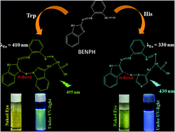
Recognition of trace level amino acids (AA), crucial in medical research and disease diagnosis is commonly based on liquid chromatography,1,2 capillary electrophoresis3,4 with pre- or post-column labeling and spectroscopic detection after suitable derivatization. Recently fluorescence sensing has become popular for operational simplicity, quick response time, non destructive methodology, high sensitivity and low operational cost.5–8
The deficiency of Trp and/or His, indispensible in human nutrition and normal metabolic functions cause negative nitrogen balance and clinical problems.9 L-Trp is vital in protein biosynthesis and serves as the precursor for various bio-molecules, viz. serotonin, melatonin, tryptamine, kynurenine, quinolinic acid and co-enzymes NAD and NADP. Thus, Trp content is closely related to the neurobehavioral effect like, appetite, sleeping-waking-rhythm, impulsivity, aggression, sexual behavior, and pain perception, HIV infections, cancers, Alzheimer's and Parkinson's disease.10–12
Highly reactive imidazole unit of L-His allows it to involve in metallo-proteins and catalytic sites in certain enzymes. L-His is essential for growth and repair of human tissues and transmission of metal ions to biological bases. L-His, a well-known neurotransmitter in the mammalian central nervous system, including retina,13–15 can minimize internal bleeding from microtrauma.16 His deficiency in plasma may cause an impaired nutritional state in patients with chronic kidney disease17 and associated with other diseases such as, epilepsy, Parkinson's disease, Friedreich ataxia and failure of normal erythropoiesis development.18
Therefore, trace level detection of Trp and His is important in biochemical analysis. Available sensors for natural AA are based on hydrogen bonding, electrostatic interaction or π-stacking.19 However, these mechanisms suffer from competitive H-bonding from solvent and hence, selectivity. Few weeks ago, a fluorescence sensor for selective detection of Trp and His is reported.20 However, it also suffers from several disadvantages, viz. use of pyrene derivative, a potential cause of malignant diseases,21 tedious, time consuming and expensive synthetic protocol. Moreover, the probe is quenched by His, incapable to detect Trp or His through naked eye.
Herein, for the first time, we report a phthalaldehyde based benzimidazole derivative for naked eye and fluorescence detection of Trp and His. The method is green, inexpensive for detection and estimation of free Trp and His in biological samples.
Results and discussions
Emission studies
Optimization of pH is necessary for best performance of the probe. BENPH (see Scheme S1 for synthesis and Fig. S1–S4, ESI† for mass, 1H NMR, 13C NMR and FTIR spectra) works best in the pH range 6.5–11, in ethanol–water = 1/1, v/v, (Fig. S5†). The physiological pH being 7.4, is chosen for the entire work. Thus, 0.1 M HEPES buffered solution (in ethanol–water, 1/1, v/v) of BENPH is used throughout. Fig. 1 shows a very weak emission of BENPH at 495 nm (λex = 410 nm) with a quantum yield, 0.0914. Concomitant addition of Trp to BENPH afforded 167 fold fluorescence and 6.2 fold quantum yield enhancement (0.5706). In case of His, corresponding values (λem = 430, λex = 330 nm, quantum yield, 0.142) are 35 fold and 5.2 fold (Fig. 2). Fig. S6† shows the plot of emission intensity vs. Trp concentration while its linear region is presented in Fig. S7.† Similarly Fig. S8 and S9† show the plot of emission intensity vs. His concentration and its linear region respectively. Linear regions are useful for determination of unknown concentrations of Trp and His in a sample.
 |
| Fig. 1 Changes in the fluorescence spectra of BENPH (20 μM, 0.1 M HEPES buffered ethanol–water, 1/1, v/v, pH 7.4) upon gradual addition of Trp (1–300 μM, λex, 410 nm, λem, 495 nm). Inset: corresponding color change under UV light (left: free BENPH). | |
 |
| Fig. 2 Changes in the fluorescence spectra of BENPH (20 μM, in 0.1 M HEPES buffered ethanol–water, 1/1, v/v, pH 7.4) upon gradual addition of His (1–300 μM, λex, 330 nm, λem, 430 nm). Inset: corresponding color change under UV light (left: free BENPH). | |
Further, enhancement of emission intensities of BENPH upon addition of Trp and His is time dependent and reaches maximum after 36 min and 34 min respectively (Fig. S10†). Fig. S11† provides respective plots of emission intensities as a function of time.
Absorption studies
After 40 min of mixing BENPH with Trp/His, changes of absorbance are recorded as a function of [Trp] and [His] (Fig. 3 and 4). Gradual addition of Trp, increases absorbance at 330 nm, 410 nm, 675 nm and 767 nm while that of His increases absorbance at 368 nm and 632 nm. Changes in the absorbance of BENPH as a function of Trp at 675 nm and 767 nm are shown in Fig. S12 and S13† while for His at 632 is shown in Fig. S14.† Moreover, for a given concentration of Trp/His, absorbance of BENPH increases up to 40 minutes. Time dependent absorbance change for Trp and His are shown in Fig. S15† and absorbance vs. time plots for Trp at 675 nm and His at 632 nm are shown in Fig. S16.† Other natural amino acids have failed to alter the emission or absorbance of BENPH under identical conditions. Further, both Trp and His can be monitored in presence of equimolar amount of other amino acids (Fig. S17 and S18†).
 |
| Fig. 3 Changes in the absorbance of BENPH (80 μM, 0.1 M HEPES buffered ethanol–water, 1/1, v/v, pH 7.4) as a function of externally added Trp (1–5000 μM). Inset: corresponding naked eye color change (left: free BENPH). | |
 |
| Fig. 4 Changes in the absorbance of BENPH (80 μM, in 0.1 M HEPES buffered ethanol–water, 1/1, v/v, pH 7.4) as a function of externally added His (1–5000 μM). Inset: corresponding naked eye color change (left: free BENPH). | |
ESI mass (Fig. S19 and S20†), FTIR (Fig. S21†) and 1H NMR (Fig. S22 and S23†) spectra support the sensing mechanism via the formation of Schiff base between BENPH and Trp/His. Job's plots (Fig. S24†) support the formation of 1
:
1 adducts between BENPH and Trp/His. The lower limit of detection of BENPH for Trp/His is 5.0 × 10⁻⁷ M (see ESI†). The binding constant values of BENPH are 1.5 × 10⁴ M−1 for Trp and 1.7 × 10⁴ M−1 for His (Fig. S25†). UV light exposed and naked eye color changes of BENPH upon addition of various amino acids (equimolar) is shown in Fig. S26.†
Weak emission of BENPH is attributed to Twisted Intra-molecular Charge Transfer (TICT) process. Addition of His/Trp leads to the formation of a new imine bond, stabilized via intra-molecular H-bonding between the sp2 N of benzimidazole of the BENPH and –NH of the heterocyclic unit of Trp/His. These reduce the entropy and enhance the rigidity of the molecular assemblies to inhibit TICT to turn on the fluorescence.
The sensing mechanism is supported by 1H NMR titrations by serial addition of Trp (Fig. S22†)/His (Fig. S23†) to the solution of BENPH in DMSO-d6–D2O. Most of the protons in the adducts have up-field shifted, relative to free BENPH. The N–H proton of benzimidazole is missing, due to its exchangeable nature. The imine proton (–CH
N–, marked 1) of free BENPH, appeared at two slightly different positions (δ 7.8, 8.0), coincides into a single peak after addition of Trp (δ 8.18) or His (δ 8.34). This is attributed to the restricted rotation around the imine (–CH
N) bond, stabilized by the formation of H-bond that changes environment of NMR active nuclei. Further, intensity of aldehyde proton (δ 10.48 ppm) of BENPH decreases upon addition of 0.5 eq. Trp/His and ultimately disappear at 1.0 eq. Trp/His, demonstrating the formation of a new imine bond between BENPH and Trp/His. The new imine proton (labeled as 11a/b for Trp/His respectively) resonates at same position with the old one (labeled as 11).
Computational studies
Density functional theoretical (DFT)23 studies have been performed (using 6-31G basis set) to optimize the energies and geometries22 of BENPH and its Trp and His adducts (Fig. 5) which strongly support the aforesaid H-bond assisted sensing mechanism.24 The respective H-bond distances for Trp and His adducts are 2.05 Å and 2.01 Å respectively. Highest Occupied Molecular Orbital (HOMO) and Lowest Unoccupied Molecular Orbital (LUMO) of both the adducts (Fig. 6) reveals that, for HOMO most of the charge resides in the heterocyclic rings of AA part while in LUMO, it reside mainly in the aromatic region of the BENPH moiety. For BENPH, charge densities in HOMO and LUMO are almost same. HOMO–LUMO energy gaps in BENPH, BENPH–Trp and BENPH–His are 3.41 eV, 2.43 eV and 3.18 eV respectively. The new CT bands in the visible region for the adducts are due to the HOMO–LUMO transitions involving benzimidazole unit and heterocyclic units of Trp/His.
 |
| Fig. 5 Probable sensing mechanism of Trp and His by BENPH. | |
 |
| Fig. 6 HOMO–LUMO energies of BENPH and its adducts with Trp and His. | |
Estimation of free Trp and His in muscle of Labeo rohita (Hamilton)
The developed fluorescence method is used for easy and fast determination of free Trp (λex = 410 nm, λem = 495 nm) and His (λex = 330 nm, λem = 430 nm) in fish muscle. Results are in close agreement to that obtained by standard biochemical methods.25,26
Table 1 indicates higher level of free His than free Trp in L. rohita muscle. Advantage of our present method is that a single sensor can measure both Trp and His level in a single experiment while other available methods in literature require individual experiments for each of Trp and His.
Table 1 Comparison of results of present method and a standard method
Amino acid (μ mole g−1 wet fish) |
Present method |
Standard method |
Trp |
2.0746 ± 0.124 |
2.897 ± 0.745 (ref. 25) |
His |
27.377 ± 1.377 |
36.974 ± 2.107 (ref. 26) |
Fig. 7 reveals that intracellular Trp/His can be visualized under fluorescence microscope using BENPH (details in ESI†).
 |
| Fig. 7 Allamandapuberula (Aapocynaceae) cells: without BENPH (1a and 2a), with BENPH under 100× objective lens (1b and 2b). BENPH stained cells pre-exposed to Trp (1c) and His (2c) for 30 min under 40× objective lens. UV filter is used; incubation temperature, 37 °C. | |
Conclusion
A novel phthalaldehyde derived colorimetric and fluorescence probe selectively detect Trp and His. DFT studies support experimental findings. BENPH can image intracellular Trp and His under fluorescence microscope. The developed method has successfully determined Trp and His in fish muscle.
Acknowledgements
AG is grateful to BU for fellowship. Help from Sisir Lohar and Sudipta Das is acknowledged. Assistance from CAS (B. U.) is gratefully acknowledged.
Notes and references
- E. Heftmann, Chromatography, Fundamentals and Applications of Chromatography and Related Differential Migration Methods, Part B: Applications, Elsevier, Amsterdam, 5th edn, 1992 Search PubMed.
- R. I. Khan, R. Onodera, M. R. Amin and N. Mohammed, Amino Acids, 2002, 22, 167 CrossRef CAS PubMed.
- D. R. Baker, Capillary Electrophoresis, Wiley, New York, 1995 Search PubMed.
- W. J. M. Underberg and J. C. M. Waterval, Electrophoresis, 2002, 23, 3922 CrossRef CAS PubMed.
- M.-Y. Chae, X. M. Cherian and A. W. Czarnik, J. Org. Chem., 1993, 58, 5797–5801 CrossRef CAS.
- A. Sahana, A. Banerjee, S. Lohar, S. Panja, S. K. Mukhopadhyay, J. S. Matalobos and D. Das, Chem. Commun., 2013, 49, 7231 RSC.
- M. Vendrell, D. Zhai, J. Cheng Er and Y.-T. Chang, Chem. Rev., 2012, 112, 4391 CrossRef CAS PubMed.
- S. Das, S. Guha, A. Banerjee, S. Lohar, A. Sahana and D. Das, Org. Biomol. Chem., 2011, 9, 7097 CAS.
- P. Fafournoux, A. Bruhat and C. Jousse, Biochem. J., 2000, 351, 1 CrossRef CAS.
-
(a) B. Y. Silber and J. A. J. Schmitt, Biobehav. Rev., 2010, 34, 387 CrossRef CAS PubMed;
(b) M. F. Murray, Lancet Infect. Dis., 2003, 3, 644 CrossRef CAS;
(c) S. Lob, A. Konigsrainer, H. G. Rammensee, G. Opelz and P. Terness, Nat. Rev. Cancer, 2009, 9, 445 CrossRef PubMed.
- M. Kia, A. Islamnezhad, S. Shariati and P. Biparva, Korean J. Chem. Eng., 2011, 28(10), 2064 CrossRef CAS PubMed.
- E. L. Sainio, K. Pulkki and S. N. Young, Amino Acids, 1996, 10, 21 CrossRef CAS PubMed.
- T. E. Creighton, Encyclopedia of Molecular Biology, Wiley, New York, 1999, vol. 2, p. 1147 Search PubMed.
- G. N. Chen, X. P. Wu, J. P. Duan and H. Q. Chen, Talanta, 1999, 49, 319 CrossRef.
- Y. Kusakari, S. Nishikawa, S. Ishiguro and M. Tamai, Curr. Eye Res., 1997, 16, 600 CrossRef CAS.
- M. Watanabe, M. E. Suliman, A. R. Qureshi, E. Garcia-Lopez, P. Barany, O. Heimburger, P. Stenvinkel and B. Lindholm, Am. J. Clin. Nutr., 2008, 87, 1860 CAS.
- D. F. Horrobin, M. S. Mandu, M. Oka, R. O. Morgan, S. C. Cunnane, A. I. Aly, T. Ghayur, M. Schweitzer and R. A. Karmali, Med. Hypotheses, 1979, 5, 969 CrossRef CAS.
- M. L. Rao, H. Stefan, C. Scheid, A. D. S. Kuttler and W. Froscher, Epilepsia, 1993, 34, 347 CrossRef CAS PubMed.
-
(a) Y. He, X. Wang, J. Zhu, S. Zhong and G. Song, Analyst, 2012, 137, 4005 RSC;
(b) M. Hariharan, S. C. Karunakaran and D. Ramaiah, Org. Lett., 2007, 9(3), 417 CrossRef CAS PubMed.
- P. G. Sutariya, A. Pandya, A. Lodha and S. K. Menon, Analyst, 2014, 139, 4794 RSC.
- L. Li, J. Lu, C. Xu, H. Li and X. Yang, Molecules, 2012, 17, 14159 CrossRef CAS PubMed.
- D. Karak, S. Das, S. Lohar, A. Banerjee, A. Sahana, I. Hauli, S. K. Mukhopadhyay, D. A. Safin, M. G. Babashkina, M. Bolte, Y. Garcia and D. Das, Dalton Trans., 2013, 6708 RSC.
- Gaussian 03, Rev. C. 02, Gaussian Inc., Wallingford CT, 2004 Search PubMed.
- S. Lohar, A. Banerjee, A. Sahana, S. Panja, I. Hauli, S. K. Mukhopadhyay and D. Das, Tetrahedron Lett., 2014, 55, 174 CrossRef CAS PubMed.
- M. K. Gaitonde and T. Dovey, Biochem. J., 1970, 117, 907 CAS.
- P. M. Newman and J. H. Turnbull, Biochem. J., 1960, 74, 379 CAS.
Footnote |
† Electronic supplementary information (ESI) available. See DOI: 10.1039/c4ra10158h |
|
This journal is © The Royal Society of Chemistry 2014 |
Click here to see how this site uses Cookies. View our privacy policy here.