A new chiral ligand exchange capillary electrophoresis system based on Zn(II)–L-leucine complexes coordinating with β-cyclodextrin and its application in screening tyrosinase inhibitors

Yuan Suab, Xiaoyu Muac and Li Qi*a
aBeijing National Laboratory for Molecular Sciences, Key Laboratory of Analytical Chemistry for Living Biosystems, Institute of Chemistry, Chinese Academy of Sciences, No. 2 Zhongguancun Beiyijie, 100190 Beijing, P. R. China. E-mail: qili@iccas.ac.cn; Tel: +86-10-82627290
bGraduate School, Shandong Agricultural University, 271018 Shandong, P. R. China
cGraduate School, University of Chinese Academy of Sciences, 19A Yuquanlu, Beijing 100049, P. R. China

Received 29th August 2014 , Accepted 17th October 2014

First published on 17th October 2014


Abstract

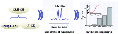
Tyrosinase plays a key role in melanin formation, and it is closely related to hyper pigmentation in animals and enzymatic browning in food. Thus, it is of great significance to screen inhibitors of tyrosinase. In this work, a new chiral ligand exchange-capillary electrophoresis (CLE-CE) system based on the coordination effect of Zn(II)–L-leucine complexes and β-cyclodextrin (β-CD) was developed for screening the inhibitors of tyrosinase. The effects of the concentration of β-CD, buffer pH, the ratio of L-leucine to Zn(II), and the complex concentration were investigated with Dns-D,L-tyrosine, Dns-D,L-valine and Dns-D,L-phenylalanine as the tested analytes. The optimum buffer conditions were composed of 100.0 mM boric acid, 5.0 mM ammonium acetate, 3.0 mM Zn(II), 6.0 mM L-leucine and 4.0 mM β-CD at pH 8.2. It has been found that six pairs of Dns-D,L-AAs could be baseline-separated and five pairs of Dns-D,L-AAs were partly enantioseparated. Then the quantitative analysis of L-tyrosine was conducted and good linearity (r2 = 0.992) was obtained with a concentration ranging from 12.95 μM to 413.3 μM. A kinetics study of tyrosinase was realized, and the Km and Vmax were 636 μM and 312 μmol min−1 mg−1. Moreover, the proposed method was applied in screening the inhibitors of tyrosinase with four kinds of chalcones as the model inhibitors. The results demonstrated that the developed CLE-CE system was favorable for screening enzyme inhibitors, and showed great potential in related drugs discovery and clinical analysis in the future.


1. Introduction

As a polyphenol oxidase, tyrosinase is a copper-containing enzyme and it is closely related to two reactions in the synthetic process of melanin: the hydroxylation of monophenols and the oxidation of o-diphenols to o-quinones.1–5 The excessive accumulation of melanin would lead to both some skin diseases (such as chloasma, age spots, and sunburn) and a change in the apparent and sensory properties of food products.6 It is reported that the formation of the melanin could be controlled by the introduction of tyrosinase inhibitors.6 Therefore, it is significant and highly desirable to screen the effective inhibitors of tyrosinase. Based on the previous studies, the inhibitors of tyrosinase are becoming more and more important in clinical treatment, cosmetic industry and food industry.7,8 Both natural and synthetic compounds have been explored as the effective inhibitors for tyrosinase, such as kojic acid, benzoic acid, hydroquinone and chalcones.9–12 Kojic acid has been widely used as the depigmenting agents in cosmetic and food industry. Meanwhile, chalcones and its derivatives, which are mainly located in plants, have displayed their great inhibition effects on tyrosinase.13–15 In the past decades, many protocols have been developed for screening the inhibitors of tyrosinase, such as UV spectrophotometry, HPLC, GC-MS and electrophoretically mediated microanalysis (EMMA).16–18 However, some of the methods still possessed some disadvantages, such as complicated sample processing, high sample consumption, and being susceptible to interference.15 Therefore, exploring new methods for efficiently screening the inhibitor of tyrosinase is pressing.

In recent years, capillary electrophoresis (CE) is turned out to be an available technique for chiral separation in life science, and this technique has various advantages, including high efficiency, high speed and low cost.19–22 Among the diverse separation modes in CE, chiral ligand exchange capillary electrophoresis (CLE-CE) has been widely applied in enantioseparation of amino acids (AAs) for its obvious advantages, such as high convenience, low cost and controllable enantiomer migration order.23–26 Nowadays, CLE-CE has been applied in the analysis of drugs,27 foods28 and enzymes.29 However, it still faces some challenges, including limited available systems and narrow applications. Thus, it is essential to construct novel CLE-CE systems and broaden their application range. Cyclodextrins (CDs) and their derivatives are the most commonly used chiral selectors in the CE separation. Meanwhile, the previous works displayed that CDs could improve the enantioseparation efficiency of CLE-CE system in some extent.29–31 Therefore, constructing the CLE-CE system based on the cooperation effect of CDs could widen the selection range of chiral ligand and further widen its application. However, to date, only a few papers have been published on the enantioseparation by combing CLE-CE system and CDs system.15,29–34 Furthermore, although it has been reported that chalcones have inhibition effect on tyrosinase,12,16 to our best knowledge, CLE-CE has not been applied in screening the inhibitors of tyrosinase with chalcones as the model inhibitors.

In this work, a new CLE-CE system with L-leucine (L-Leu) as the chiral ligand, Zn(II) as the central ion and β-CD as the additive was developed. Various pairs of D,L-AAs, including the substrate of tyrosinase, D,L-tyrosine (D,L-Tyr), were well enantioseparated under the optimized conditions. In addition, the inhibitory efficiency of tyrosinase was studied. Moreover, the proposed CLE-CE method has been applied in screening the inhibitors of tyrosinase with chalcones as the model compounds, indicating its great potential in clinical analysis and drug discovery in the future.

2. Materials and methods

2.1 Reagents and chemicals

Chalcone, 4-hydroxychalcone (4-HC), 4′-hydroxychalcone (4′-HC), 2-hydroxychalcone (2-HC), 2′-hydroxychalcone (2′-HC) were purchased from J&K Chemistry Company (Beijing, China). Boric acid, ammonium acetate, zinc sulfate, sodium hydroxide, lithium carbonate, tris(hydroxymethyl) aminomethane (Tris), acetone, β-cyclodextrin (β-CD), γ-cyclodextrin (γ-CD), α-cyclodextrin (α-CD) and other reagents were obtained from Beijing Chemical Factory (Beijing, China). Tyrosinase (source: mushrooms) was bought from Express Technology Co., Ltd. (565 U mg−1, Beijing, China). Dansyl chloride (Dns-Cl), D,L-Tyr, D,L-Leu, D,L-valine (D,L-Val), D,L-phenylalanine (D,L-Phe) and other D,L-AAs were from Sigma-Aldrich Chemical Company (St. Louis, USA).

2.2 Instruments and separation conditions

The CE apparatus used in this work consisted of a 1229-High Performance Capillary Electrophoresis (HPCE) analyzer (Beijing Institute of New Technology and Application, Beijing, China), a UV detector (Rilips Photoelectricity Factory, Beijing, China), an uncoated fused-silica capillary (Yongnian Optical Fiber Factory, Hebei, China) and a HW-2000 chromatography workstation (Qianpu Software, Nanjing, China).

The bare fused-silica capillary (60.0 cm × 75.0 μm id, effective length 45.0 cm to detector) was washed by 0.1 M NaOH and water for 30.0 min in turn. Prior to sample injection, the capillary was rinsed with 0.1 M HCl, 0.1 M NaOH, water and running buffer for 2.0 min, respectively. Samples were siphoned into the capillary at 15.0 cm height for 8.0 s and separated at −21.0 kV. Sample detection was performed at 254 nm. All operations were conducted at 25 °C. Generally, the running buffer system was composed of 100.0 mM boric acid, 5.0 mM ammonium acetate, 3.0 mM zinc sulfate, 6.0 mM L-Leu and 4.0 mM β-CD at pH 8.2. All of CLE-CE performance were operated for multiple measurements (n = 3).

2.3 Sample preparation and AAs derivatization

All aqueous solutions were prepared with triply distilled water and stored at 4 °C. Standard sample solutions were prepared by dissolving 2.0 mg of D-, and L-AAs in 40.0 mM lithium carbonate buffer (adjusted to pH 9.5 with 0.1 M HCl), and they were diluted to the required concentrations with lithium carbonate solution in the range of 10–104-fold.

The D,L-AAs were derivatized as described in the previous study.30 Briefly, 20.0 μL D,L-AAs (2.0 mg mL−1), 20.0 μL 40.0 mM lithium carbonate and 20.0 μL Dns-Cl (1.5 mg mL−1 in acetone) were mixed in a 200.0 μL vial and kept at room temperature for 30 min. In order to terminate the reaction, 5.0 μL 2% ethylamine was added. All Dns-D,L-AAs samples were stored at 4 °C before use.

2.4 Enzyme incubation

All enzymatic reactions were carried out in 100 mM NaH2PO4 buffer (pH 6.8) at 25 °C with a final tyrosinase concentration of 565 U mg−1. The enzyme incubation experiments were conducted as following: 40.0 μL of different concentrations of l-Tyr, 40.0 μL NaH2PO4 buffer and 40.0 μL tyrosinase (565 U mg−1) were mixed at 25 °C and incubated for 15 min, then the reactions were ended by heating in boiling water for 10 min, followed by centrifugation for 10 min at 10[thin space (1/6-em)]000 rpm. After that, the supernatant were gathered and derivatized with Dns-Cl by the same processes as that of the standard AAs.

In order to screening the inhibitors of tyrosinase, IC50 (concentration of a substance with half of the maximal inhibitory) of different inhibitors was investigated. The experiments were conducted as following: different concentrations of inhibitors (4-HC, 4′-HC, 2-HC, 2′-HC, chalcone) were mixed with equivoluminal L-Tyr solution (333 μM), then the mixture was incubated with tyrosinase solution for 15 min at 25 °C. After that, the reactions were treated with the same steps as described above.

2.5 Calculation

The inhibition efficiency was calculated according to the following equation:
I = (C2C1)/(C0C1)
where I is the inhibition efficiency, C0 represents the concentration of L-Tyr in the absence of tyrosinase and inhibitor, C1 and C2 are the remaining concentration of L-Tyr in the absence of the inhibitor and in the presence of the inhibitor, respectively.

3. Results and discussion

3.1 Optimization of separation condition

A new CLE-CE system with L-Leu as the chiral ligand and Zn(II) as the central ion was developed in this work. In this CLE-CE system, β-CD was used as the additive. Several key parameters including pH, the ratio of central ion to ligand, the concentrations of the complex and β-CD were investigated in detail with Dns-D,L-Val, Dns-D,L-Tyr and Dns-D,L-Phe (which represented the aromatic D,L-AAs and aliphatic D,L-AAs) as the test analytes to get the optimal separation conditions.
3.1.1 Effect of CDs. CDs have been widely applied in chiral separation owing to its intrinsic chirality.30 The separation performance of different kinds of CDs was investigated by the chiral separation efficiency (Rs) and the plate numbers. As shown in Table S1, it could be observed that α-CD displayed no enantioseparation ability for the test analytes. Meanwhile, other CDs were all suitable for enantioseparation. Although the Rs and plate numbers of γ-CD were higher than that of β-CD, the migration times were prolonged (Table S1). Thus, taking the key factors (Rs, migration times, plate numbers) into consideration, β-CD was the best choice for further study.

It has been reported that the enantioseparation of D,L-AAs in CLE-CE system could be improved in cooperation with β-CD.28,29 Therefore, the influence of the concentration of β-CD (0–5.0 mM) on the chiral recognition was studied. As displayed in Fig. 1, the separation efficiency obviously became better with the addition of β-CD in the buffer solution. We found that when the concentration of β-CD was 4.0 mM, the resolution (Rs) of the three test pairs of Dns-D,L-AAs was higher than 1.5. However, the migration times of the test analytes were greatly prolonged with increasing the concentration of β-CD (Fig. S1A). Based on the CLE-CE requirement of high Rs and shorter migration times, 4.0 mM β-CD was finally chosen for further study.


image file: c4ra09433f-f1.tif
Fig. 1 Influence of β-CD concentration on Rs. Buffer conditions: 100.0 mM boric acid, 5.0 mM ammonium acetate, 3.0 mM Zn(II), 6.0 mM L-Leu, adjusted pH to 8.2, and different concentrations of β-CD ranging from 0–5.0 mM. Capillary: 60.0 cm, 45.0 cm effective × 75.0 μm id; injection: siphoned for 8.0 s at 15.0 cm; voltage: −21.0 kV; UV detection: 254 nm; temperature: 25 °C. Rs1: resolution of Dns-D,L-Val; Rs2: resolution of Dns-D,L-Tyr; Rs3: resolution of Dns-D,L-Phe.
3.1.2 Effect of pH. According to the mechanism of CLE-CE, the ionization equilibrium between the analytes and chiral ligands is closely related to pH of the running buffer. In addition, pH of the running buffer can obviously affect the silicon hydroxyl dissociation on the capillary wall and the electroosmotic flow (EOF) in CLE-CE system. Therefore, it is greatly necessary for studying the influence of pH on Rs in this work. As shown in Fig. 2, it could be observed that the Rs increased obviously with the increasing of pH from 7.9 to 8.3. Meanwhile, the migration times of the test analytes were greatly prolonged (Fig. S1B). Moreover, the current of the CLE-CE system was increased to a higher pH value, which would affect the Rs and peak shapes. Thus, taking all of the factors into consideration, the buffer pH at 8.2 was finally selected for further analysis.
image file: c4ra09433f-f2.tif
Fig. 2 Influence of pH on Rs. Buffer conditions: 100.0 mM boric acid, 5.0 mM ammonium acetate, 3.0 mM Zn(II), 6.0 mM L-Leu and 4.0 mM β-CD at different pH values. Other conditions are the same as Fig. 1.
3.1.3 Effect of Zn(II)–L-Leu. The complexation reaction in CLE-CE is between the chiral ligand and the central ion, thus the ratio of the central ion to the ligand should have a significant effect on the Rs of the test analytes. Therefore, it is essential to investigate the effect of the ratio of Zn(II) to L-Leu on Rs. In this work, the concentration of Zn(II) was fixed at 3.0 mM and the concentration of L-Leu was changed from 1.5 mM to 9.0 mM. As exhibited in Fig. 3, it could be found that the Rs increased when the ratio of Zn(II) to L-Leu was changed from 2[thin space (1/6-em)]:[thin space (1/6-em)]1 to 1[thin space (1/6-em)]:[thin space (1/6-em)]2.5. Moreover, the migration times of the test analytes were prolonged with the ratio increasing (Fig. S1C). In order to get higher Rs in short migration time, the concentration ratio of Zn(II) to L-Leu at 1[thin space (1/6-em)]:[thin space (1/6-em)]2 was chosen for further investigation.
image file: c4ra09433f-f3.tif
Fig. 3 Influence of concentration ratio of Zn(II) to L-Leu on Rs. Buffer conditions: 100.0 mM boric acid, 5.0 mM ammonium acetate, 4.0 mM β-CD, the concentration ratio of Zn(II) to L-Leu was from 2[thin space (1/6-em)]:[thin space (1/6-em)]1 to 1[thin space (1/6-em)]:[thin space (1/6-em)]2.5 with Zn(II) kept at 3.0 mM, adjusted pH to 8.2. Other conditions are the same as Fig. 1.
3.1.4 Effect of complex concentration. The complex concentration was also an important parameter in the formation of ternary mixed metal complexes. Thus, when the ratio of Zn(II) to L-Leu was kept at 1[thin space (1/6-em)]:[thin space (1/6-em)]2, the influence of the complex concentration on Rs was investigated with the concentration of Zn(II) ranging from 2.0 mM to 6.0 mM. Fig. 4 demonstrated that the separation efficiency decreased with the concentration of Zn(II) increasing. Meanwhile, the migration times of the test analytes were also shortened (Fig. S1D). In view of the higher resolution and the shorter migration time, 3.0 mM Zn(II) and 6.0 mM L-Leu were selected as the finally complex concentration for further work.
image file: c4ra09433f-f4.tif
Fig. 4 Influence of complex concentration on Rs. Buffer conditions: 100.0 mM boric acid, 5.0 mM ammonium acetate, 4.0 mM β-CD and different concentrations of Zn(II) complex with L-Leu in the range of 2.0–6.0 mM, the ratio of Zn(II) to L-Leu was kept at 1[thin space (1/6-em)]:[thin space (1/6-em)]2, adjusted pH to 8.2. Other conditions are the same as Fig. 1.

Based on the above experimental results, the optimum buffer was composed of 100.0 mM boric acid, 5.0 mM ammonium acetate, 3.0 mM Zn(II), 6.0 mM L-Leu and 4.0 mM β-CD at pH 8.2. As shown in Table 1 and Fig. 5, we observed that six pairs of Dns-D,L-AAs (containing Dns-D,L-Val, Dns-D,L-Tyr, Dns-D,L-Phe) were baseline-separated, and five pairs of Dns-D,L-AAs were partly separated. It should be noted that under the optimum conditions, although eleven pairs of Dns-D,L-AAs could be well enantioseparated, six pairs of the analytes still could not be enantioseparated as exhibited in Table 1. The reason is unclear, and further exploration should be conducted in the future.

Table 1 Enantioseparation of Dns-D,L-AAs under the optimum conditiona
Dns-D,L-AAs Rsb tLc/min tDc/min
a Running buffer: 100.0 mM boric acid, 5.0 mM NH4 AC, 3.0 mM Zn(II), 6.0 mM L-Leu and 4 mM β-CD at pH 8.2.b Rs = 2(tDtL)/(WD + WL).c t: migration time.
Dns-D,L-Thr 2.31 48.67 51.40
Dns-D,L-Phe 2.05 53.96 55.95
Dns-D,L-Val 1.96 38.46 40.08
Dns-D,L-Tyr 1.84 47.60 49.44
Dns-D,L-Met 1.65 62.41 65.08
Dns-D,L-Asn 1.54 65.01 67.34
Dns-D,L-Trp 1.05 86.12 89.50
Dns-D,L-Ala 1.03 56.23 57.90
Dns-D,L-Pro 0.87 37.23 38.35
Dns-D,L-Ser 0.45 62.70 63.29
Dns-D,L-Asp 0.37 27.99 28.22
Dns-D,L-Glu 0 26.97 26.97
Dns-D,L-Ile 0 42.57 42.57
Dns-D,L-Cys 0 24.97 24.97
Dns-D,L-His 0 21.71 21.71
Dns-D,L-Leu 0 24.99 24.99
Dns-D,L-Arg 0 19.75 19.75


image file: c4ra09433f-f5.tif
Fig. 5 Entioseparation of the three test Dns-D,L-AAs under the optimum conditions. (A) Dns-D,L-Val (5.69 mM); (B) Dns-D,L-Tyr (3.68 mM); (C) Dns-D,L-Phe (4.04 mM). Buffer conditions: 100.0 mM boric acid, 5.0 mM ammonium acetate, 3.0 mM Zn(II), 6.0 mM L-Leu and 4.0 mM β-CD at pH 8.2.

It has been reported that the enantiomer migration order (EMO) of D,L-AAs was related to the different chiral ligand.30,35 As shown in Fig. S2 in this study, Dns-L-Tyr always migrated ahead of Dns-D-Tyr, no matter the ligand was D-Leu or L-Leu, which is different from the reported experiments.30,35 Thus it was speculated that although the stabilization of the formed ternary complexes with L-Leu or D-Leu as the chiral ligand was different, the enantiorecognition ability of the ternary complexes was much improved by coordinating with β-CD. Meanwhile, the EMO of Dns-D,L-Tyrs was also much dependent on β-CD in the proposed CLE-CE system. In other words, the EMO of Dns-D,L-AAs should be Dns-L-AAs > Dns-D-AAs, which could not be tuned if β-CD was used as the coordinators in CLE-CE system (Fig. 2, Table 1 and Fig. S2).

3.2 Discussion on chiral recognition mechanism

In this study, the results exhibited in Fig. 1 and Table S2 clearly displayed that the three test analytes could not be enantioseparated without β-CD. On the other hand, only Dns-D,L-Tyr could be partly enantioseparated among the three test analytes (Table S2) while only β-CD was added in the buffer solution. Therefore, we assumed that the coordination reaction between Zn(II)–L-Leu complex and β-CD could achieve the chiral resolution.

The separation mechanism of ligand exchange is based on the formation of diastereomeric ternary complex between the analytes and the mental–ligand complexes. The principle of CLE-CE is shown as follows:

Zn(L-Sel)2 + L-A → (L-Sel)Zn(L-A) + L-Sel

Zn(L-Sel)2 + D-A → (L-Sel)Zn(D-A) + L-Sel Sel: selector; A: analyte

Resolution is due to the difference in complex stability constants of the mixed complexes with analyte enantiomers. In this work, the three test analytes could not be baseline separated when the CLE-CE system with Zn(II)-L-Leu as the chiral selector worked alone. The results indicated the differences in the stability of the formed complexes were limited and the satisfactory enantioseparation result could not be obtained.26,36 Thus, other kinds of chiral selectors should be added into the CLE-CE system as the chiral additives to improve the chiral separation efficiency.

It's well known that β-CD is the traditional chiral additive as its intrinsic chirality and unique structure.37,38 The analytes could be contained to this special structure of β-CD, and then react with the hydroxyl of β-CD, forming the inclusion complexes. The different formation constants in the formation of the inclusion complexes for enantiomers make the analytes separate.39–41 However, as displayed in Table S2, none of the three test analytes could be baseline separated when β-CD worked alone.

It has been reported that CDs could act as an additional ligand toward the central metal ion of the complexes in CLE-CE.42 Therefore, we assumed that the coordinating ability of CDs could be utilized for the CLE-CE system. As shown in Table 1, the coordinating ability of β-CD in the CLE-CE system was clearly proved. It should be mentioned that in the proposed CLE-CE system, although β-CD depicted its well effect in assisting enantioseparation, there were still six pairs of Dns-D,L-AAs could not be enantioseparated (Table 1). The reason was remained unknown and further study on the chiral recognition mechanism should be carried out in the future.

3.3 Application in screening the inhibitors of tyrosinase

3.3.1 Quantitative analysis of L-Tyr. Since L-Tyr was selected as the substrate of tyrosinase, it is essential to conduct the quantitative analysis of L-Tyr. A series of standard solutions of Dns-D,L-Tyr ranging from 12.95 μM to 413.3 μM were examined. The linearity was investigated by plotting the peak area versus analyte concentration. The obtained equation of linearity regression of L-Tyr was shown as follows: y = 5648x + 5816 (R2 = 0.992). The limit of detection (LOD, S/N = 3) of the substrate was 6.5 μM.

The good linearity of L-Tyr demonstrated that the proposed CLE-CE method is available to determine the enzymatic activity of tyrosinase and further screen its inhibitors.

3.3.2 Kinetics study of tyrosinase. Then the enzyme kinetics was estimated by the Michaelis–Menten equation as shown following:
V0 = Vmax [S]/(Km + [S])
where, V0 is the initial rate of enzyme reaction, [S] is the substrate concentration, Vmax is the maximum rate, and Km is the Michaelis constant.43 As shown in Fig. S3, the Line-weaver Burk plot was obtained with good linearity (R2 = 0.994). It was calculated that the Km and Vmax were 636 μM and 312 μmol min−1 mg−1, respectively.
3.3.3 Inhibitors screening. Based on the experimental results, the proposed CLE-CE method could be applied in screening the inhibitors of tyrosinase. Meanwhile, it has been reported that the chalcones and its derivatives, which are mainly located in plants, have displayed their great inhibition effects on tyrosinase.13–15 Therefore, the obtained IC50 of the five inhibitors were conducted to screening the inhibitors of tyrosinase. As displayed in Fig. S4 and Table 2, it could be found that the IC50 of 4-HC was the lowest one among the five inhibitors, indicating the best inhibition efficiency. In addition, the inhibition efficiency order of the selected inhibitors was shown as follows: 4-HC ≫ 4′-HC > 2′-HC > chalcone > 2-HC, which was coincident with the previous work.12 Moreover, the results demonstrated that the proposed CLE-CE method based on cooperation effect of β-CD is indeed suitable for screening the inhibitors of tyrosinase.
Table 2 IC50 of the five inhibitors of tyrosinasea
Inhibitions IC50/mM
a Incubation conditions: 333 μM L-Tyr as the substrate and different inhibitors were incubated with tyrosinase for 15.0 min at 25 °C.
4-HC 0.82
4′-HC 2.65
2′-HC 16.99
Chalcone 23.91
2-HC 43.14


4. Conclusion

In this work, a new CLE-CE system based on Zn(II)–L-Leu complex with the cooperation of β-CD was developed. The system has been successfully explored for enantioseparation of Dns-D,L-AAs and further applied in screening the inhibitiors of tyrosinase with chalcones as the model compounds. The obtained results demonstrated the method is feasible for enatioseparation of Dns-D,L-AAs and screening the inhibitors of tyrosinase. Moreover, this proposed CLE-CE system can provide a new strategy for screening the inhibitors of tyrosinase, and it has good potential for relevant drug discovery and clinical analysis in future.

Acknowledgements

We are grateful for the financial support from National Natural Science Foundation of China (no. 21175138, no. 21375132 and no. 21321003).

Notes and references

  1. I. Kubo, Q. X. Chen and K. Nihei, Food Chem., 2003, 81, 241–247 CrossRef CAS.
  2. Q. K. Chen and I. Kubo, J. Agric. Food Chem., 2002, 50, 4108–4112 CrossRef CAS PubMed.
  3. H. S. Lee, J. Agric. Food Chem., 2002, 50, 1400–1403 CrossRef CAS PubMed.
  4. J. Z. Zhang, B. Li, G. B. Xu, G. J. Cheng and S. J. Dong, Analyst, 1999, 124, 699–703 RSC.
  5. X. Chen, G. J. Cheng and S. J. Dong, Analyst, 2001, 126, 1728–1732 RSC.
  6. M. Jiménez, S. Chazarra, J. Escribano, J. Cabanes and F. García-Carmona, J. Agric. Food Chem., 2001, 49, 4060–4063 CrossRef.
  7. I. Kubo, I. Kinst-Hori and Y. Yokokawa, J. Nat. Prod., 1994, 57, 545–551 CrossRef CAS.
  8. I. Kubo, Y. Yokokawa and I. Kinst-Hori, J. Nat. Prod., 1995, 58, 739–743 CrossRef CAS.
  9. Z. P. Zheng, K. W. Cheng, Q. Zhu, X. C. Wang, Z. X. Lin and M. F. Wang, J. Agric. Food Chem., 2010, 58, 5368–5373 CrossRef CAS PubMed.
  10. S. Parvez, M. Kang, H. S. Chung and H. Bae, Phytother. Res., 2007, 21, 805–816 CrossRef CAS PubMed.
  11. O. Nerya, R. Musa, S. Khatib, S. Tamir and J. Vaya, Phytochemistry, 2004, 65, 1389–1395 CrossRef CAS PubMed.
  12. F. Sonmez, S. Sevmezler, A. Atahan, M. Ceylan, D. Demir, N. Gencer, O. Arslan and M. Kucukislamoglu, Bioorg. Med. Chem., 2011, 21, 7479–7482 CrossRef CAS PubMed.
  13. J. B. Liu, C. H. Chen, F. Y. Wu and L. Z. Zhao, Chem. Biol. Drug Des., 2013, 82, 39–47 CAS.
  14. X. D. Zhang, X. Hu, A. J. Hou and H. Y. Wang, Biol. Pharm. Bull., 2009, 32, 86–90 CAS.
  15. B. B. Sun, L. Qi, X. Y. Mu, J. Qiao and M. L. Wang, Talanta, 2013, 116, 1121–1125 CrossRef CAS PubMed.
  16. S. Khatib, O. Nerya, R. Musa, M. Shmuel, S. Tamir and J. Vaya, Bioorg. Med. Chem., 2005, 13, 433–441 CrossRef CAS PubMed.
  17. N. Jun, G. Hong and K. Jun, Bioorg. Med. Chem., 2007, 15, 2396–2402 CrossRef CAS PubMed.
  18. W. H. Zhang, Z. H. Lv, T. F. Jiang, Y. H. Wang and L. H. Guo, J. Food Drug Anal., 2012, 20, 159–163 Search PubMed.
  19. Z. L. Chen, K. Uchiyama and T. Hobo, Anal. Chim. Acta, 2004, 523, 1–7 CrossRef CAS.
  20. Z. Q. Li, C. C. Liu, X. M. Dou, Y. Ni, J. X. Wang and Y. Yamaguchi, J. Chromatogr. A, 2014, 1331, 100–107 CrossRef CAS PubMed.
  21. Z. Q. Li, S. X. Chen, C. C. Liu, D. W. Zhang, X. M. Dou and Y. Yamaguchi, J. Chromatogr. A, 2014, 1361, 286–290 CrossRef CAS PubMed.
  22. Z. Q. Li, X. M. Dou, Y. Ni, Q. M. Chen, S. Y. Cheng and Y. Yamaguchi, J. Chromatogr. A, 2012, 1229, 274–279 CrossRef CAS PubMed.
  23. Z. X. Zheng, J. M. Lin and F. Qu, J. Chromatogr. A, 2003, 1007, 189–196 CrossRef CAS PubMed.
  24. T. D. Wang and J. W. Kang, Electrophoresis, 2009, 30, 1349–1354 CrossRef CAS PubMed.
  25. P. T. T. Ha, J. Hoogmartens and A. V. Schepdael, J. Pharm. Biomed. Anal., 2006, 41, 1–11 CrossRef CAS PubMed.
  26. M. G. Schmid, N. Grobuschek, O. Lecnik and G. Gübitz, J. Biochem. Biophys. Methods, 2001, 48, 143–154 CrossRef CAS PubMed.
  27. H. Hödl, A. Krainer, K. Holzmüller, J. Koidl, M. G. Schmid and G. Gübitz, Electrophoresis, 2007, 28, 2675–2682 CrossRef PubMed.
  28. S. Kodama, A. Yamamoto, A. Matsunaga and K. Hayakawa, J. Chromatogr. A, 2001, 932, 139–143 CrossRef CAS PubMed.
  29. L. Qi, J. Qiao, G. L. Yang and Y. Chen, Electrophoresis, 2009, 30, 2266–2272 CrossRef CAS PubMed.
  30. L. Qi, Y. L. Han, M. Zuo and Y. Chen, Electrophoresis, 2007, 28, 2629–2634 CrossRef CAS PubMed.
  31. L. Qi, G. L. Yang, H. Z. Zhang and J. Qiao, Talanta, 2010, 81, 1554–1559 CrossRef CAS PubMed.
  32. Z. X. Zheng, F. Qu and J. M. Lin, Chin. J. Chem., 2003, 21, 1478–1484 CrossRef CAS.
  33. A. Giuffrida, V. Cucinotta, G. Maccarrone, M. Messina, E. Rizzarelli and G. Vecchio, Eur. J. Inorg. Chem., 2014, 2014, 377–383 CrossRef CAS.
  34. T. Horimai, M. Ohara and M. Ichinose, J. Chromatogr. A, 1997, 760, 235–244 CrossRef CAS.
  35. H. Z. Zhang, L. Qi, J. Qiao and L. Q. Mao, Anal. Chim. Acta, 2011, 691, 103–109 CrossRef CAS PubMed.
  36. M. G. Schmid and G. Gübitz, Anal. Bioanal. Chem., 2011, 400, 2305–2316 CrossRef CAS PubMed.
  37. W. X. Zhu, Q. Y. Wang, K. F. Du, S. Yao and H. Song, Chin. Sci. Bull., 2013, 58, 3390–3397 CrossRef CAS.
  38. J. Wang and I. M. Warner, J. Chromatogr. A, 1995, 711, 297–304 CrossRef CAS PubMed.
  39. G. Gübitz and M. G. Schmid, Electrophoresis, 2000, 21, 4112–4135 CrossRef.
  40. M. Blanco and I. Valverde, Trends Anal. Chem., 2003, 22, 428–439 CrossRef CAS.
  41. C. M. Boone, J. C. M. Waterval, H. Lingeman, K. Ensing and W. J. M. Underberg, J. Pharm. Biomed. Anal., 1999, 20, 831–863 CrossRef CAS PubMed.
  42. B. D. Wu, Q. Q. Wang, L. Guo, R. Shen, J. W. Xie, L. H. Yun and B. H. Zhong, Anal. Chim. Acta, 2006, 558, 80–85 CrossRef CAS.
  43. T. S. Chang, H. Y. Ding and H. C. Lin, Biosci., Biotechnol., Biochem., 2005, 69, 1999–2001 CrossRef CAS PubMed.

Footnote

Electronic supplementary information (ESI) available. See DOI: 10.1039/c4ra09433f

This journal is © The Royal Society of Chemistry 2014
Click here to see how this site uses Cookies. View our privacy policy here.