
Contribution of polytetrafluoroethylene to the atmosphere-dependent synthesis of Cu-based nanomaterials through ion–dipole interactions†
Li Zhaoa,
Le Xin Song*ab,
Juan Xia*b,
Yue Tengb,
Zheng Kun Yangc and
Qing Shan Wanga
aDepartment of Chemistry, University of Science and Technology of China, Hefei 230026, P. R China
bCAS Key Laboratory of Materials for Energy Conversion, Department of Materials Science and Engineering, University of Science and Technology of China, Jin Zhai Road 96, Hefei 230026, P. R China
cDivision of Nanomaterials and Chemistry, Hefei National Laboratory for Physical Sciences at Microscale, University of Science and Technology of China, Hefei 230026, P. R China. E-mail: solexin@ustc.edu.cn; xiajuan@mail.ustc.edu.cn; Fax: +86-551-63601592; Tel: +86-551-63492002
First published on 15th October 2014
Abstract
A simple, but efficient, method for the preparation of a series of Cu-based nanomaterials including Cu, CuCl, CuO, Cu@CuCl and CuCl@CuO was developed. Initially, we prepared a novel precursor consisting of a Cu(II)–urea complex (CUC, a coordination compound) and polytetrafluoroethylene (PTFE, a water-insoluble polymer) using the technique of solid–solid grinding. Subsequently, the Cu-based materials were synthesized by sintering the precursor at different atmospheres and temperatures. This approach provides an environmentally friendly and green alternative towards creating Cu-based materials by generating complex–polymer binary systems without the use of solvents. Our results indicated that the effect of PTFE played a crucial role in the formation of the materials. Further, the role of PTFE was found to be associated with sintering atmospheres: the reduction of CuCl to Cu in nitrogen was delayed; contrarily its oxidation to CuO in air was advanced. The nature of this role was ascribed to an ion–dipole interaction between the partial negative charges on the fluorine atoms (Fδ−) on the PTFE chains and the Cu(II) ions in the coordination centers [Cu(II)–urea] induced by a solid–solid mixing process, leading to the CUC complex decomposing later in nitrogen and earlier in air. Also, the presence of CUC drastically affects the thermal pyrolysis pattern of PTFE, resulting in a remarkable decrease, and even disappearance of the etching signals of PTFE. Finally, our data suggested that the as-prepared Cu@CuCl nanomaterial induced a strong surface-enhanced Raman scattering effect for several model compounds, including rhodamine 6G and crystal violet. We believe that this study represents a significant step towards the development of practical synthesis of Cu-based nanomaterials and opens up exciting opportunities for the design and construction of a wide range of transition metal-based nanomaterials through the link between polymers and coordination compounds.
1. Introduction
Experimental observations made in the past few years have shown that the addition of an inorganic compound such as ammonium molybdate tetrahydrate1 and a copper complex of ethylenediaminetetraacetic acid2 into a polymer matrix such as polyethylene glycol3 and polytetrafluoroethylene (PTFE)4 by mixing them in solvents and then removing the solvents can significantly effect the thermal stability of both the polymer and the compound, thus providing a possibility of practical application.5–7 It was found that solvents played an important role in promoting the liquid–liquid or liquid–solid mixing process.8 However, the introduction of solvents will lead to some problems, for example, causing the polymer to undergo conformational changes,9 and requiring extra processes for the removal of solvents and the drying of products. On the other hand, solid–solid mixing, a common operation in particulate processes, have been widely used in many fields such as solid-phase synthesis10,11 and pharmaceutical industry.12,13 This encourages us to investigate whether such a solid–solid mixing process between a polymer component such as water-insoluble PTFE and an inorganic component such as Cu(II)–urea complex (CUC) could be used as a strategy that improves their thermal stability significantly, thereby producing different decomposition patterns to create a series of metal-based materials. To the best of our knowledge, this is the first experimental study to exploit this approach.Initially, we prepared a precursor formed by CUC and PTFE through a solid–solid grinding process and compared the difference of decomposition patterns between CUC and the CUC in the precursor at different sintering parameters. Our results demonstrated that PTFE produced a significant effect on the decomposition pattern of CUC, and the effect was related to the sintering atmospheres: an earlier decomposition in air and a later decomposition in nitrogen, as shown in Scheme 1.
 | ||
| Scheme 1 Effects of PTFE on the decomposition pattern of CUC in different atmospheres. | ||
To understand the origin of this effect, we proposed an ion–dipole interaction mode between the fluorine atoms (Fδ−) on PTFE chains and the Cu(II) ions in the coordination centers [Cu(II)–urea] based on the results of Fourier transformation infrared (FTIR) spectroscopy and X-ray photoelectron spectroscopy (XPS). Several control experiments support this interpretation.
Finally, a series of Cu-based nanomaterials including Cu, CuCl, CuO, Cu@CuCl and CuCl@CuO with different shapes and sizes were prepared by this approach. Also, we found that the as-obtained Cu@CuCl composite nanomaterial exhibits a large enhancement of surface-enhanced Raman scattering (SERS) activity for rhodamine 6G (R6G) and crystal violet (CV).
In a word, the results of the present work provide important new insights into better understanding of the interaction between a transition metal coordination compound and a polymer. In particular, this interaction can be easily realized by a simple solid–solid grinding process (no use of solvents and templates) and may be used to create metal-based composite nanomaterials.
2. Experimental
2.1 Materials
CuCl2·2H2O (99.0% + pure), urea (99.0% + pure), CuCl (97.0% + pure) and commercial Cu powder (99.7% + pure) with a particle size of about 200 mesh were purchased from Sinopharm Chemical Reagent Co. Ltd and used as received without further purification. PTFE and CV were purchased from Aladdin Chemistry Co. Ltd. R6G was purchased from Sigma-Aldrich. Polyvinyl chloride (PVC) and β-cyclodextrin (β-CD) were obtained from Shanghai Jingchun Corp., China. All other reagents are of analytical-reagent grade, unless stated otherwise.2.2 Preparation of CUC
CUC was prepared according to the procedure of Radovanovic, Premovic and Mizushima.14 CuCl2·2H2O (0.02 mol, 3.42 g) and urea (0.04 mol, 2.40 g) were resolved in 200 mL of anhydrous ethanol. Subsequently, this solution was stirred for 1 h at 300 K in a round-bottom flask. After the ethanol was removed from the solution at reduced pressure, a green product with the chemical formula:15 [Cu(urea)4]Cl2 was obtained in yield of about 33.1% (1.36 g). FTIR (2% KBr pellet): 3455 (w), 3386 (w), 1638 (s), 1583 (m), 1476 (s), 1163 (w), 584 (w), 530 (w), 430 cm−1 (w).2.3 Preparation of PTFE/CUC and PTFE/CuCl2
The precursor PTFE/CUC was prepared by grinding the mixture of PTFE (500 mg, 1.0 mmol, ten CF2 units) and CUC (375 mg, 1.0 mmol) for 30 min with an agate mortar and pestle at room temperature. FTIR (2% KBr pellet): 3456 (w), 3385 (w), 1640 (s), 1582 (m), 1477 (m), 1229 (s), 1159 (s), 637 (m), 505 (s), 426 cm−1 (m). The precursor PTFE/CuCl2 was obtained by the same method as described above, except for the substitution of CUC with CuCl2. FTIR (2% KBr pellet): 1223 (s), 1154 (s), 634 (m), 505 cm−1 (s).2.4 Preparation of PVC/CUC and β-CD/CUC
The precursors formed by CUC with PVC and β-CD were prepared by grinding the mixtures of CUC (375 mg, 1.0 mmol) with PVC (62.5 mg, 1.0 mmol, one C2H3Cl unit) and β-CD (1.16 g, 1.0 mmol) for 30 min with an agate mortar and pestle at room temperature. FTIR (2% KBr pellet) of PVC/CUC: 3457 (w), 3388 (w), 1638 (s), 1580 (m), 1478 (m), 1161 (w), 765 (w), 690 (w), 630 (w), 530 (w), 428 cm−1 (m). FTIR (2% KBr pellet) of β-CD/CUC: 3456 (w), 3388 (w), 2917 (m), 1638 (s), 1582 (w), 1445 (w), 1335 (w), 1254 (w), 1155 (s), 1030 (s), 858 (w), 760 (w), 706 (w), 582 (m), 530 cm−1 (w).2.5 Sintering experiments
All samples to be sintered were initially dried to a constant mass at 323 K under vacuum (76 Torr) to remove moisture. Pressureless sintering experiments of the dried samples were performed in a muffle furnace under an air atmosphere and in a tube furnace (Nabertherm, M7/11, with a program controller) under a nitrogen atmosphere, respectively, at 673 and 873 K for 2 h. After sintering, a series of Cu-based nanomaterials were produced and allowed to be slowly cooled to room temperature and kept in an evacuated desiccator over P2O5 until further use.2.6 Instruments and methods
X-ray diffraction (XRD) analyses were carried out in a Philips X'Pert Pro X-ray diffractometer. Samples were irradiated with monochromatized Cu Kα and analyzed in the 2θ range from 5 to 75°. Tube voltage and current were 40 kV and 40 mA, respectively. Crystal morphologies were determined using a Supra 40 field emission scanning electron microscope (FE-SEM) operated at 5 kV. FTIR spectra were recorded on a Bruker Equinox 55 spectrometer with KBr pellets in the range of 400–4000 cm−1 with a resolution of less than 0.09 cm−1.XPS analysis were carried out in a ESCALAB 250 electron spectrometer with a VG Scienta R3000 electron energy analyzer, using Al Kα radiation (1486.6 eV) in ultra-high vacuum (2.00 × 10⁻⁹ Torr) at room temperature. The energy resolution of the instrument is 0.16 eV. The C 1s peak (284.8 eV) was used as the internal standard for binding-energy calibration.
Thermogravimetric (TG) analyses were conducted on a Shimadzu TGA-50 analyser with temperature programmed at a constant heating rate of 10.0 K min−1 under nitrogen and air atmosphere with a gas flow of 25 mL min−1.
Gas chromatography-time of flight mass spectrometry (GC-TOF-MS) experiments were done on a Micromass GCT-MS spectrometer with a controlled heating device. The heating program of samples was the same as in the previous study.16
SERS measurements were made using a LABRAM-HR Confocal Laser Micro Raman spectrometer operated with a 514.5 nm laser excitation in the range of 100–2000 cm−1 and laser power of 2.5 mW at the sample for an exposure time of 10 s at room temperature, with a resolution of 0.6 cm−1. R6G and CV were used as probe molecules. The Cu-based materials were dispersed on a silica substrate (1 cm × 1 cm) after suspended in alcohol. Then, the R6G and CV probe solutions (20 μL, 10−3 M) were dropped onto a freshly prepared silica substrate. Spectroscopy data were background corrected and normalized for comparison.
3. Results and discussion
The purpose of this study is to investigate whether the existence of PTFE has a significant effect on the decomposition of CUC into Cu-based materials. Therefore, we designed and performed several sintering experiments of CUC and PTFE/CUC in different parameters (atmospheres and temperatures) to explore the relationship between the formation of the Cu-based materials, the role of PTFE and the effect of sintering parameters.3.1 Formation of Cu, CuCl and Cu@CuCl crystals in nitrogen
Fig. 1 exhibits the XRD patterns of the sintering products of CUC and PTFE/CUC at 673 and 873 K for 2 h in nitrogen in a tube furnace. The patterns reveal several interesting phenomena, which have not been detected before. | ||
| Fig. 1 XRD patterns of CuCl (a), the mixture of CuCl and PTFE (b), Cu (c) and Cu@CuCl (d) obtained by sintering CUC at 673 K, PTFE/CUC at 673 K, CUC at 873 K and PTFE/CUC at 873 K in nitrogen, respectively. Diffraction peaks due to CuCl, Cu and PTFE were shown by red, blue and green asterisks, respectively. | ||
At 673 K, the free CUC was completely decomposed to generate a pure face-centered cubic phase of CuCl, having diffraction peaks at 28.6, 47.5 and 56.2° due to the (111), (220) and (311) planes (JCPDS: 82-2114),17 respectively. This CuCl material presents a flake-like structure with an average thickness of ca. 20 nm (Fig. 2a). When the temperature was increased to 873 K, the cubic phase disappeared; instead, two new strong peaks at 43.4 and 50.4° emerged, belonging to the (111) and (200) planes of face-centered cubic crystal structure of Cu according to the XRD database (JCPDS 89-2838).18 This Cu material shows a microspherical morphology (1–5 μm diameter). The formation and disappearance of the CuCl nanoflakes can be represented by eqn (1)–(4). Initially, the CUC was decomposed into CuCl2 and urea (eqn (1))15 at a lower temperature, and then the urea was further decomposed to produce NH3 (eqn (2)).19 Subsequently, the CuCl2 was reduced into CuCl by the NH3 (eqn (3)). Finally, the CuCl was completely decomposed into Cu at a high temperature (eqn (4)).
 | (1) |
 | (2) |
 | (3) |
 | (4) |
 | ||
| Fig. 2 FE-SEM images of CuCl (a), the mixture of CuCl and PTFE (b), Cu (c) and Cu@CuCl (d) obtained by sintering CUC at 673 K, PTFE/CUC at 673 K, CUC at 873 K and PTFE/CUC at 873 K in nitrogen, respectively. | ||
As shown in Fig. 1b, the PTFE in PTFE/CUC did not decompose at 673 K (Fig. S1, ESI†), and the CUC in it, like free CUC, was decomposed into a cubic phase of CuCl. This mixture formed by the CuCl and the PTFE indicates a large blocky structure (Fig. 2b). However, we also noticed that, when the PTFE was completely composed at 873 K, a small amount of the cubic phase CuCl still persisted (Fig. 1d), forming a composite of Cu and CuCl (Cu@CuCl). This finding strongly suggests that the decomposition and carbonization of PTFE can partially inhibit the decomposition reaction of CuCl into Cu. In other words, the introduction of PTFE into the CUC significantly delayed the self-redox reaction of CuCl in nitrogen.
The surface morphology of the Cu@CuCl composite was shown to be dominated by a nanometer-scale fiber-like network structure (Fig. 2d). This spider-net like structure is unique in that it differs both from the nanoflakes of CuCl and from the microspheres of Cu, implying a role of PTFE in the regulation of the formation of Cu-based materials. To better understand this role, we examined and compared the decomposition behaviour of CUC and the PTFE/CUC in air.
3.2 Formation of CuO and CuCl@CuO nanocrystals in air
Fig. 3 illustrates XRD patterns of sintering products of CUC and PTFE/CUC at 673 and 873 K in air for 2 h in a muffle furnace. Clearly, the free CUC was sintered at 673 K to produce a crystal composite (CuCl@CuO) consisting of a face-centered cubic phase CuCl and a monoclinic crystalline phase CuO with diffraction peaks at 35.5, 38.7 and 48.8°, corresponding to the (![[1 with combining macron]](https://www.rsc.org/images/entities/char_0031_0304.gif) 11), (111) and (
11), (111) and (![[2 with combining macron]](https://www.rsc.org/images/entities/char_0032_0304.gif) 02) planes (JCPDS 89-2529), respectively.20,21
02) planes (JCPDS 89-2529), respectively.20,21
 | ||
| Fig. 3 XRD patterns of CuCl@CuO (a), the mixture of CuO and PTFE (b), CuO-1 (c) and CuO-2 (d) obtained by sintering CUC at 673 K, PTFE/CUC at 673 K, CUC at 873 K and PTFE/CUC at 873 K in air, respectively. Diffraction peaks due to CuCl, CuO and PTFE were shown by red, blue and green asterisks, respectively. | ||
The presence of CuCl in this composite still can be described by eqn (1)–(3). At the same time, some of the CuCl was oxidized into CuO by oxygen in air at this temperature (eqn (5)). Fig. 4a shows that the CuCl@CuO material has a simple stacked-layer nanostructure with a thickness of about 20 nm.
 | (5) |
 | ||
| Fig. 4 FE-SEM images of CuCl@CuO (a), the mixture of CuO and PTFE (b), CuO-1 (c) and CuO-2 (d) obtained by sintering CUC at 673 K, PTFE/CUC at 673 K, CUC at 873 K and PTFE/CUC at 873 K in air, respectively. | ||
Also, we noticed that such a composite could not be formed in the presence of PTFE; instead, only a mixture of PTFE and a monoclinic phase CuO emerged at the same temperature (Fig. 3b), reflecting a complete disappearance of CuCl. This is a rare example of a physical mixing mechanism of a coordination compound with a polymer playing a constructive role in mediating the decomposition route of the coordination compound. Fig. 4b indicates that this mixture has a uniform nanorod-like structure with a diameter of less than 100 nm.
At a higher temperature (873 K), both CUC and the PTFE/CUC produced a pure monoclinic phase of CuO (CuO-1 and CuO-2, see Fig. 3c and d, respectively) materials, but the shape and size of the CuO materials are very different as shown in Fig. 4c and d. Unlike the CuCl@CuO, the CuO-1 presents a column-like structure with a large diameter of approximately 2 to 4 μm. Meanwhile, the CuO-2 still adopts a similar rod-like structure (diameter: 200 nm) as that in the mixture of PTFE and CuO.
To further demonstrate the role of PTFE, CuCl2 was used to substitute CUC as the Cu source. Interestingly, we find that free CuCl2 and the CuCl2 in the PTFE/CuCl2 indicate completely different decomposition patterns: the former exhibiting the formation of an intermediate Cu2OCl2 derived from the partial decomposition of CuCl2, and the latter exhibiting the formation of CuO (Fig. S2, ESI†) at 673 K.
These observations suggest two important conclusions: (1) PTFE seems to play a promoting role in the complete oxidation of CuCl in air, and (2) PTFE can contribute to the creation and maintenance of the well-ordered nanorod structure of CuO.
This, taken together with the analysis of the results obtained in nitrogen, we conclude that the contribution of PTFE to the formation of Cu-based composite nanomaterials is dependent on two key parameters: sintering atmospheres and sintering temperatures. That is to say, in nitrogen atmosphere PTFE serves to promote the creation of the Cu@CuCl composite at a higher temperature. On the contrary, in air atmosphere PTFE is able to prevent the construction of the CuCl@CuO composite even at a lower temperature. We think that the role mechanism of PTFE is mainly associated with two factors: the solid–solid grinding process between PTFE and CUC and the pyrolysis pattern of PTFE.
3.3 Ion–dipole interactions between PTFE and CUC
Fig. 5 indicates FTIR spectra of PTFE and PTFE/CUC. Apparently, the vibrations of CF2 groups at 505 and 1155 cm−1 of PTFE22,23 have slightly shifted to higher wavenumbers (indicated by the red arrows) in the presence of CUC. We consider that such shift can be due essentially to an ion–dipole interaction between the partial negative charges on the fluorine atoms (Fδ−) on the PTFE chains and the Cu(II) ions in the coordination centers [Cu(II)–urea]. XPS analysis further confirms the existence of this interaction. | ||
| Fig. 5 FTIR spectra of PTFE (a) and PTFE/CUC (b). | ||
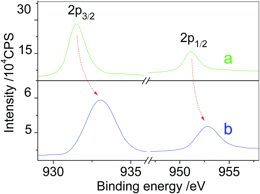
Fig. 6 exhibits the binding energies of core levels of Cu in CUC and PTFE/CUC. The binding energies of Cu 2p1/2 at 951.3 eV and 2p3/2 at 931.5 eV in the free CUC are dramatically increased to 952.7 and 933.1 eV in the PTFE/CUC, respectively, as indicated by the red arrows. Such large decreases in electron densities of the 2p inner-shell region of the Cu(II) in the coordination centers strongly suggest that the presence of PTFE has caused a significant change in the inner electronic configuration of Cu atoms.
 | ||
| Fig. 6 XPS-(Cu 2p) spectra of CUC (a) and PTFE/CUC (b). | ||
Based on the structure feature of PTFE, we propose an ion–dipole interaction scheme (see Fig. 7) between the fluorine atoms (Fδ−) on the PTFE chains and the Cu(II) in the coordination ions [Cu(urea)4]2+. In our opinion, this ion–dipole interaction is likely to be a result of the interface interaction between PTFE and CUC induced by a solid-solid grinding process. Also, a similar change trend was observed in FTIR (Fig. S3, ESI†) and XPS (Fig. S4, ESI†) spectra for the binary mixing system of PTFE and CuCl2. This result provides further support for the validity of the physical mixing process. Further, there is an unexpected result when PTFE was replaced by PVC and β-CD. PVC plays the same role of improving the oxidation of CuCl as PTFE (Fig. S5, ESI†) in air, while β-CD do not affect the decomposition of CUC (Fig. S6, ESI†). This may be due to the Cl atoms with a highly negative electron affinity on PVC chains play a similar role like the F atoms on PTFE chains. It gives support to the model of the ion–dipole interaction mode.
 | ||
| Fig. 7 Proposed ion–dipole interaction scheme between the fluorine atoms on the PTFE chains and the Cu(II) in the coordination ions [Cu(urea)4]2+. | ||
Although the contribution of PTFE to the formation of Cu-based composite nanomaterials still cannot be directly seen from the ion–dipole interaction between PTFE and CUC, we believe that such interaction will directly affect the thermal stability of the two mixed components, thereby changing their decomposition properties.
3.4 Effect of PTFE on the decomposition of CUC
To investigate the effect of PTFE on the decomposition behavior of CUC, we performed thermal analysis of the binary mixing system of PTFE and CUC. Fig. 8 and 9 show TG and DTG profiles of CUC and PTFE/CUC in nitrogen and in air, respectively.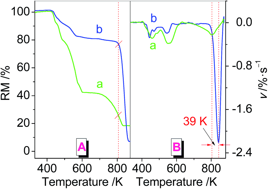 | ||
| Fig. 8 TG (A) and DTG (B) profiles of CUC (a) and PTFE/CUC (b) in nitrogen. | ||
 | ||
| Fig. 9 TG (A) and DTG (B) profiles of CUC (a) and PTFE/CUC (b) in air. | ||
As seen from the DTG curves, PTFE seems to play a completely opposite role in nitrogen than in air; in the former the decomposition of the CUC upon mixing with PTFE was delayed by about 39 K, while in the latter it was advanced by about 42 K. A comparison of the TG curves also provides similar information, as illustrated in Fig. 8A and 9A. It means that the CUC in the precursor PTFE/CUC generated by an ion–dipole interaction exhibits a higher stability in nitrogen and a lower stability in air in comparison with free CUC (an ionic crystal). In other words, the precursor is much more stable in an inert atmosphere than in an air atmosphere. This may be explained by the joint effect of the three factors together: (1) the weakened static stability caused by a grinding process; (2) a protection of CUC by PTFE chains; and (3) the involvement of oxygen both in the oxidation of CuCl and in the impairment of the ion–dipole interaction.
Although TG measurements and sintering experiments have completely different heating mechanisms, the observations above allow us to stress the fact that the physical mixing between a polymer and a coordination compound can significantly change the thermal property of the coordination compound. Such a change also occurred in the polymer as described below.
3.5 Effect of CUC on the pyrolysis pattern of PTFE
To understand the thermal performance of the complex–polymer precursors, it is necessary to investigate how the complex influences the chain structure of the polymer. Fig. 10 shows the mass spectra of PTFE and PTFE/CUC at the maximum degradation rates (approximately corresponding to 773 K, 35.8 min, determined based on total ion current curves i.e., TIC curves, Fig. S7, ESI†). There are two important phenomena that we can observe in this figure. | ||
| Fig. 10 Mass spectra of PTFE (a) and PTFE/CUC (b) at 35.8 min. | ||
First, the molecular ion peaks at m/z 30.999 (CF+), 49.997 (CF2+) and 80.994 (C2F3+), which resulted from the degradation of PTFE, exhibit completely different relative abundances (RA, %) in the two PTFE samples. In particular, the molecular ion peaks at m/z 84.970 (SiF3+) and 43.990 (CO2+), due to the etching effect24,25 of the activated F components (CF+, CF2+ and C2F3+) on the quartz component (SiO2, see eqn (6) and (7)), were either seriously weakened (indicated by the green arrow), or had even disappeared completely (indicated by the red arrow) in the presence of CUC. This result strongly implies that CUC produces a great effect on the scission of PTFE chains. A similar effect also was observed in the presence of CuCl2 (Fig. S8, ESI†). Our data indicate that the effect of CUC is more pronounced than that of CuCl2. We consider that the activated Cu ions generated in the decomposition process of CUC may have better opportunities to interact with the F atoms than the Cu ions in the CuCl2, thereby prohibiting the occurrence of etching effects.

Second, we notice that a strong signal with RA of 80.1% at m/z 99.995 due to a basic repeat unit of the PTFE structure: C2F4+ occurs in the case of CuCl2 (Fig. S8, ESI†). However, it is not observed in Fig. 10a and b. From a comparative point of view, this is a very significant result, since it implies that the structure of Cu salts is involved in the incision mechanism of PTFE chains. That is to say, different Cu(II) centers can induce distinctly different cleavage patterns of PTFE. Thus, this result allows us to make the assumption that the ions (Cu2+ and Cl− ions) in the ionic crystal of CuCl2 are able to directly attack the main chain of PTFE leading to backbone fragmentation. It is worth emphasizing that the presence of the monomer fragment C2F4+ in such a high RA would be useful in the recycling of waste PTFE materials.
In a word, the fact that CUC produces different influences on etching effects and cleavage patterns of PTFE from CuCl2 could be a consequence of different ion–dipole interactions induced by the interface interaction between the polymer and the salts caused by grinding.
3.6 SERS of the Cu@CuCl composite material
Nanostructured transition metals such as gold, silver and copper are usually used as general substrates of SERS for detecting chemical or biological substances.26–28 Here, we hope to carry out SERS studies of a composite system consisting of a nanostructured transition metal and its compound to widen the scope of the existing research. Therefore, the Cu@CuCl composite with a nanometer-scale fiber-like network structure was selected as a model to represent its SERS response for organic molecules.Fig. 11 presents Raman spectra of R6G and CV on the Cu@CuCl composite material deposited onto a silica substrate. As shown in curve a, no Raman signal of R6G and CV on a silica glass substrate was observable. However, in the presence of the Cu@CuCl, both R6G and CV exhibit very strong Raman signals: R6G29 at 581, 730, 1154, 1330 and 1637 cm−1 and CV30 at 914, 1178, 1370 and 1618 cm−1 (curve b). Such a large enhancement in Raman intensity has not been demonstrated previously for composites of Cu or CuCl. Since CuCl itself does not have SERS activity for R6G and CV, this enhancement effect mainly has been due to the contribution of Cu in the composite.
 | ||
| Fig. 11 SERS spectra of (A) R6G and (B) CV on the silica substrate (a), the Cu@CuCl (b). Experimental conditions are as follows: λex = 514.5 nm, power = 2.5 mW, integration time = 10 s. | ||
Control experiments show that the Cu@CuCl composite presents an improved SERS effect for R6G compared with commercial Cu powder with a particle size of about 200 mesh (Fig. S9, ESI†). In particular, a physical mixture (grinding the commercial Cu powder and commercial CuCl with a mass ratio of 2![[thin space (1/6-em)]](https://www.rsc.org/images/entities/char_2009.gif) :1 for 30 min in an agate mortar) of commercial Cu powder and commercial CuCl gives a much higher enhancement than the commercial Cu (Fig. S9, ESI†). This result may signify that the doping of a small amount of CuCl into Cu by mixing or compositing will lead to an improvement of SERS activity of metallic Cu. This may be due to the coupling between the localized surface plasmon resonances of different particles, inducing a significantly higher electromagnetic field enhancement.
:1 for 30 min in an agate mortar) of commercial Cu powder and commercial CuCl gives a much higher enhancement than the commercial Cu (Fig. S9, ESI†). This result may signify that the doping of a small amount of CuCl into Cu by mixing or compositing will lead to an improvement of SERS activity of metallic Cu. This may be due to the coupling between the localized surface plasmon resonances of different particles, inducing a significantly higher electromagnetic field enhancement.
4. Conclusions
The present work provides strong evidence that a simply physical mixing process between PTFE (a polymer) and CUC (a coordination compound) can induce an ion–dipole interaction between the F atoms on PTFE chains and the Cu(II) within CUC, leading to a significant change in decomposition patterns of CUC. This change was found to be closely related to sintering temperatures and sintering atmospheres, such as for CuCl an earlier oxidation in air and a delayed reduction in nitrogen. Therefore, it can be used to design and construct Cu-based composite nanomaterials. According to this, a series of Cu-based materials including Cu, CuCl, CuO, Cu@CuCl and CuCl@CuO were successfully prepared. Such a change also occurred in the thermal behaviour of PTFE, for example, the decline and even disappearance of etching effects. Furthermore, the presence of PTFE can greatly affect the morphologies and sizes of the Cu-based materials. Finally, the as-obtained Cu@CuCl nanomaterial exhibits strong SERS activity. We believe that this study provides a simple but useful framework for understanding the link between coordination compounds and polymers in the preparation of transition metal oxide nanoparticles.Note added after first publication
This article replaces the version published on 15th October 2014, which contained the wrong graphic for Fig. 2.Acknowledgements
The authors are grateful to NSFC of China (no. 21071139) for financial support of this work.References
- (a) N. K. Richtmyer and C. Hudson, J. Am. Chem. Soc., 1951, 73, 2249–2250 CrossRef CAS; (b) L. X. Song, M. Wang, Z. Dang and F. Y. Du, J. Phys. Chem. B, 2010, 114, 3404–3410 CrossRef CAS PubMed; (c) L. X. Song, J. Xia, Z. Dang, J. Yang, L. B. Wang and J. Chen, CrystEngComm, 2012, 14, 2675–2682 RSC.
- (a) S. Z. Pan, L. X. Song, J. Chen, F. Y. Du, J. Yang and J. Xia, Dalton Trans., 2011, 40, 10117–10124 RSC; (b) S. Chaberek Jr and A. Martell, J. Am. Chem. Soc., 1952, 74, 6228–6231 CrossRef.
- (a) H. D. Wagner and R. A. Vaia, Mater. Today, 2004, 7, 38–42 CrossRef CAS; (b) K. I. Winey and R. A. Vaia, MRS Bull., 2007, 32, 314–322 CrossRef CAS.
- (a) W. G. Sawyer, K. D. Freudenberg, P. Bhimaraj and L. S. Schadler, Wear, 2003, 254, 573–580 CrossRef CAS; (b) L. X. Song, M. Wang, S. Z. Pan, J. Yang, J. Chen and J. Yang, J. Mater. Chem., 2011, 21, 7982–7989 RSC.
- (a) S.-Y. Park, Y.-H. Cho and R. A. Vaia, Macromolecules, 2005, 38, 1729–1735 CrossRef CAS; (b) S. Noel, B. Leger, R. Herbois, A. Ponchel, S. Tilloy, G. Wenz and E. Monflier, Dalton Trans., 2012, 41, 13359–13363 RSC.
- (a) R. Herbois, S. Noel, B. Leger, L. Bai, A. Roucoux, E. Monflier and A. Ponchel, Chem. Commun., 2012, 48, 3451–3453 RSC; (b) P. Du, L. X. Song, J. Xia, Y. Teng and Z. K. Yang, J. Mater. Chem. A, 2014, 2, 11439–11447 RSC.
- (a) J. Xia, L. X. Song, W. Liu and Y. Teng, Soft Matter, 2013, 9, 9714–9722 RSC; (b) R. A. Vaia, K. D. Jandt, E. J. Kramer and E. P. Giannelis, Chem. Mater., 1996, 8, 2628–2635 CrossRef CAS.
- (a) J. Yuan, H. Schmalz, Y. Xu, N. Miyajima, M. Drechsler, M. W. Moller, F. Schacher and A. H. Mueller, Adv. Mater., 2008, 20, 947–952 CrossRef CAS; (b) L. Bai, F. Wyrwalski, J.-F. Lamonier, A. Y. Khodakov, E. Monflier and A. Ponchel, Appl. Catal., B, 2013, 138, 381–390 CrossRef PubMed.
- (a) L. Ding, E. Y. Chi, K. S. Schanze, G. P. Lopez and D. G. Whitten, Langmuir, 2009, 26, 5544–5550 CrossRef PubMed; (b) E. Ruiz-Hitzky, M. Darder and P. Aranda, J. Mater. Chem., 2005, 15, 3650–3662 RSC.
- (a) E. Ruiz-Hitzky, Adv. Mater., 1993, 5, 334–340 CrossRef CAS; (b) A. Villanueva, M. Morales-Varela and E. Ruiz-Hitzky, Eur. J. Inorg. Chem., 2004, 2004, 949–952 CrossRef.
- S. Diamanti, S. Arifuzzaman, A. Elsen, J. Genzer and R. A. Vaia, Polymer, 2008, 49, 3770–3779 CrossRef CAS PubMed.
- (a) D. Braga and F. Grepioni, Angew. Chem., Int. Ed., 2004, 43, 4002–4011 CrossRef CAS PubMed; (b) M. Gohel and S. A. Nagori, Pharm. Dev. Technol., 2009, 14, 679–686 CrossRef CAS PubMed.
- (a) S. Bisaillon and R. Tawashi, J. Pharm. Sci., 1971, 60, 1874–1877 CrossRef CAS; (b) I. Krycera and J. A. Hersey, Int. J. Pharm., 1980, 6, 119–129 CrossRef.
- (a) B. Radovanovic, Thermochim. Acta, 1991, 186, 171–177 CrossRef; (b) R. B. Penland, S. Mizushima, C. Curran and J. Quagliano, J. Am. Chem. Soc., 1957, 79, 1575–1578 CrossRef CAS; (c) P. I. Premovic and P. R. West, Can. J. Chem., 1974, 52, 2919–2922 CrossRef CAS.
- B. S. Radovanovic and P. Premovic, J. Therm. Anal., 1992, 38, 715–719 CrossRef.
- (a) L. X. Song, J. Yang, L. Bai, F. Y. Du, J. Chen and M. Wang, Inorg. Chem., 2011, 50, 1682–1688 CrossRef CAS PubMed; (b) L. X. Song, S. Z. Pan, L. H. Zhu, M. Wang, F. Y. Du and J. Chen, Inorg. Chem., 2011, 50, 2215–2223 CrossRef CAS PubMed.
- (a) L. Tsao and C. Chen, Corros. Sci., 2012, 63, 393–398 CrossRef CAS PubMed; (b) K. Chen and D. Xue, J. Phys. Chem. C, 2013, 117, 22576–22583 CrossRef CAS; (c) T. Xie, M. Gong, Z. Niu, S. Li, X. Yan and Y. Li, Nano Res., 2010, 3, 174–179 CrossRef CAS PubMed.
- (a) S. Vettivel, N. Selvakumar and N. Leema, Mater. Des., 2013, 45, 323–335 CrossRef CAS PubMed; (b) X. Jia, J. Li and E. Wang, Small, 2013, 9, 3873–3879 CrossRef CAS PubMed; (c) Q. Cai, F. Liao, F. Hu, Y. Li, T. Wang and M. Shao, RSC Adv., 2014, 4, 6424–6429 RSC.
- (a) A. Lundstrom, B. Andersson and L. Olsson, Chem. Eng. J., 2009, 150, 544–550 CrossRef PubMed; (b) S. D. Yim, S. J. Kim, J. H. Baik, I.-S. Nam, Y. S. Mok, J.-H. Lee, B. K. Cho and S. H. Oh, Ind. Eng. Chem. Res., 2004, 43, 4856–4863 CrossRef CAS.
- (a) C. Karunakaran, P. Anilkumar and P. Gomathisankar, Chem. Cent. J., 2011, 5, 1–9 CrossRef PubMed; (b) M. N. Rashin and J. Hemalatha, Int. J. Eng. Appl. Sci., 2012, 6, 216–220 Search PubMed.
- (a) Y. Ni, Y. Zhu and X. Ma, Dalton Trans., 2011, 40, 3689–3694 RSC; (b) J. Yang, L. X. Song, J. Yang, Z. Dang and J. Chen, Dalton Trans., 2012, 41, 2393–2398 RSC.
- (a) H. S. Salapare III, F. Guittard, X. Noblin, E. Taffin de Givenchy, F. Celestini and H. J. Ramos, J. Colloid Interface Sci., 2013, 396, 287–292 CrossRef PubMed; (b) J. Su, G. Wu, Y. Liu and H. Zeng, J. Fluorine Chem., 2006, 127, 91–96 CrossRef CAS PubMed.
- (a) A. Gruger, A. Régis, T. Schmatko and P. Colomban, Vib. Spectrosc., 2001, 26, 215–225 CrossRef CAS; (b) M. Womack, M. Vendan and P. Molian, Appl. Surf. Sci., 2004, 221, 99–109 CrossRef CAS.
- (a) S. Marais, Y. Hirata, C. Cabot, S. Morin-Grognet, M.-R. Garda, H. Atmani and F. Poncin-Epaillard, Surf. Coat. Technol., 2006, 201, 868–879 CrossRef CAS PubMed; (b) T. K. Lin, S. J. Wu, C. K. Peng and C. H. Yeh, Polym. Int., 2009, 58, 46–53 CrossRef CAS.
- (a) K. Su and O. Tabata, Microsyst. Technol., 2002, 9, 11–16 CrossRef CAS PubMed; (b) T. Shi, M. Shao, H. Zhang, Q. Yang and X. Shen, Appl. Surf. Sci., 2011, 258, 1474–1479 CrossRef CAS PubMed.
- (a) R. Woods, G. A. Hope and K. Watling, J. Appl. Electrochem., 2000, 30, 1209–1222 CrossRef CAS; (b) L. Y. Chen, J. S. Yu, T. Fujita and M. W. Chen, Adv. Funct. Mater., 2009, 19, 1221–1226 CrossRef CAS.
- (a) J. Liu, I. White and D. L. DeVoe, Anal. Chem., 2011, 83, 2119–2124 CrossRef CAS PubMed; (b) Y. Wang and T. Asefa, Langmuir, 2010, 26, 7469–7474 CrossRef CAS PubMed.
- (a) A. Campion and P. Kambhampati, Chem. Soc. Rev., 1998, 27, 241–250 RSC; (b) Z.-Q. Tian, B. Ren and D.-Y. Wu, J. Phys. Chem. B, 2002, 106, 9463–9483 CrossRef CAS.
- (a) Z. Xie, J. Tao, Y. Lu, K. Lin, J. Yan, P. Wang and H. Ming, Opt. Commun., 2009, 282, 439–442 CrossRef CAS PubMed; (b) A. A. Moore, M. L. Jacobson, N. Belabas, K. L. Rowlen and D. M. Jonas, J. Am. Chem. Soc., 2005, 127, 7292–7293 CrossRef CAS PubMed.
- (a) K. W. Kho, Z. X. Shen, H. C. Zeng, K. C. Soo and M. Olivo, Anal. Chem., 2005, 77, 7462–7471 CrossRef CAS PubMed; (b) L. Y. Chen, J. S. Yu, T. Fujita and M. W. Chen, Adv. Funct. Mater., 2009, 19, 1221–1226 CrossRef CAS.
Footnote |
| † Electronic supplementary information (ESI) available: (1) TG and DTG profiles of PTFE in nitrogen; (2) XRD patterns of the materials obtained by sintering the CuCl2 and PTFE/CuCl2 at 673 K, CuCl2 and PTFE/CuCl2 at 873 K in air; (3) FTIR spectrum of PTFE/CuCl2; (4) XPS-(Cu2p) spectra of CuCl2 and PTFE/CuCl2; (5) XRD patterns of the CuO materials obtained by sintering the PVC/CUC at 673 and 873 K for 2 h in air; (6) XRD patterns of CuCl@CuO and CuO obtained by sintering the β-CD/CUC at 673 and 873 K for 2 h in air; (7) TIC curves of PTFE, PTFE/CuCl2 and PTFE/CUC; (8) mass spectra of PTFE/CuCl2 at 35.8 min; (9) SERS spectra of R6G on the commercial Cu and the commercial mixture of Cu and CuCl. See DOI: 10.1039/c4ra09682g |
| This journal is © The Royal Society of Chemistry 2014 |