Designing triphenylamine derivative dyes for highly effective dye-sensitized solar cells with near-infrared light harvesting up to 1100 nm

Zhenqing Yangab, Di Wangab, Xiaohui Baiab, Changjin Shaob and Dapeng Cao*a
aState Key Laboratory of Organic–Inorganic Composites, Beijing University of Chemical Technology, Beijing 100029, China. E-mail: caodp@mail.buct.edu.cn
bCollege of Science, China University of Petroleum, Beijing 102249, China

Received 29th August 2014 , Accepted 15th September 2014

First published on 15th September 2014


Abstract

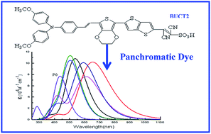
Designing highly efficient sensitizers for dye-sensitized solar cells (DSSCs) is an urgent task because it is closely related to the practical application of DSSCs. In this work, we designed and screened a series of triphenylamine derivative dyes with donor–π–acceptor (D–π–A) structure using different electron donors, π bridges and electron acceptors, and further used density functional theory (DFT) and time-dependent DFT (TDDFT) approaches to investigate the molecular orbital energy levels, absorption spectra, and light harvesting efficiency of these newly designed dyes. Results indicate that the donor group in D2, π bridges in Pi10–Pi12 and the acceptor group in A7 are promising functional groups for D–π–A structure. Using the above screened functional groups as donors, π bridges and acceptors, we designed six novel D–π–A structures of BUCT1–BUCT6. The results indicate that BUCT1–BUCT6 dyes show smaller HOMO–LUMO energy gaps, higher molar extinction coefficients and obvious redshifts compared to the experimentally synthesized P0 dye. In particular, the newly designed BUCT2 dye exhibits not only a 215 nm redshift and a higher molar extinction coefficient with an increment of 32.4% compared to P0 dye, but also has an extremely broad absorption spectrum covering the entire visible range up to the near-IR region of 1100 nm. Therefore, the BUCT2 dye is a very promising candidate for highly effective DSSCs with near-infrared light harvesting up to 1100 nm. We also found that the dyes with two –CN groups and a sulfonic acid group as the electron acceptor are more efficient than dyes with one –CN group and a sulfonic acid group.

1. Introduction

Because of the shortage of traditional fossil fuels caused by the fast consumption by a developing global society, it is an urgent task to develop renewable and clean energy sources, such as power from wind and water, biomass and solar energy. Currently, the sun provides our planet with approximately 104 times more energy than our global daily consumption.1,2 Photovoltaic cells are the most direct way to convert sunlight into electricity. Moreover, photovoltaic cells require very little maintenance because of the absence of moving components, and this makes them attractive for long term outdoor installation. Therefore, developing highly effective solar cells is very significant for the wide application of solar energy.

Since the Grätzel research group used the high specific surface area of nanoporous TiO2 film as a semiconductor electrode to develop dye-sensitized solar cells (DSSCs) with photoelectric conversion efficiency (PCE) of 7.1% in 1991,3 DSSCs have become a hot topic and attracted extensive attention. In 2005, Grätzel et al. used N719 dye as sensitizers to produce DSSCs with PCE of up to 11.18%.4 In 2011, they further improved the efficiency of solar cells based on porphyrin dyes to 12.3%.5 Recently, Grätzel6 and Oxford University research teams7 independently developed a solid-state DSSCs whose PCE exceeded 15%, further creating a new record.

In DSSCs, the dye sensitizers have been considered as one of the most important components to exhibit high performance, and various types of dyes have been explored to improve overall device performance. Ruthenium dyes have limitations for DSSCs because of the presence of noble metals and relatively large environmental pollution. Therefore, organic photosensitive dyes without noble metals have attracted considerable attention, owing to many advantages, such as low cost, variety structure, strong light-harvesting ability, convenient synthesis and minimal harm to the environment. Recently, a series of new organic dyes with donor–π–acceptor (D–π–A) structure were synthesized.8–14 Previous studies have shown that the D–π–A molecular structure is not only beneficial to the intramolecular charge transfer from the electron donor to the electron acceptor moiety, but also facilitated the regulation of molecule performance by modifying the donor, π bridge and acceptor groups.9 At present, investigators have identified some relatively good electron donors (such as triphenylamine, indole, dimethyl fluorene, phenothiazine) and π bridges (such as methylidyne, thiophene, furan, pyrrole and benzene).2 Cyanoacrylic acid is the most widely used electron acceptor, which also acts as an anchoring group, because the dye molecule can be adsorbed on the surface of the TiO2 nanocrystal. Currently, designing the D–π–A molecular structure to further improve the PCE of DSSC devices is still a great challenge.15

The PCE of a DSSC device is mainly determined by short circuit current density (Jsc), open circuit photovoltage (Voc) and fill factor (FF). Improving the short circuit current density can significantly enhance the PCE of DSSCs. The most critical factor for improving the short circuit current density is to increase the electron injection (Φinject)16 and the light harvesting efficiency (LHE),17 as well as the molar extinction coefficients of the dyes, and broaden the absorption spectrum scope in the visible light and near-infrared regions. Φinject is related to the driving force ΔGinject of the electrons injecting from the excited states of dye molecules to the semiconductor conduction band. LHE can be calculated by the equation LHE = 1–10−ƒ, where ƒ represents the oscillator strength of the adsorbed dye molecules.17

In order to improve the short circuit current density, Park et al. introduced triphenylamine as donor, benzene ring as the π bridge and cyanoacrylic acid as the acceptor to synthesize the TA–St–CA dye, and further applied it to DSSCs. The short circuit current density of the TA–St–CA-sensitized DSSC device reached 18.1 mA cm−2 and exhibited a PCE of 9.1%, and it has attracted significant attention because of its simple structure and outstanding photoelectric performance.18 Triphenylamine derivatives as sensitizers of DSSCs can expand the light absorption ability of the dye, and its non-planar structure can also inhibit aggregation of the dye on the TiO2 surface.19 Triphenylamine has been widely used as an electron donor in the sensitizer. The three benzene rings of the triphenylamine group are mutually twisted, and the triphenylamine and π-conjugated groups also have certain distortion angles. Therefore, the non-planar structure of the dye molecule effectively prevents intermolecular π–π aggregation, which is beneficial because the aggregation would cause electron transfer between the molecules, which leads to loss of energy and therefore affects the PCE of DSSCs. Accordingly, triphenylamine compounds have been widely used as photovoltaic materials because of their excellent hole transport capability.20–39

In order to design highly efficient photosensitive dyes, in this work, we first used density functional theory (DFT) and time-dependent DFT (TDDFT) to calculate the molecular orbital energy levels, absorption spectra, LHE and electron injection driving force of the experimentally synthesized TA–St–CA dyes.18 Using TA–St–CA dye as a prototype, we further designed and screened a series of new D–π–A structure dyes by adopting different donors, π conjugate bridges and acceptor groups. Finally, we recommended a type of novel triphenylamine derivative dyes with near-infrared light harvesting for highly effective DSSCs.

2. Computational details

2.1 D–π–A molecular models

The experimentally synthesized TA–St–CA dye is shown in Fig. 1. It is also named P0 (R-bit of P0 is H in Fig. 1) for simplification. The dye P1 (also named D5)40–42 and P2 (ref. 43) were obtained using furan and thiophene groups to replace the benzene group of P0. The dyes P1 and P2 have been experimentally synthesized and the corresponding P1 and P2-sensitized DSSC devices exhibit PCEs of approximately 6.0% and 7.36%, respectively. In order to explore considerably higher efficient triphenylamine derivative dyes, we designed a series of dyes using different π-conjugated bridge groups, Pi1–Pi12, shown in Fig. 1. Pi1–Pi3 were obtained by replacing the benzene group of P0 with EDOT, bithiophene and s-DTT, respectively. Simultaneously, Pi4–Pi9 were constructed using two combinations of benzene, thiophene, furan and EDOT to replace the π-conjugated bridge of P0, and Pi10–12 were designed using any two of EDOT, bithiophene and s-DTT as substituting groups. In addition, we adopted the orthogonal design to modify the donor and acceptor groups of P0 using different electron-donating groups (i.e. –CH3,–O–CH3,–C4H9, and –NH2) and different electron-accepting groups (A1–A7). All the abovementioned constructed D–π–A molecular structures are shown in Fig. 1.
image file: c4ra09444a-f1.tif
Fig. 1 Molecular structures of the triphenylamine P0 dye and derivatives.

2.2 Computational methods

The geometries of these dye molecules were optimized by DFT calculations using Becke's three-parameters and Lee–Yang–Parr gradient-corrected correlation hybrid functional (B3LYP) and 6-31G(d) basis sets. The vibrational frequency calculations were also carried out at the same level to confirm that the triphenylamine derivative structures are local minima (no imaginary frequencies) on potential energy surfaces.

Previous investigations indicate that TDDFT is highly efficient and accurate in the calculation of vertical excitation energy, electric properties and optical absorption.44,45 However, different exchange–correlation (XC) functionals for charge-transfer excitations often show significant effects. To select suitable functionals, we adopted different XC functionals, including B3LYP, CAM-B3LYP and WB97XD, in TDDFT to calculate the vertical excitation energies of P0, P1 and P2. The calculated results and the experimental data are shown in Table 1. It was found that the vertical excitation energies of dyes were severely underestimated by the B3LYP functional, while the long range-corrected (LC) functionals, such as CAM-B3LYP and WB97XD, are in satisfactory agreement with the experimental values. Considering computational cost, we first screened the donors, π-conjugated bridges and acceptors using the CAM-B3LYP functional with 6-311+g(d, p) basis sets, combining a CPCM46 solvation model in acetonitrile solution for these newly designed dye molecules in Fig. 1. Then, we adopted the more accurate WB97XD functional at the same level to predict the optical properties of six newly designed dyes of BUCT1–BUCT6. All the calculations were performed using a suite of Gaussian09 packages.47

Table 1 The vertical excitation energy in eV of dyes P0–P2 calculated with TDDFT using different exchange–correlation functionals in acetonitrile and the experimental data
  B3LYP CAM-B3LYP WB97XD Exp.
P0 2.04 2.84 2.91 3.21 (ref. 18)
P1 1.96 2.49 2.59 2.61–2.90 (ref. 40−42)
P2 1.93 2.57 2.66 2.64 (ref. 43)


3. Results and discussion

3.1 Screening of the electron donor

Fig. 2 shows the energy levels for P0 and D1–D4 in acetonitrile solution. The results indicate that the LUMO energies of D1–D4 are higher than the conduction band edge (CBE) of the TiO2 electrode (approximately −4.0 eV),2 shown as a red dashed line in Fig. 2. Thus, these excited state molecules could successfully inject electrons into the TiO2 electrode. In addition, the HOMO energies of D1–D3 are lower than the potential (approximately −4.8 eV) of the I/I3 redox electrolyte.2 Therefore, those dye molecules that lose electrons could quickly obtain electrons from the electrolyte. The HOMO energy of D4 is higher than the potential of the I/I3 redox electrolyte, and may not be suitable for sensitizer. Compared to the band gap of P0, D1 and D2 may be suitable candidates for DSSCs.
image file: c4ra09444a-f2.tif
Fig. 2 Energy levels for P0 and D1–D4 dyes, where the horizontal dashed line indicates the levels (−4.0 eV) of the conduction band edge (CBE) of the anatase TiO2 in electrolyte solutions and the redox energy level (−4.8 eV) of redox electrolyte (I/I3).2

Fig. 3 shows the UV-visible absorption spectra of D1–D4 dyes. The maximum absorption peaks of D1–D4 are 445, 462, 456 and 465 nm, which exhibit 7, 24, 18 and 27 nm redshifts compared to that of P0 (438 nm), respectively. The molar extinction coefficients of these dyes also slightly increase with the exception of D4 (see Table 2). The optical absorption spectra redshift and the molar extinction coefficient definitely indicate that D2 may be a good candidate as a photosensitizer for a DSSC device, which is consistent with the band gap results mentioned above. This suggests that introducing the strongly electron-donating –O–CH3 group into the electron donor of the dye is favorable for light absorption, which has been confirmed in previous experiments. The Grätzel research group adopted –O–CH3 to replace the R-bit in P1 to synthesize the dye D9,42 and found that the dye D9 shows a redshift of approximately 30 nm compared to dye P1, and the PCE of a D9-sensitized device significantly increases to 6.9% from 5.9% for the P1-sensitized device. Both the experimental and simulation results indicate that introducing the –O–CH3 group into the donor of the dye may improve the Jsc and PCE of DSSCs.


image file: c4ra09444a-f3.tif
Fig. 3 The UV-visible absorption spectra of P0 and D1–D4 dyes.
Table 2 The calculated maximum absorption wavelengths λ (nm) and corresponding vertical excitation energies Eex (eV) (in parentheses), molar extinction coefficient ε (104 M−1 cm−1), oscillator strengths ƒ and light harvesting efficiency (LHE) for triphenylamine derivative dyes
Dye λ(Eex) ε f LHE
P0 438(2.84) 7.71 1.9060 0.988
D1 445(2.79) 7.75 1.9140 0.988
D2 462(2.68) 7.75 1.9123 0.988
D3 456(2.72) 7.83 1.9326 0.988
D4 465(2.67) 7.58 1.8706 0.987
P1 498(2.49) 6.17 1.5613 0.973
P2 482(2.57) 4.40 1.0972 0.920
Pi1 483(2.56) 7.10 1.7726 0.983
Pi2 489(2.53) 9.06 2.2741 0.995
Pi3 493(2.51) 8.23 2.0311 0.991
Pi4 447(2.78) 9.19 2.2684 0.995
Pi5 447(2.78) 9.22 2.2721 0.995
Pi6 447(2.78) 9.35 2.3013 0.995
Pi7 495(2.51) 7.72 1.8811 0.987
Pi8 504(2.46) 8.21 2.0278 0.991
Pi9 504(2.46) 6.91 1.7065 0.980
Pi10 525(2.36) 10.44 2.5768 0.997
Pi11 501(2.44) 9.99 2.4570 0.997
Pi12 510 (2.43) 10.87 2.6649 0.998
A1 495(2.51) 5.44 1.3433 0.955
A2 397(3.12) 7.87 1.9437 0.989
A3 363(3.41) 7.03 1.7346 0.983
A4 375(3.30) 6.65 1.6420 0.977
A5 435(2.85) 7.05 1.7361 0.983
A6 445(2.79) 7.58 1.8705 0.987
A7 501(2.48) 6.70 1.6535 0.977
BUCT1 537(2.31) 10.82 2.6717 0.998
BUCT2 653(1.90) 10.21 2.5210 0.997
BUCT3 505(2.46) 10.52 2.5837 0.997
BUCT4 609(2.04) 7.65 1.8848 0.987
BUCT5 505(2.45) 11.36 2.778 0.998
BUCT6 592(2.09) 10.11 2.4907 0.997


3.2 Screening of the π-conjugated bridge

The π-conjugated bridge in D–π–A structure dye can accelerate electron transport from donor to acceptor, and thus speed up electron injection from the dye molecule to the conduction band of the semiconductor. Therefore, the π-conjugated bridge is a key factor of high performance in D–π–A structure. In order to screen excellent π-conjugated bridges of triphenylamine derivative dyes, we investigated the performance of a series of newly designed dyes (Pi1–Pi12 in Fig. 1).

The molecular orbital energy levels of P0–P2 and Pi1–Pi12 in acetonitrile solution are shown in Fig. 4. The LUMO energies of Pi1–Pi12 are higher than the CBE (red dashed line in Fig. 4) of the TiO2 electrode,2 which ensures that the excited electrons smoothly inject into the TiO2 electrode. Moreover, their HOMO energies are sufficiently lower than the potential of the I/I3 redox electrolyte.2 Therefore, these lost electron molecules could quickly be restored by the electrolyte. The energy gaps of Pi1–Pi12 are basically smaller than that of P0, which causes the redshift of absorption spectra. In particular, the energy gaps of Pi10–Pi12 are 2.01, 2.07 and 1.95 eV, respectively, which is considerably lower than that of P0.


image file: c4ra09444a-f4.tif
Fig. 4 Energy levels for P0–P2 and Pi1–Pi12 dyes.

The UV-visible absorption spectra of P1, P2 and Pi1–Pi12 dyes in acetonitrile solution are shown in Fig. 5. For clear observation, they are divided into Fig. 5(a) and (b). Compared to P0, the absorption maxima of these dyes are red-shifted 44–87 nm (see Fig. 5(a) and (b)), except that Pi4-Pi6 exhibits a redshift of only 8 nm. Their maximal absorption wavelengths and corresponding vertical excitation energies are listed in Table 2. Compared to P0, the optical properties of Pi10–Pi12 show obvious advantages. In particular, as shown in Fig. 5(b), the absorption peaks of Pi10–Pi12 are 525, 501 and 510 nm, which exhibit redshifts of 87, 63 and 72 nm, respectively. The absorption spectra of Pi10–Pi12 cover a broader solar spectrum, exceeding 700 nm for Pi12. Interestingly, it is found from the optimized geometric structures (Fig. S1) that the π-conjugated bridges of Pi10–Pi12 have planar structure, which is greatly beneficial for the photoinduced transfer of electrons from the electron donor to the electron acceptor.


image file: c4ra09444a-f5.tif
Fig. 5 The UV-visible absorption spectra of different dyes. (a) P0–P2 and Pi1–Pi3, (b) P0 and Pi4–Pi12.

The maximum molar extinction coefficient is a very important factor for light-absorbing dyes, and its magnitude directly demonstrates the light absorption ability of the dye. The maximum molar extinction coefficients of Pi1–Pi12 are listed in Table 2. It was found that the maximum molar extinction coefficients of Pi2, Pi4–Pi6 and Pi10–Pi12 show a significant increase from 17.5% to 40.9%, and other dyes also show increases except for Pi9. In particular, the maximum molar extinction coefficients of Pi10–Pi12 are 10.44, 9.99 and 10.87 × 10⁴ M−1 cm−1 with increases of 35.4%, 29.7% and 40.9%, respectively, compared to P0 (Table 2). Moreover, the LHEs of the Pi10–Pi12 dyes are also considerably higher than that of P0.

From the above π-conjugated bridge screening, we found that a π-conjugated bridge with planar structure enables a more efficient intramolecular charge transfer from the electron donor to the electron acceptor. By considering the energy gap, absorption spectra (including molar extinction coefficient, absorption peak redshift and the broad extent of light absorption) and LHE of P0 and Pi1–Pi12 we believe that the Pi10–Pi12 may be excellent candidates for DSSC devices. This observation is in excellent agreement with previous experiments, in which the Wang research group adopted EDOT and bithiophene groups as π-conjugated bridges to synthesize dye C217 (similar to Pi10),48 and found that the dye C217 shows a redshift of approximately 114 nm compared to dye P0, and that the PCE of the C217-sensitized device significantly increases to 9.8%. These findings are expected to provide a promising method to design new organic dyes and improve the Jsc and PCE of DSSCs.

3.3 Screening of the electron acceptor

Currently, researchers have mainly focused on the modifications of the electron donor and π-bridge structures for highly effective D–π–A dyes. However, the electron acceptor, which connects the dye to the semiconductor, plays an important role in anchoring on the TiO2/ZnO semiconductor surface, optical absorption, as well as electron injection processes. Currently, cyanocyclic acid is very popular for DSSCs because its functionality largely fulfills the need of the electron acceptor.49 It is beneficial to improve the injection efficiency by inserting the electron-withdrawing –CN group into the electron acceptor. Therefore, we designed a series of dyes A1–A7 (Fig. 1) in order to screen acceptors by exploring the influence of electron-withdrawing chemical group substitution, such as –F, –CH3, –CF3 and –NO2, and the sulfonic acid anchoring group.

The molecular orbital energy levels of A1–A7 in acetonitrile solution are shown in Fig. 6. The LUMO energies of A1–A7 are higher than the CBE of the TiO2 electrode and their HOMO energies are lower than the potential of the I/I3 redox electrolyte.2 The energy gaps of A1 and A7 are 1.76 and 1.72 eV, respectively, which are smaller than P0 because of the significant decrease of the LUMO energy level. However, the energy gaps of A2–A4 are larger than that of P0, which results in the blue shifts.


image file: c4ra09444a-f6.tif
Fig. 6 Energy levels for P0 and A1–A7 dyes.

The UV-visible absorption spectra of A1–A7 in acetonitrile solution are shown in Fig. 7. The maximum absorption peaks of A2–A4 are blue-shifted by 41–75 nm. Thus, the visible light utilization for A2–A4 is relatively low because the maximal absorption peaks are located in the violet light range. A5–A6 and P0 have very similar optical properties (Table 2). The maximum absorption peaks of A1 and A7 are red-shifted by 57 and 63 nm, respectively. However, the molar extinction coefficient of A1 shows a decrease of nearly 30%, and that of A7 is decreased by approximately 9%. Thus, the decrease of the molar extinction coefficient of dye molecules can be effectively prevented using a sulfonic acid group to replace the carboxylic acid group as the electron acceptor.


image file: c4ra09444a-f7.tif
Fig. 7 The UV-visible absorption spectra of P0 and A1–A7 dyes.

3.4 Design of novel D–π–A triphenylamine derivatives

On the basis of the above screening of the D–π–A structure, we further designed six novel dyes, BUCT1–BUCT6, by employing 4,4′-dimethoxy triphenylamine as the electron donor, any two of EDOT, bithiophene and s-DTT groups as the π-conjugated bridge, and –CN and sulfonic acid groups as electron acceptors. The structures of BUCT1–BUCT6 are presented in Fig. 8.
image file: c4ra09444a-f8.tif
Fig. 8 Molecular structures of newly designed D–π–A triphenylamine derivatives.

The molecular orbital energy levels of P0 and BUCT1–BUCT6 dyes in acetonitrile solution are shown in Fig. 9. The LUMO energies of BUCT1–BUCT6 are higher than the CBE of TiO2.2 The HOMO energies of BUCT2 and BUCT4 are below the I/I3 redox potential, while those of BUCT1, BUCT3, BUCT5 and BUCT6 are higher than the redox potential of the I/I3 electrolyte.2 Thus, BUCT1, BUCT3, BUCT5 and BUCT6 are not suitable as sensitizers in DSSCs because they could not be quickly restored by the electrolyte after they lose their electrons and the sensitizer was oxidized. The LUMO levels of BUCT2 and BUCT4 are −3.45 and −3.44 eV, and the energy gaps are also reduced to 1.36 and 1.34 eV, respectively.


image file: c4ra09444a-f9.tif
Fig. 9 Energy levels of BUCT1–BUCT6 and P0 dyes.

Fig. 10 shows the absorption spectra of BUCT1–BUCT6 in acetonitrile solution. The maximum molar extinction coefficients of BUCT2 and BUCT4 dyes are 10.21 and 7.65 × 10⁴ M−1 cm−1, respectively, representing an increment of 32.4% and −0.8% compared with P0. The maximum absorption peaks of BUCT2 and BUCT4 are 653 and 609 nm, which have extremely large redshifts of 215 and 171 nm, compared to P0. Obviously, the newly designed BUCT2 dye has the highest molar extinction coefficient and the largest redshift among all the dyes studied. Interestingly, the absorption spectrum of BUCT2 covers the entire visible range and extends into the near-IR region up to 1100 nm, which could be considered as a full light absorbing dye. Therefore, BUCT2 dye is a promising candidate for panchromatic absorption, which may significantly improve the PCE of DSSC devices.


image file: c4ra09444a-f10.tif
Fig. 10 Absorption spectra of BUCT1–BUCT6 dyes.

Interestingly, we find that the band gaps of BUCT2, BUCT4 and BUCT6, where the acceptors have two –CN groups, are noticeably smaller than that of BUCT1, BUCT3 and BUCT5, in which the acceptors have only one –CN group. As a result, the maximum absorption peak of dyes with two –CN groups also exhibits apparent redshifts. In particular, the maximum absorption peak of BUCT2 shows a 116 nm redshift compared to that of BUCT1. The maximum absorption peaks of BUCT4 and BUCT6 also exhibit similar phenomena compared to those of BUCT3 and BUCT5. Therefore, we believe that the dyes with two –CN groups and a sulfonic acid group as the electron acceptor are superior to the dyes with one –CN group and a sulfonic acid group.

4. Conclusions

We have systematically investigated the molecular orbital energy levels, absorption spectra, and light harvesting efficiency of D–π–A type dyes designed by adopting different electron donors, π bridges and electron acceptors. The results indicate that the donor group in D2, π bridges in Pi10–Pi12 and the acceptor group in A7 are promising functional groups for D–π–A structure, because these functional groups significantly improve the absorption of visible light and the molar extinction coefficient of the dyes, and speed up intramolecular charge transfer from the electron donor to the electron acceptor.

By using the above screened promising functional groups as the donor, π bridge and acceptor, we reconstructed six novel D–π–A structures, BUCT1–BUCT6. The calculated results indicate that the BUCT1–BUCT6 dyes show smaller HOMO–LUMO energy gaps, higher molar extinction coefficients and obvious redshifts compared to the experimentally synthesized P0 dye. In particular, the newly designed BUCT2 dye exhibits not only a 215 nm redshift and a higher molar extinction coefficient with an increment of 32.4% compared to P0, but also a extremely broad absorption spectrum covering the entire visible range up to the near-IR region of 1100 nm. Therefore, the BUCT2 dye is a very promising candidate for highly effective DSSCs with near-infrared light harvesting up to 1100 nm. Interestingly, we also found that the dyes with two –CN groups and a sulfonic acid group as the electron acceptor are more efficient than the ones with one –CN group and a sulfonic acid group. It is expected that this work can provide a new strategy for the design of new dyes for highly effective DSSCs, and can also encourage scientists to synthesize the new dyes with two –CN groups and a sulfonic acid group as the electron acceptor, such as BUCT2, and further confirm the excellent performance of this type of the dye-sensitized device.

Acknowledgements

This work is supported by National 863 Program (2013AA031901), NSF foundation (no. 91334203, 21274011, 21121064), National 973 Program (2011CB706900), National Scientific Research Funding (ZZ1304), Outstanding Talent Funding (RC1301) from BUCT and Science Foundation (KYJJ2012-06-026) from CUP.

References

  1. M. Grätzel, Acc. Chem. Res., 2009, 42, 1788–1798 CrossRef PubMed.
  2. A. Hagfeldt, G. Boschloo, L. Sun, L. Kloo and H. Pettersson, Chem. Rev., 2010, 110, 6595–6663 CrossRef CAS PubMed.
  3. B. O'regan and M. Grätzel, Nature, 1991, 353, 737–740 CrossRef.
  4. M. K. Nazeeruddin, F. De Angelis, S. Fantacci, A. Selloni, G. Viscardi, P. Liska, S. Ito, B. Takeru and M. Grätzel, J. Am. Chem. Soc., 2005, 127, 16835–16847 CrossRef CAS PubMed.
  5. A. Yella, H.-W. Lee, H. N. Tsao, C. Yi, A. K. Chandiran, M. K. Nazeeruddin, E. W.-G. Diau, C.-Y. Yeh, S. M. Zakeeruddin and M. Grätzel, Science, 2011, 334, 629–634 CrossRef CAS PubMed.
  6. J. Burschka, N. Pellet, S.-J. Moon, R. Humphry-Baker, P. Gao, M. K. Nazeeruddin and M. Grätzel, Nature, 2013, 499, 316–319 CrossRef CAS PubMed.
  7. M. Liu, M. B. Johnston and H. J. Snaith, Nature, 2013, 501, 395–398 CrossRef CAS PubMed.
  8. P. Shen, Y. Liu, X. Huang, B. Zhao, N. Xiang, J. Fei, L. Liu, X. Wang, H. Huang and S. Tan, Dyes Pigm., 2009, 83, 187–197 CrossRef CAS PubMed.
  9. A. Mishra, M. K. Fischer and P. Bäuerle, Angew. Chem., Int. Ed., 2009, 48, 2474–2499 CrossRef CAS PubMed.
  10. P. Shen, X. Liu, S. Jiang, Y. Huang, L. Yi, B. Zhao and S. Tan, Org. Electron., 2011, 12, 1992–2002 CrossRef CAS PubMed.
  11. X. Ren, S. Jiang, M. Cha, G. Zhou and Z.-S. Wang, Chem. Mater., 2012, 24, 3493–3499 CrossRef CAS.
  12. M. Liang and J. Chen, Chem. Soc. Rev., 2013, 42, 3453–3488 RSC.
  13. N. Cai, J. Zhang, M. Xu, M. Zhang and P. Wang, Adv. Funct. Mater., 2013, 23, 3539–3547 CrossRef CAS.
  14. Z. Wan, C. Jia, Y. Duan, X. Chen, Y. Lin and Y. Shi, Org. Electron., 2013, 14, 2132–2138 CrossRef CAS PubMed.
  15. H. Tian, X. Yang, J. Cong, R. Chen, C. Teng, J. Liu, Y. Hao, L. Wang and L. Sun, Dyes Pigm., 2010, 84, 62–68 CrossRef CAS PubMed.
  16. R. Katoh, A. Furube, T. Yoshihara, K. Hara, G. Fujihashi, S. Takano, S. Murata, H. Arakawa and M. Tachiya, J. Phys. Chem. B, 2004, 108, 4818–4822 CrossRef CAS.
  17. H. S. Nalwa, Handbook of Advanced Electronic and Photonic Materials and Devices, Academic Press, San Diego, CA, 2001, p. 1 Search PubMed.
  18. S. Hwang, J. H. Lee, C. Park, H. Lee, C. Kim, C. Park, M.-H. Lee, W. Lee, J. Park and K. Kim, Chem. Commun., 2007, 4887–4889 RSC.
  19. C. Karthikeyan, K. Peter, H. Wietasch and M. Thelakkat, Sol. Energy Mater. Sol. Cells, 2007, 91, 432–439 CrossRef PubMed.
  20. Y. Zhang, S.-L. Lai, Q.-X. Tong, M.-F. Lo, T.-W. Ng, M.-Y. Chan, Z.-C. Wen, J. He, K.-S. Jeff and X.-L. Tang, Chem. Mater., 2011, 24, 61–70 CrossRef.
  21. S. P. Singh, M. Roy, K. J. Thomas, S. Balaiah, K. Bhanuprakash and G. Sharma, J. Phys. Chem. C, 2012, 116, 5941–5950 CAS.
  22. D. Gudeika, A. Michaleviciute, J. V. Grazulevicius, R. Lygaitis, S. Grigalevicius, V. Jankauskas, A. Miasojedovas, S. Jursenas and G. Sini, J. Phys. Chem. C, 2012, 116, 14811–14819 CAS.
  23. N. Metri, X. Sallenave, C. d. Plesse, L. Beouch, P.-H. Aubert, F. Goubard, C. Chevrot and G. Sini, J. Phys. Chem. C, 2012, 116, 3765–3772 CAS.
  24. X. Yang, H. Huang, B. Pan, M. P. Aldred, S. Zhuang, L. Wang, J. Chen and D. Ma, J. Phys. Chem. C, 2012, 116, 15041–15047 CAS.
  25. Y.-K. Fang, C.-L. Liu, G.-Y. Yang, P.-C. Chen and W.-C. Chen, Macromolecules, 2011, 44, 2604–2612 CrossRef CAS.
  26. K. Tsuchiya, T. Sakakura and K. Ogino, Macromolecules, 2011, 44, 5200–5208 CrossRef CAS.
  27. A. Leliège, P. Blanchard, T. Rousseau and J. Roncali, Org. Lett., 2011, 13, 3098–3101 CrossRef PubMed.
  28. S. Gómez Esteban, P. de la Cruz, A. Aljarilla, L. M. Arellano and F. Langa, Org. Lett., 2011, 13, 5362–5365 CrossRef PubMed.
  29. Z. Xiang, X. Zhou, G. Wan, G. Zhang and D. Cao, ACS Sustainable Chem. Eng., 2014, 2, 1234–1240 CrossRef CAS.
  30. J. Zhao, X. Yang, M. Cheng, S. Li and L. Sun, J. Phys. Chem. C, 2013, 117, 12936–12941 CAS.
  31. M. W. Lee, J. Y. Kim, D. H. Lee and M. J. Ko, ACS Appl. Mater. Interfaces, 2014, 6, 4102–4108 CAS.
  32. W. H. Nguyen, C. D. Bailie, J. Burschka, T. Moehl, M. Grätzel, M. D. McGehee and A. Sellinger, Chem. Mater., 2013, 25, 1519–1525 CrossRef CAS.
  33. L. E. Polander, A. Yella, J. Teuscher, R. Humphry-Baker, B. F. E. Curchod, N. Ashari Astani, P. Gao, J.-E. Moser, I. Tavernelli, U. Rothlisberger, M. Grätzel, M. K. Nazeeruddin and J. Frey, Chem. Mater., 2013, 25, 2642–2648 CrossRef CAS.
  34. K. C. Robson, K. Hu, G. J. Meyer and C. P. Berlinguette, J. Am. Chem. Soc., 2013, 135, 1961–1971 CrossRef CAS PubMed.
  35. L. Yang, B. Xu, D. Bi, H. Tian, G. Boschloo, L. Sun, A. Hagfeldt and E. M. Johansson, J. Am. Chem. Soc., 2013, 135, 7378–7385 CrossRef CAS PubMed.
  36. H. J. Son, C. Prasittichai, J. E. Mondloch, L. Luo, J. Wu, D. W. Kim, O. K. Farha and J. T. Hupp, J. Am. Chem. Soc., 2013, 135, 11529–11532 CrossRef CAS PubMed.
  37. J. Yang, P. Ganesan, J. Teuscher, T. Moehl, Y. J. Kim, C. Yi, P. Comte, K. Pei, T. W. Holcombe, M. K. Nazeeruddin, J. Hua, S. M. Zakeeruddin, H. Tian and M. Gratzel, J. Am. Chem. Soc., 2014, 136, 5722–5730 CrossRef CAS PubMed.
  38. Z. Wu, Z. An, X. Chen and P. Chen, Org. Lett., 2013, 15, 1456–1459 CrossRef CAS PubMed.
  39. Y. S. Tingare, M.-T. Shen, C. Su, S.-Y. Ho, S.-H. Tsai, B.-R. Chen and W.-R. Li, Org. Lett., 2013, 15, 4292–4295 CrossRef CAS PubMed.
  40. D. P. Hagberg, T. Edvinsson, T. Marinado, G. Boschloo, A. Hagfeldt and L. Sun, Chem. Commun., 2006, 2245–2247 RSC.
  41. D. P. Hagberg, T. Marinado, K. M. Karlsson, K. Nonomura, P. Qin, G. Boschloo, T. Brinck, A. Hagfeldt and L. Sun, J. Org. Chem., 2007, 72, 9550–9556 CrossRef CAS PubMed.
  42. D. P. Hagberg, J.-H. Yum, H. Lee, F. De Angelis, T. Marinado, K. M. Karlsson, R. Humphry-Baker, L. Sun, A. Hagfeldt and M. Grätzel, J. Am. Chem. Soc., 2008, 130, 6259–6266 CrossRef CAS PubMed.
  43. J. T. Lin, P.-C. Chen, Y.-S. Yen, Y.-C. Hsu, H.-H. Chou and M.-C. P. Yeh, Org. Lett., 2008, 11, 97–100 CrossRef PubMed.
  44. D. Casanova, F. o. P. Rotzinger and M. Grätzel, J. Chem. Theory Comput., 2010, 6, 1219–1227 CrossRef CAS.
  45. S. Meng, E. Kaxiras, M. K. Nazeeruddin and M. Grätzel, J. Phys. Chem. C, 2011, 115, 9276–9282 CAS.
  46. V. Barone and M. Cossi, J. Phys. Chem. A, 1998, 102, 1995–2001 CrossRef CAS.
  47. M. J. Frisch, G. W. Trucks, H. B. Schlegel, G. E. Scuseria, M. A. Robb, J. R. Cheeseman, G. Scalmani, V. Barone, B. Mennucci, G. A. Petersson, H. Nakatsuji, M. Caricato, X. Li, H. P. Hratchian, A. F. Izmaylov, J. Bloino, G. Zheng, J. L. Sonnenberg, M. Hada, M. Ehara, K. Toyota, R. Fukuda, J. Hasegawa, M. Ishida, T. Nakajima, Y. Honda, O. Kitao, H. Nakai, T. Vreven, J. A. Montgomery Jr, J. E. Peralta, F. Ogliaro, M. Bearpark, J. J. Heyd, E. Brothers, K. N. Kudin, V. N. Staroverov, R. Kobayashi, J. Normand, K. Raghavachari, A. Rendell, J. C. Burant, S. S. Iyengar, J. Tomasi, M. Cossi, N. Rega, J. M. Millam, M. Klene, J. E. Knox, J. B. Cross, V. Bakken, C. Adamo, J. Jaramillo, R. Gomperts, R. E. Stratmann, O. Yazyev, A. J. Austin, R. Cammi, C. Pomelli, J. W. Ochterski, R. L. Martin, K. Morokuma, V. G. Zakrzewski, G. A. Voth, P. Salvador, J. J. Dannenberg, S. Dapprich, A. D. Daniels, O. Farkas, J. B. Foresman, J. V. Ortiz, J. Cioslowski and A. D. J. Fox, Gaussian 09, Gaussian Inc., Wallingford, CT, 2009 Search PubMed.
  48. G. Zhang, H. Bala, Y. Cheng, D. Shi, X. Lv, Q. Yu and P. Wang, Chem. Commun., 2009, 2198–2200 RSC.
  49. M. Pastore, S. Fantacci and F. De Angelis, J. Phys. Chem. C, 2013, 117, 3685–3700 CAS.

Footnote

Electronic supplementary information (ESI) available. See DOI: 10.1039/c4ra09444a

This journal is © The Royal Society of Chemistry 2014
Click here to see how this site uses Cookies. View our privacy policy here.