A pyrene-functionalized Zinc(II)–BPEA complex: sensing and discrimination of ATP, ADP and AMP†
Qin-chao Xua,
Hai-juan Lva,
Zi-qi Lva,
Min Liua,
Yang-jie Lia,
Xiang-feng Wangb,
Yuan Zhangac and
Guo-wen Xing*a
aDepartment of Chemistry, Beijing Normal University, Beijing 100875, China. E-mail: gwxing@bnu.edu.cn
bAnalytical and Testing Center, Beijing Normal University, Beijing, 100875, China
cKey Laboratory of Theoretical and Computational Photochemistry, Ministry of Education, College of Chemistry, Beijing Normal University, Beijing 100875, China
First published on 22nd September 2014
Abstract
A tailor-made ratiometric fluorescent probe for nucleoside polyphosphates was fabricated based on a pyrene-functionalized zinc(II)–BPEA complex. The significant difference in both the fluorescence and 1H NMR signals of the probe on addition of ATP, ADP and AMP enables efficient discrimination between ATP, ADP and AMP.
Introduction
Over the past few decades, there has been tremendous progress towards molecular recognition and sensing in supramolecular chemistry owing to its significant roles in biology, medicine and environmental science.1 Particular attention has been paid to the design of diverse tailor-made fluorescent probes for phosphorylated anions involved in many specific biological events due to the simplicity, effectiveness, high sensitivity and low detection limit of fluorescence analysis.2Nucleoside polyphosphates are ubiquitous, as they play pivotal roles in various biological processes including energy storage and transduction, information processing, and signal transmission in biological systems.3 For example, adenosine-5′-triphosphate (ATP), as “the molecular currency” of energy in living cells, is responsible for energy production and storage,3b signal transmission3c and modulating ion channels.3d Energy output is generally accomplished by releasing one or two phosphate groups from ATP leading to adenosine-5′-diphosphate (ADP) and adenosine-5′-monophosphate (AMP), respectively. It is well-known that ADP is the most important component contributing to the platelet aggregation in the internal environment.3e Moreover, cAMP, derived from ATP in organisms and in general regulated by epinephrine or glucagon, serves an important role in signal transduction.3f Given the central roles nucleotides play in biology, it is urgent to achieve the selective binding molecules to sense and distinguish a certain nucleoside polyphosphate among various phosphate-containing anions.
To date, diverse recognition interactions, such as hydrogen bonding, electrostatic interactions, π–π interactions and metal cation coordination have been exploited alone or in concert to fabricate efficient probes for phosphorylated anion recognition, including ATP,4 GTP,5 TTP,6 UTP,7 ADP,8 AMP9 and PPi2c,10b etc. However, the reported nucleotide probes are either not easily prepared, or lack of high selectivity to individual species. Therefore, the development of high sensitive and selective receptors for nucleoside polyphosphates remains a challenging and active research field in supramolecular chemistry.
Results and discussion
It has been reported that synergistic effect of different non-covalent interactions leads to a more efficient recognition for some phosphate-containing anions.2 Given that, we aimed at devising a new fluorescent probe to discriminate ATP, ADP and AMP selectively by employing the synergistic effect of various recognition interactions. Firstly, we need to pick a suitable anion host which can effectively sense and recognize phosphate groups of nucleotide in aqueous solution. Zn(II)–BPEA (BPEA: N,N-bis(2-pyridylmethyl)ethylenediamine) complex was chosen as the ionophore for phosphate based on our previous study.10 Secondly, pyrenes have been considered to be inclined to form a π–π stacked sandwich-type dimmer under proper conditions.4b,l,5c,11 It is possible to choose pyrene as the fluorophore to detect target phosphate-containing anions via the fluorescent signal changes in the ratio of emission intensities of excimer to monomer at two wavelengths. More importantly, based on our previous work,10a we found that synergistic effect of Zn2+–ligand coordination and hydrogen bonding can further improve the sensitivity of the sensors. Consequently, an amide group, as the hydrogen bond acceptor to the target phosphate-containing anions, is introduced into the sensing system.Scheme 1 shows the structure of the ratiometric fluorescent sensor 1–Zn2+ devised in this work. 1–Zn2+ consists of three parts. The first is a zinc(II)–BPEA complex as the ionophore. The next is an amide residue which serves as a hydrogen bond acceptor. The third part is pyrene aromatic rings as fluorophore.
Compound 1 was facilely synthesized from 1-pyrenebutyric acid and BPEA via a one-step condensation with EDC as the coupling agent in high yield (80%). The structure of 1 was confirmed by 1H and 13C NMR spectroscopy (Fig. S1†) and mass spectrometry. 1 has low solubility in water and was dissolved in DMSO as stock solution (1.0 mM) for further use. Then, the complex 1–Zn2+ was prepared in situ by mixing 1 with Zn(ClO4)2 at a 1![[thin space (1/6-em)]](https://www.rsc.org/images/entities/char_2009.gif) :
:![[thin space (1/6-em)]](https://www.rsc.org/images/entities/char_2009.gif) 1 ratio in HEPES buffer (10 mM, pH 7.4) (Scheme 1). As shown in ESI-MS spectrum (Fig. S2†), the major peak at 575.2 corresponding to [1 + Zn − H]+ was observed, which is consistent with the reports that zinc ion usually causes deprotonation during the complexation with probes.10c,d,12
1 ratio in HEPES buffer (10 mM, pH 7.4) (Scheme 1). As shown in ESI-MS spectrum (Fig. S2†), the major peak at 575.2 corresponding to [1 + Zn − H]+ was observed, which is consistent with the reports that zinc ion usually causes deprotonation during the complexation with probes.10c,d,12
The emission output of 1–Zn2+ in the presence of pyrophosphate (PPi), AMP, ADP and ATP was performed to elucidate the effect of different anions in the aqueous solution of 1–Zn2+. Upon addition of 0.5 equiv. PPi, a drastic increase in the intensity of the long-wavelength emission at 482 nm was achieved, which can be attributed to the excimer emission peak indicating the formation of 1–Zn2+–PPi adduct in a 2![[thin space (1/6-em)]](https://www.rsc.org/images/entities/char_2009.gif) :
:![[thin space (1/6-em)]](https://www.rsc.org/images/entities/char_2009.gif) 1 stoichiometry. Follow-up input of another 0.5 equiv. of PPi resulted in a small decrease of excimer fluorescence intensity. Moreover, further addition of another 2.0 equiv. of PPi did not lead to the complete excimer fluorescence quenching, suggesting that the 1–Zn2+–PPi complex of 2
1 stoichiometry. Follow-up input of another 0.5 equiv. of PPi resulted in a small decrease of excimer fluorescence intensity. Moreover, further addition of another 2.0 equiv. of PPi did not lead to the complete excimer fluorescence quenching, suggesting that the 1–Zn2+–PPi complex of 2![[thin space (1/6-em)]](https://www.rsc.org/images/entities/char_2009.gif) :
:![[thin space (1/6-em)]](https://www.rsc.org/images/entities/char_2009.gif) 1 stoichiometry is the main species in the aqueous solution (Fig. 1a).
1 stoichiometry is the main species in the aqueous solution (Fig. 1a).
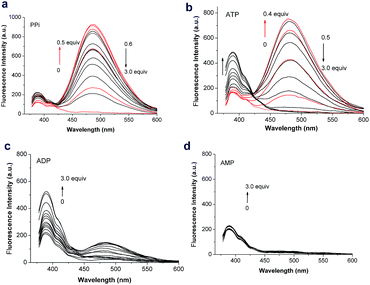 | ||
| Fig. 1 Fluorescence spectra (λex = 350 nm) of 1–Zn2+ (10 μM) upon addition of (a) PPi (0–3.0 equiv.) (b) ATP (0–3.0 equiv.) (c) ADP (0–3.0 equiv.) (d) AMP (0–3.0 equiv.) (10 mM HEPES buffer, pH 7.4). | ||
Notably, ATP not only induced a significant excimer emission peak at 482 nm, but also gave rise to a remarkable increase in the intensity of the short-wavelength monomer emission at 390 nm (Fig. 1b). When less than 0.5 equiv. of ATP is added, an excimer emission of 1–Zn2+ sharply increased, but no changes occurred in the monomer emission, which indicates that adenine is outside the pyrene dimer of 2(1–Zn2+)–ATP complex (Scheme 2). Upon continuous addition of another 0.5 equiv. of ATP, the excimer emission of 1–Zn2+ decreased gradually, but the monomer emission increased obviously. In contrast, ADP with diphosphate moiety can hardly induce the excimer fluorescence, but cause a large enhancement in the monomer emission peak of 1–Zn2+ (Fig. 1c). The results suggest that the adenine aromatic ring should be inside the pyrene dimer to afford a fully intramolecular sandwich structure of 2(1–Zn2+)–ADP complex (Scheme 2). In the case of AMP, there is no fluorescent signal changes with 0–3.0 equiv. of AMP adding into the probe solution (Fig. 1d), indicating that no obvious interaction occurs between AMP and the probe. These unique and different fluorescent responses on the individual nucleotides enable 1–Zn2+ to effectively discriminate ATP from the structurally similar ADP and AMP, as well as PPi.
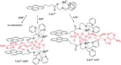 | ||
| Scheme 2 A proposed schematic representation of the fluorescent chemosensor recognition of ATP, ADP and AMP. | ||
Job's plot for the binding between 1–Zn2+ and ATP (ADP) indicates a 2![[thin space (1/6-em)]](https://www.rsc.org/images/entities/char_2009.gif) :
:![[thin space (1/6-em)]](https://www.rsc.org/images/entities/char_2009.gif) 1 stoichiometry (Fig. S3 and 4†), which further confirms the intramolecular sandwich structures were formed in the aqueous solution. Quantum yield of probe 1–Zn2+ in HEPES buffer (10 mM, pH 7.4) was determined to be 0.34 with pyrene (Φ = 0.32 in cyclohexane) as reference.11,13 Upon addition of 0.5 equivalent of ATP into the aqueous solution of 1–Zn2+, the quantum yield of monomer of 1–Zn2+–ATP complex is 0.20, and the excimer of 1–Zn2+–ATP has a large quantum yield (0.78). For nucleotide ADP used for the analysis, higher quantum yield (0.64) of monomer of 1–Zn2+–ADP and much lower quantum yield (0.05) of excimer of 1–Zn2+–ADP were obtained. These results are consistent with the observations in fluorescent determinations (Fig. 1b and c).
1 stoichiometry (Fig. S3 and 4†), which further confirms the intramolecular sandwich structures were formed in the aqueous solution. Quantum yield of probe 1–Zn2+ in HEPES buffer (10 mM, pH 7.4) was determined to be 0.34 with pyrene (Φ = 0.32 in cyclohexane) as reference.11,13 Upon addition of 0.5 equivalent of ATP into the aqueous solution of 1–Zn2+, the quantum yield of monomer of 1–Zn2+–ATP complex is 0.20, and the excimer of 1–Zn2+–ATP has a large quantum yield (0.78). For nucleotide ADP used for the analysis, higher quantum yield (0.64) of monomer of 1–Zn2+–ADP and much lower quantum yield (0.05) of excimer of 1–Zn2+–ADP were obtained. These results are consistent with the observations in fluorescent determinations (Fig. 1b and c).
Upon addition of TTP, UTP or CTP (0–1.0 equiv.), 1–Zn2+ displayed the similar excimer fluorescent response with that of PPi. Further addition of another 2.0 equiv. of TTP, UTP or CTP brought on a complete excimer fluorescence quenching (Fig. 2a–c). Interestingly, both excimer and monomer emission changes of 1–Zn2+ were observed with the GTP concentration varying from 0–3.0 equiv., which is alike with the case of ATP. However, there are still some differences when 1–Zn2+ was titrated by excessive nucleotide (>3 equiv.). As shown in Fig. 3, the ratiometric sensing deals with the simultaneous measurement of two fluorescence signals at 482 nm (excimer emission) and 390 nm (monomer emission), and the ratiometric response at I390/I482 gives following orders: ATP > ADP ≅ GTP > TTP ≅ CTP > AMP > UPT ≅ PPi. Generally, 1–Zn2+ is an ATP-specific ratiometric fluorescent probe to discriminate ATP from various nucleotides and PPi.
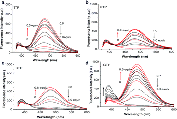 | ||
| Fig. 2 Fluorescence spectra (λex = 350 nm) of 1–Zn2+ (10 μM) upon addition of (a) TTP (0–3.0 equiv.) (b) UTP (0–3.0 equiv.) (c) CTP (0–3.0 equiv.) (d) GTP (0–3.0 equiv.) (10 mM HEPES buffer, pH 7.4). | ||
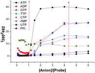 | ||
| Fig. 3 Ratiometric calibration curve I390/I482 as a function of the concentration of UTP, TTP, CTP, GTP, ATP, ADP, AMP or PPi (0–6.0 equiv.). | ||
To further illuminate the binding pattern of 1–Zn2+ with ATP, ADP and AMP, we performed the 1H NMR spectroscopy and 2D NOESY (dimensional nuclear Overhauser effect spectroscopy) titration experiments of 1–Zn2+ with ATP, ADP and AMP, respectively. Fig. 4 shows the 1H NMR signals of 1–Zn2+ with 0.5 equiv. of ADP and ATP, respectively. Addition of 0.5 equiv. of ADP(ATP) to the 1–Zn2+ solution in DMSO-d6 promoted obvious upfield shifts of the protons in the methylene (H5), pyridyl (H2, H3, H4), and pyrenyl (H7, H8) groups. These results indicate the formation of 1–Zn2+–ADP(ATP) complex, which is consistent with the observations by fluorescence determination. No obvious 1H NMR signal changes were obtained when AMP was mixed with the probe solution (Fig. S5†), which further confirms that there is no remarkable interaction of AMP with 1–Zn2+.
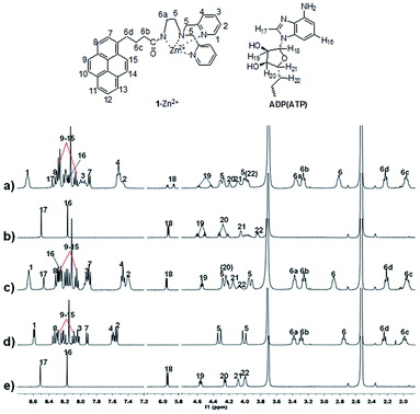 | ||
| Fig. 4 Regions from the 400 MHz 1H NMR spectra for (a) 1–Zn2+ + ADP (0.5 equiv.), (b) ADP, (c) 1–Zn2+ + ATP (0.5 equiv.), (d) 1–Zn2+, and (e) ATP in DMSO-d6. | ||
Particularly, as Fig. 4 shown, there is great difference in 1H NMR signals of protons (H18) between ADP and ATP, when they are coordinated with 1–Zn2+, respectively. The H18 peak of ribose moiety in 1–Zn2+–ADP complex is split into two peaks while the corresponding proton in 1–Zn2+–ATP complex did not undergo this change (Fig. 4a and c), which renders the probe 1–Zn2+ an easy way to discriminate ATP from ADP. Furthermore, in the case of 1–Zn2+–ADP complex, the NOE correlations between the proton (H19) of ribose and the pyrene hydrogen atoms (Hpyrene) reveal that the ribose moiety of ADP is located closely to at least one pyrene moiety (Fig. 5d). These results confirm the hypothesis that, in the 1–Zn2+–ADP complex, the hydrogen-bonding net work is formed between the amide C![[double bond, length as m-dash]](https://www.rsc.org/images/entities/char_e001.gif) O and H–O of the five-carbon sugar in ADP, which induces a long-range coupling of H21 to H18 with the appearance of doublet of doublets peaks of H18 in the 1H NMR spectrum of 1–Zn2+–ADP (Fig. 4a). On the other hand, the significant NOE correlations from H19 of ATP to the methylene (H5) protons (Fig. 5a), as well as the weak cross-peaks between pyridyl H atom (H1) and the hydrogens in ribose ring (H20, H21) (Fig. 5b), indicate that the ribose moiety of ATP is located closely to at least one BPEA–Zn2+ moiety of the 1–Zn2+–ATP complex. In contrast, there was no cross-peak between H19 of ADP and the methylene (H5) protons (Fig. 5c). The results further suggest that 1–Zn2+ should interact with ADP and ATP in different binding patterns.
O and H–O of the five-carbon sugar in ADP, which induces a long-range coupling of H21 to H18 with the appearance of doublet of doublets peaks of H18 in the 1H NMR spectrum of 1–Zn2+–ADP (Fig. 4a). On the other hand, the significant NOE correlations from H19 of ATP to the methylene (H5) protons (Fig. 5a), as well as the weak cross-peaks between pyridyl H atom (H1) and the hydrogens in ribose ring (H20, H21) (Fig. 5b), indicate that the ribose moiety of ATP is located closely to at least one BPEA–Zn2+ moiety of the 1–Zn2+–ATP complex. In contrast, there was no cross-peak between H19 of ADP and the methylene (H5) protons (Fig. 5c). The results further suggest that 1–Zn2+ should interact with ADP and ATP in different binding patterns.
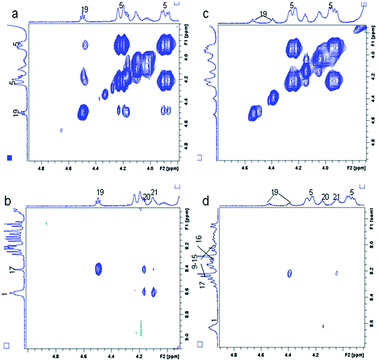 | ||
| Fig. 5 Regions from the 400 MHz NOESY spectra for (a and b) 1–Zn2+ + ATP (0.5 equiv.), (c and d) 1–Zn2+ + ADP (0.5 equiv.) in DMSO-d6. | ||
Based on above investigations on fluorescence and NMR spectroscopy, the hypothetical binding modes of 1–Zn2+ with ATP and ADP were illustrated in Scheme 2. A fully intramolecular sandwich structure was formed between two molecule 1–Zn2+ and one ADP through three synergistic effects: (i) coordination of BPEA–Zn2+ with diphosphate anion, (ii) hydrogen bonding between amide and ribose, and (iii) π–π stacking interaction among pyrenes and adenine. As a comparison, the assembly of two molecule 1–Zn2+ and one ATP provided a partial intramolecular sandwich structure by two synergistic interactions: coordination of BPEA–Zn2+ with triphosphate anion and π–π stacking of two pyrenes. These distinct binding patterns of 1–Zn2+ with ATP and ADP support 1–Zn2+ is an excellent fluorescent probe to discriminate ATP and ADP. In addition, the binding affinity (Ka) of 1–Zn2+ with ATP is estimated as 3.6 × 10¹⁰ M−2 from the fluorescence titration,10a which is comparable with that (7.1 × 10⁹ M−2) of 1–Zn2+ with ADP, suggesting that the intramolecular sandwich structures of 1–Zn2+ with ATP and ADP have similar stabilities in water solution.
Conclusion
In summary, we have reported a tailor-made pyrene-functionalized zinc(II)–BPEA complex as an ATP-specific ratiometric sensor among various nucleotides (ADP, AMP, UTP, CTP, TTP) under physiological conditions. Meanwhile, 1–Zn2+ is also an efficient fluorescent probe to discriminate ATP, ADP and AMP. The probe exhibits distinctive fluorescence response and vivid 1H NMR signal changes in the presence of ATP and ADP. The results reported in this study may help tracking multiple metabolic processes involved in nucleoside polyphosphates, such as numerous enzymatic processes in cells.Acknowledgements
The project was financially supported by the National Natural Science Foundation of China (21272027, 51403020), Beijing National Natural Science Foundation (2122031), and Beijing Municipal Commission of Education.Notes and references
- (a) Fluorescent Chemosensors for Ion and Molecular Recognition, ed. A. W. Czarnik, American Chemical Society, Washington, DC, 1993 Search PubMed; (b) Supramolecular Chemistry of Anions, ed. A. Bianchi, K. Bowman-James and E. García-España, Wiley-VCH, New York, 1997 Search PubMed; (c) Supramolecular Chemistry of Anionic Species thematic issue, Guest ed. P. A. Gale and T. Gunnlaugsson, Chem. Soc. Rev., 2010, 39, 3581–4008 RSC; (d) Z. Xu, S. K. Kim and J. Yoon, Chem. Soc. Rev., 2010, 39, 1457–1466 RSC; (e) M. Wenzel, J. R. Hiscock and P. A. Gale, Chem. Soc. Rev., 2012, 41, 480–520 RSC.
- For recent reviews, see: (a) Y. Zhou, Z. Xu and J. Yoon, Chem. Soc. Rev., 2011, 40, 2222–2235 RSC; (b) A. E. Hargrove, S. Nieto, T. Zhang, J. L. Sessler and E. V. Anslyn, Chem. Rev., 2011, 111, 6603–6782 CrossRef CAS PubMed; (c) S. K. Kim, D. H. Lee, J.-I. Hong and J. Yoon, Acc. Chem. Res., 2009, 42, 23–31 CrossRef CAS PubMed.
- (a) B. Alberts, A. Johnson, J. Lewis, M. Raff and K. Roberts, P. Wlater, in Molecular Biology of the Cell, Garland Science, New York, 4th edn, 2002 Search PubMed; (b) J. R. Knowles, Annu. Rev. Biochem., 1980, 49, 877–919 CrossRef CAS PubMed; (c) F. M. Ashcroft and F. M. Gribble, Diabetologia, 1999, 42, 903–919 CrossRef CAS PubMed; (d) P. B. Dennis, A. Jaeschke, M. Saitoh, B. Fowler, S. C. Kozma and G. Thomas, Science, 2001, 294, 1102–1105 CrossRef CAS PubMed; (e) J. L. Daniel, C. Dangelmaier, J. Jin, B. Ashby and J. B. Smith, J. Biol. Chem., 1998, 273, 2024–2029 CrossRef CAS PubMed; (f) F. Valsecchi, C. Konrad and G. Manfredi, Biochim. Biophys. Acta, 2014 DOI:10.1016/j.bbadis.2014.05.035.
- For recent papers about ATP sensors, see: (a) M. Zhang, W.-J. Ma, C.-T. He, L. Jiang and T.-B. Lu, Inorg. Chem., 2013, 52, 4873–4879 CrossRef CAS PubMed; (b) Z. Xu, N. J. Singh, J. Lim, J. Pan, H. N. Kim, S. Park, K. S. Kim and J. Yoon, J. Am. Chem. Soc., 2009, 131, 15528–15533 CrossRef CAS PubMed; (c) A. S. Rao, D. Kim, H. Nam, H. Jo, K. H. Kim, C. Ban and K. H. Ahn, Chem. Commun., 2012, 48, 3206–3208 RSC; (d) M. Strianese, S. Milione, A. Maranzana, A. Grassia and C. Pellecchiaa, Chem. Commun., 2012, 48, 11419–11421 RSC; (e) A. J. Moro, P. J. Cywinski, S. Körstena and G. J. Mohrac, Chem. Commun., 2010, 46, 1085–1087 RSC; (f) P. Mahato, A. Ghosh, S. K. Mishra, A. Shrivastav, S. Mishra and A. Das, Chem. Commun., 2010, 46, 9134–9136 RSC; (g) Y. Kurishita, T. Kohira, A. Ojida and I. Hamachi, J. Am. Chem. Soc., 2010, 132, 13290–13299 CrossRef CAS PubMed; (h) A. Ojida, I. Takashima, T. Kohira, H. Nonaka and I. Hamachi, J. Am. Chem. Soc., 2008, 130, 12095–12101 CrossRef CAS PubMed; (i) Z. Yao, X. Feng, W. Hong, C. Li and G. Shi, Chem. Commun., 2009, 4696–4698 RSC; (j) J. Kaur and P. Singh, Chem. Commun., 2011, 47, 4472–4474 RSC; (k) Z. Zeng, A. A. J. Torriero, A. M. Bond and L. Spiccia, Chem.–Eur. J., 2010, 16, 9154–9163 CrossRef CAS PubMed; (l) Z. Xu, D. R. Spring and J. Yoon, Chem.–Asian J., 2011, 6, 2114–2122 CrossRef CAS PubMed; (m) A. J. Moro, J. Schmidt, T. Doussineau, A. Lapresta-Fernandéz, J. Wegenere and G. J. Mohrb, Chem. Commun., 2011, 47, 6066–6068 RSC; (n) P. Mahato, A. Ghosh, S. K. Mishra, A. Shrivastav, S. Mishra and A. Das, Inorg. Chem., 2011, 50, 4162–4170 CrossRef CAS PubMed; (o) Y. Kurishita, T. Kohira, A. Ojida and I. Hamachi, J. Am. Chem. Soc., 2012, 134, 18779–18789 CrossRef CAS PubMed; (p) J. H. Lee, A. R. Jeong, J.-H. Jung, C.-M. Park and J.-I. Hong, J. Org. Chem., 2011, 76, 417–423 CrossRef CAS PubMed; (q) H. N. Kim, J. H. Moon, S. K. Kim, J. Y. Kwon, Y. J. Jang, J. Y. Lee and J. Yoon, J. Org. Chem., 2011, 76, 3805–3811 CrossRef CAS PubMed; (r) Z. Xu, N. R. Song, J. H. Moon, J. Y. Lee and J. Yoon, Org. Biomol. Chem., 2011, 9, 8340–8345 RSC; (s) D. Wang, X. Zhang, C. He and C. Duan, Org. Biomol. Chem., 2010, 8, 2923–2925 RSC; (t) J. Kong, C. He, X. Zhang, Q. Meng and C. Duan, Dyes Pigm., 2014, 101, 254–260 CrossRef CAS PubMed.
- (a) P. P. Neelakandan, M. Hariharan and D. Ramaiah, J. Am. Chem. Soc., 2006, 128, 11334–11335 CrossRef CAS PubMed; (b) S. Wang and Y.-T. Chang, J. Am. Chem. Soc., 2006, 128, 10380–10381 CrossRef CAS PubMed; (c) J. Y. Kwon, N. J. Singh, H. N. Kim, S. K. Kim, K. S. Kim and J. Yoon, J. Am. Chem. Soc., 2004, 126, 8892–8893 CrossRef CAS PubMed.
- (a) Z. Zeng and L. Spiccia, Chem.–Eur. J., 2009, 15, 12941–12944 CrossRef CAS PubMed; (b) T.-H. Kwon, H. J. Kim and J.-I. Hong, Chem.–Eur. J., 2008, 14, 9613–9619 CrossRef CAS PubMed.
- (a) X. Chen, M. J. Jou and J. Yoon, Org. Lett., 2009, 11, 2181–2184 CrossRef CAS PubMed; (b) F. Schmidt, S. Stadlbauer and B. König, Dalton Trans., 2010, 39, 7250–7261 RSC.
- A. Kumar, P. Prasher and P. Singh, Org. Biomol. Chem., 2014, 12, 3071–3079 CAS.
- (a) Y. Hisamatsu, K. Hasada, F. Amano, Y. Tsubota, Y. Wasada-Tsutsui, N. Shirai, S. Ikeda and K. Odashima, Chem.–Eur. J., 2006, 12, 7733–7741 CrossRef CAS PubMed; (b) X. Shang, H. Su, H. Lin and H. Lin, Inorg. Chem. Commun., 2010, 13, 999–1003 CrossRef CAS PubMed.
- (a) Q. C. Xu, X. F. Wang, G. W. Xing and Y. Zhang, RSC Adv., 2013, 3, 15834–15841 RSC; (b) Q. C. Xu, C. Jin, X. H. Zhu and G. W. Xing, Chin. J. Org. Chem., 2014, 34, 647–661 Search PubMed; (c) J. Jia, Q. C. Xu, R. C. Li, X. Tang, Y. F. He, M. Y. Zhang, Y. Zhang and G. W. Xing, Org. Biomol. Chem., 2012, 10, 6279–6286 RSC; (d) Q. C. Xu, X. H. Zhu, C. Jin, G. W. Xing and Y. Zhang, RSC Adv., 2014, 4, 3591–3596 RSC; (e) J. Jia, Z. Y. Gu, R. C. Li, M. H. Huang, C. S. Xu, Y. F. Wang, G. W. Xing and Y. S. Huang, Eur. J. Org. Chem., 2011, 4069–4615 Search PubMed.
- H. K. Cho, D. H. Lee and J.-I. Hong, Chem. Commun., 2005, 1690–1692 RSC.
- (a) L. Xue, C. Liu and H. Jiang, Org. Lett., 2009, 11, 1655–1658 CrossRef CAS PubMed; (b) C. Lu, Z. Xu, J. Cui, R. Zhang and X. Qian, J. Org. Chem., 2007, 72, 3554–3557 CrossRef CAS PubMed.
- I. B. Berlman, Handbook of Fluorescence Spectra of Aromatic Molecules, Academic Press, New York, 2nd edn, 1971 Search PubMed.
Footnote |
| † Electronic supplementary information (ESI) available: Detailed experimental procedures and additional spectral data. See DOI: 10.1039/c4ra07923j |
| This journal is © The Royal Society of Chemistry 2014 |

