Peptide-decorated gold nanoparticles via strain-promoted azide–alkyne cycloaddition and post assembly deprotection

Xiaoxiao Wang a, Pierangelo Gobboab, Mojmir Suchya, Mark S. Workentin*ab and Robert H. E. Hudson*ab
aDepartment of Chemistry, The University of Western Ontario, London, Ontario N6A 5B7, Canada. E-mail: robert.hudson@uwo.ca; mworkent@uwo.ca; Fax: +1-519-661-3022
bCentre for Advanced Materials and Biomaterials Research, The University of Western Ontario, London, Ontario N6A 5B7, Canada

Received 24th July 2014 , Accepted 2nd September 2014

First published on 2nd September 2014


Abstract

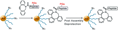
A new method combining an interfacial strain-promoted azide–alkyne cycloaddition and post assembly deprotection (SPAAC-PAD) has been developed for the well-defined functionalization of small, water-soluble gold nanoparticles with oligopeptides.


Due to their excellent biocompatibility, physical properties, and easily modified surfaces, gold nanoparticles (AuNPs) are being investigated for their use in medical applications such as imaging, clinical diagnosis and drug delivery systems.1–3 The progress of AuNPs in these fields has led to the Food and Drug Administration (FDA) approval for AuNP-based in vitro diagnostic systems in clinical trials for cancer treatments.4 To develop effective systems for targeted cancer diagnosis and therapies, it is necessary to functionalize AuNPs with biomolecules such as DNA, proteins and peptides. Peptide–AuNP conjugates have been exploited recently as contrast agents and nanocarriers in targeted imaging for early stage cancer diagnosis and in the pharmaceutical field for targeted intracellular drug/gene delivery, respectively.2,5

The main challenge of conjugation of peptides to AuNPs lies in understanding and controlling the interfacial chemistry between the peptide and the nanomaterial surface on the molecular scale. The reported strategies for bioconjugation of AuNPs with peptides include sulfur–gold bond formation by the direct reaction of cysteine-terminated peptides with the gold surface and electrostatic interactions between a particle and peptides that have been coupled to bovine serum albumin (BSA).6 Although the first method is operationally easy to perform, it is difficult to achieve a high degree of substitution. AuNP bioconjugation via electrostatic interactions has good stability in aqueous solution, but the layer-by-layer assembly results in large hydrodynamic radii of nanoparticles, which limits their application under certain conditions, and offers poor control over the degree of functionalization. Alternatively, a strategy via amide bonds formation involving a water-soluble carbodiimide, N-(3-dimethylaminopropyl)-N-ethylcarbodiimide hydrochloride (EDC), was developed for bioconjugation of AuNPs. Carboxylic groups can be activated by EDC and reacted with primary amines through a condensation reaction to yield amide bonds. Moreover, sulfo-NHS (N-hydroxy sulfosuccinimide) is used to increase the stability of active intermediates in coupling reactions via the formation of active ester functional groups with carboxylates. EDC/NHS coupling has been widely used in preparation of AuNP–peptide conjugates.7 However, this method is still a challenge as quite often, the peptides do not couple efficiently to AuNPs.8,9 Additionally, EDC/NHS coupling method is not very selective as it involves the reaction between an amine and carboxylic acid groups, which widely exist in peptides.10

In order to overcome the aforementioned difficulties, recent effort has focused on the use of “click chemistry” for conjugation of AuNPs with biomolecules.8 For example, the copper-catalyzed azide–alkyne cycloaddition has been utilized to conjugate large azido-containing AuNPs with alkyne functionalized horseradish peroxidase (HRP).9 However, the requirement of using copper ion as catalyst complicates the reaction and could increase in vivo biological toxicity. The strain-promoted azide–alkyne cycloaddition (SPAAC), also known as Cu-free alkyne–azide cycloaddition, can be used to overcome these problems. The SPAAC reaction has been developed as a powerful bioconjugation tool that displays outstanding chemoselectivity, excellent biocompatibility and takes place under very mild reaction conditions. Since its first development,11 much effort has been directed toward the synthesis of cycloalkynes with increased ring strain (to accelerate the reaction) and higher hydrophilic character (for in vivo applications). The challenge behind the syntheses of these interesting molecules relies on finding the right balance between reactivity and chemoselectivity. Indeed, the greater the ring strain, the more susceptible the triple bond is to nucleophilic attack (i.e., addition reactions or rearrangement of cyclooctynes) thus lowering the chemoselectivity towards the cycloaddition with an azide. For example, it has been reported that highly strained biarylazacyclooctynone and analogous bioconjugation reagents undergo rearrangement and addition reactions leading to tetracyclic products.12 The rate of rearrangement was found to be accelerated as the concentration of acid increased. This is problematic because concentrated TFA, such as 95% TFA with scavengers, is the typical reagent for cleavage of peptides from solid supports like Wang resin, Rink Amide resin, etc. together with the removal of the side chain protecting groups. This limits the applications of strained alkynes for preparation of clickable peptides. Additionally, strained alkynes have been shown to undergo side reactions with cellular cysteines and other nucleophilic residues.13,14 However, nucleophilic side chain functionalities, such as hydroxyl (Tyr, Ser, Thr), amino (Lys), guanidine (Arg) or sulfhydryl (Cys), widely exist in bioactive peptides and may react with highly reactive cyclooctynes through a nucleophilic attack.15 This also limits the application of SPAAC. Interestingly, the application of interfacial SPAAC for bioconjugation of AuNPs with peptides has not yet been disclosed.

Herein, we report a novel general method for the synthesis of AuNP-bioconjugates that takes into account the side reactivity of the strained alkynes and therefore combines an interfacial SPAAC with a post assembly deprotection (PAD) for the conjugation of dibenzocyclooctyne (DBCO)-oligopeptides to small, water-soluble azide–AuNPs. We show how this general approach overcomes the aforementioned drawbacks and extends the application of SPAAC to the bioconjugation of metallic nanoparticles in a quick, reliable and facile way. Importantly, we show that the undesired side reactions of the strained alkyne in the presence of concentrated TFA and with nucleophilic sites on peptides are avoided through mild cleavage condition and retention of side-chain protecting groups on the peptides during the SPAAC reaction. The protecting groups can then be easily removed, i.e. post assembly deprotection or PAD, under reaction conditions that preserve the integrity of the gold core, and the AuNP bioconjugate can be easily purified by dialysis. Finally, the amount of conjugated peptide can be easily calculated.

To showcase this SPAAC-PAD strategy we employed azide–AuNPs based on tri- and tetra-ethylene glycol ligands that impart both water- and organic solvent-solubility to final AuNPs. The amphilicity is useful for manipulating the AuNP in organic solvents to facilitate both the SPAAC reaction and the deprotection reaction, as well as maintaining solubility in water for further in vivo applications.

The azide–AuNPs were prepared and characterized following our previously reported procedure.16 Briefly, triethylene glycol–monomethyl ether AuNP (Me-EG3-AuNP) with a gold core diameter of 3 ± 0.5 nm were synthesized via a modified Brust–Schiffrin method. The azide ligand N3-EG4-SH was synthesized and introduced onto the surface of Me-EG3-AuNP using a place-exchange reaction. In a typical synthesis 42.5 mmol of N3-EG4-SH were stirred for 20 min in acetone and in presence of 50 mg of Me-EG3-AuNP. The free thiols were subsequently removed by trituration of the dried AuNP film first with hexanes and followed by isopropanol. Azide–AuNPs were characterized by thermogravimetric analysis (TGA), transmission electron microscopy (TEM) and FT-IR spectroscopy. The IR spectrum of resulting AuNPs showed the appearance of asymmetrical stretching of the azide group at 2110 cm−1 (Fig. S3a, ESI). TGA data indicated that the corona of the AuNP is composed of 35% of azide ligands (Fig. S5, ESI) and thus these nanoparticles contain azide functionalities at a concentration of 0.745 μmol mg−1. Based on 1H NMR spectrum and TGA data, and assuming that the nanoparticles have a spherical shape and that their size is monodispersed, we can estimate a nanoparticle formula of Au1000(Me-EG3-S)455(N3-EG4-S)245 (calculation reported in ESI).

The effectiveness of our SPAAC-PAD approach is shown through the example of bioconjugation of the arginylglycylaspartic acid (RGD) peptide. This peptide was chosen because it has targeting relevance- it is well-known to be specifically recognized by the integrin αvβ3 receptor and can act as tumour and angiogenesis marker.17,18 In Scheme 1 two potential routes are illustrated for bioconjugation of the azide–AuNP with the RGD peptide. The first approach (Route 1, Scheme 1), involves preparing a “naked” peptide consisting of a dibenzocyclooctyl (DBCO) moiety connected via a polyethylglycol (PEG) linker to the RGD tripeptide (hereafter named “DBCO”-RGD). After preparation PEG-containing RGD, DBCO was constructed to the peptide via standard peptide coupling reaction. The resulting peptide, “DBCO”-RGD, was cleaved from the resin and deprotected by using 95/5 TFA/TES. After purification, it was then directly reacted with the azide–AuNP. The second approach (Route 2, Scheme 1) applies a construct possessing a side-chain protected peptide fragment (DBCO-(PG)RGD). It is noteworthy that dilute TFA (5/5/90 TFA/TES/DCM) was used as the reagent for cleavage of the peptide from the resin and for retention of protecting groups on peptide side chains. The peptide reacted with the azide–AuNPs via an I-SPAAC and all protecting groups were removed in a post-assembly deprotection step.


image file: c4ra07574a-s1.tif
Scheme 1 Illustration of the attempted routes for the bioconjugation of azide–AuNPs with RGD peptide via SPAAC-PAD. (a) Use of an unprotected peptide (Route 1) or use of a protected peptide (Route 2), (b) and (c) molecular structures of “DBCO”-RGD and DBCO-(PG)RGD. Protecting groups (PG) are Pbf and tBu.

To undertake the approach described by Route 1, RGD peptide was synthesized via standard Fmoc solid-phase peptide synthesis (SPPS) procedure using rink amide resin as the solid support (Scheme S1, ESI) and DBCO acid was coupled to N-terminus of the peptide. Cleavage from the resin and protecting group removal was done in one step by treatment with 95/5 TFA/TES. The resulting native peptide was purified by HPLC and characterized by ESI-MS (Fig. S1a and S2a, ESI). The observed molecular mass of 849.3472 was in agreement with the calculated mass (849.3657). In spite of this, close examination of the UV-spectrum of the peptide (Fig. S2a, ESI) indicated that the chromophore was changed from DBCO (Fig. S2d, ESI), thus implying decomposition of DBCO had occurred.

A control experiment was carried out to determine if the standard Fmoc-based oligomerization chemistry cleavage condition of concentrated TFA induced decomposition of DBCO. Starting with DBCO amine, we measured its purity by HPLC before and after treatment with 95% TFA/DCM (rt, 1 h). HPLC analysis of the reaction mixture, along with subsequent 1H NMR and high resolution mass spectral analysis, clearly showed the complete consumption of starting material and the production of several products (Fig. S2d and e, ESI). The two major components of the mixture, i.e. the peaks with retention time of 20–20.5 min and of 26–26.5 min (Fig. S2e) do not correspond to DBCO. On-line monitoring showed significant changes the UV spectrum indicating structural changes in the chromophore. The 1H NMR spectra of the two isolated compounds (Fig. S7b and c, ESI) confirmed neither was DBCO (Fig. S7a, ESI). Mass spectral analysis showed that the earlier eluting peak retained the same mass as DBCO implying a molecular rearrangement had occurred,12 while the later eluting peak possessed a greater mass. It's also noteworthy that the UV spectrum of the first peak in Fig. S2e showed strong absorbance at 344 nm, which has also been detected in UV spectrum of “DBCO”-RGD (Fig. S2a, ESI), indicating that DBCO on peptide RGD had decomposed under the standard cleavage condition of 95% TFA. To study the stability of the DBCO moiety toward TFA, DBCO was exposed to a series of solutions of 0–50% TFA/DCM for 1 h at room temperature. Under these conditions, DBCO was found to be stable when the TFA concentration was less than 30%.

Another control experiment was performed to model the interfacial SPAAC reaction by using “DBCO”-RGD peptide with an azide, namely ω-azido-triethylene glycol-monomethyl ether (Me-EG3-N3) as the reactive partner. The reactions were carried out in either water or acetonitrile solutions and the course of the reaction was monitored by UPLC/ESI-MS. Only the two starting materials were detected over 4 hours, indicating that the “DBCO”-RGD peptide and Me-EG3-N3 were not undergoing the SPAAC. Independently we determined that the Me-EG3-N3 was capable of participating in SPAAC with DBCO. Therefore, “DBCO”-RGD lost the ability of click and cannot be used as clickable peptide for bioconjugation of AuNPs. As a control for further study, the peptide “DBCO”-RGD was still added into azide–AuNPs (azide group:peptide, 1[thin space (1/6-em)]:[thin space (1/6-em)]1.2 ratio). The mixture was stirred in water at room temperature for 1 h. After removal of solvent, AuNPs were purified by centrifugal filtration (Millipore centrifugal filter units MWCO 10 kDa). As expected, characterization of these AuNPs by IR spectroscopy showed the absorption of the azide group at 2110 cm−1 was still present with similar intensity to the starting material, which indicated lack of SPAAC reactivity, and showed no stretching at 1685 cm−1 corresponding to the amide functional groups. To circumvent the problem of decomposition of DBCO, Route 2 was devised. First we prepared DBCO-(PG)RGD by using a mild cleavage condition, it was then clicked to the azide–AuNP and subsequently the peptide was deprotected (Scheme 1). To synthesize the DBCO-(PG)RGD, the acid sensitive 2-chlorotritylchloride resin was used as solid support. The synthesis of DBCO-containing peptide followed the above procedure, except that the cleavage was performed with TFA/TES/DCM (5/5/90). This treatment effected the cleavage of the peptide from the resin but left the protecting groups intact and did not destroy DBCO (Scheme S2, ESI). The resulting protected peptide was purified by HPLC and characterized by ESI-MS (Fig. S1b and S2b, ESI). The UV spectrum of DBCO-(PG)RGD showed the agreement with DBCO with absorbance at 290 nm and 308 nm. A model click was carried out with DBCO-(PG)RGD and Me-EG3-N3 in acetonitrile. Characterization of the reaction by UPLC/ESI-MS showed successful click, indicating that DBCO was not destroyed in the cleavage step. Then SPAAC between azide–AuNPs and DBCO-(PG)RGD was simply performed by adding them (azide:peptide, 1[thin space (1/6-em)]:[thin space (1/6-em)]1.2) in acetonitrile and stirring the resultant mixture at room temperature for 1 h (Fig. 1). After reaction, the AuNPs were purified by using centrifugal filtration (Millipore centrifugal filter units MWCO 10 kDa) and washed with 60% MeOH/H2O until no unreacted peptide was observed in the UPLC/ESI-MS analysis of the filtrate. The unreacted peptide was recovered from the filtrate and washings and the amount indicates that approximately 31% of the azide groups on AuNPs had reacted. Therefore, the approximate nanoparticle formula can be calculated as Au1000((PG)RGD-linker-S)75(N3-EG4-S)170(MeEG3S)450. The IR spectrum of the purified (PG)RGD–AuNPs showed that the azide stretching mode at 2110 cm−1 had markedly decreased while it had not changed in Route 1 (Fig. S3c, ESI). Additionally, concomitant with the azide disappearance the appearance of stretching at 1685 cm−1 corresponding to the amide functional groups was observed, indicating that the protected peptide was covalently bonded to the AuNP via the Cu-free cycloaddition.


image file: c4ra07574a-f1.tif
Fig. 1 Bioconjugation of AuNPs with RGD peptide via SPAAC-PAD, (1) acetonitrile, room temperature, 1 h; (2) 90% TFA/DCM, room temperature, overnight; and AuNP size distributions obtained from the corresponding TEM image of RGD-functionalized AuNPs via SPAAC-PAD.

To remove the protecting groups of the peptide, the AuNPs were then treated with 90% TFA/DCM at room temperature overnight to ensure complete deprotection. It is important to note that these small oligo-ethylene glycol-based AuNP are perfectly stable under these acidic conditions. TEM images recorded before and after this acidic treatment confirmed that the relatively harsh condition for PAD did not adversely affect the AuNP size distribution (Fig. 1). The obtained RGD–AuNPs were then purified by dialysis using cellulose ester dialysis membranes (6–8 KDa MWCO).

To further confirm that the interfacial SPAAC reaction had proceeded, the deprotected RGD–AuNPs were reacted with molecular iodine. Oxidation of AuNPs with iodine dissolved the gold into a mixture of Au(I) and Au(III) complexes and released the corona ligands as disulphides.19 The structure of the most possible disulphide molecule produced by this reaction is shown in Fig. 2. The mixture of disulphides was characterized by ESI-MS and the mass 1365.6084 [M + H]+ m/z agrees well with the calculated mass of 1364.5992 for the proposed disulphide 1. Importantly the mass for the corresponding disulphide with the protected peptide was not observed, indicating that the complete deprotection was achieved. This confirms the successful bioconjugation of AuNPs with a short oligopeptide via SPAAC-PAD without interference with side chains of the peptide. Moreover, efficient substitution was achieved on the surface of small size AuNPs with 31% of the azide groups reacting with the RGD peptide. This result is comparable with the reported conjugation of AuNPs with peptides via covalent bonding,20 with the difference that this AuNP-bioconjugate was obtained through a simple and rapid pour-and-mix chemistry under ambient temperature and atmosphere while the purification only involved centrifugation and dialysis. The final calculated molecular raw formula of this AuNP is Au1000(RGD-linker-S)75(N3-EG4-S)170(MeEG3S)450.


image file: c4ra07574a-f2.tif
Fig. 2 Re-oxidation of RGD–AuNPs by iodine. Disulfide 1 was characterized by ESI-MS. Calculated 1364.5992 and found 1365.6084 [1+] m/z.

To test the versatility of this method we employed a peptide with different nucleophilic residues. In particular because it has been reported that in presence of reduced peptidylcysteine residues, strained cyclooctynes mainly react via thiol–yne addition instead of SPAAC reaction,15 we decided to synthesize a CRGDK peptide and conjugate it to the azide–AuNPs via our SPAAC-PAD strategy. This peptide has an additional interest because it is a neuropilin-1 (Nrp-1) receptor-targeted peptide for cancer treatment.7,21,22

For these experiments, we followed the same procedure as previously discussed with the noted exception. The CRGDK peptide was synthesized via standard Fmoc SPPS method and DBCO acid was coupled to the N-terminus. A change in the acid strength for resin cleavage (0.5% TFA/DCM) was made to preserve the trityl thioether protecting group of cysteine. After SPAAC-PAD, the CRGDK-functionalized AuNPs were characterized by IR (Fig. S3e, ESI). The azide stretch in the IR spectrum became weaker compared to the azide–AuNPs before conjugation and the stretching at 1685 cm−1 corresponded to the functional groups of peptide bond appeared, suggesting successful conjugation through the I-SPAAC-PAD. Also, a new absorbance at 2692 cm−1 indicated the existence of the thiol group of cysteine and shows that the side chain of cysteine that would otherwise react with cyclootyne via thiol–yne addition did not interfere with the I-SPAAC reaction with the aid of PAD. Decomposition of the AuNPs by treatment with excess I2 followed by ESI-MS analysis showed the two most possible disulfide molecules expected after a successful I-SPAAC-PAD (Fig. S1e, ESI), and no trace of protected peptide was found. Therefore, our strategy showed to be also effective in creating small AuNP–oligopeptide bioconjugates in presence of nucleophilic residues that could otherwise attack the strained alkyne bond.

In summary, we have created a new, efficient, easy method to implement general approach to the synthesis of AuNP–peptide bioconjugates that combines the I-SPAAC reaction with post assembly deprotection. Using this strategy, peptides with protected side chains can be conjugated onto gold nanoparticles via I-SPAAC and all protecting groups can then be easily removed by PAD. The mild reaction conditions preserve the integrity of the alkyne so that it may perform the SPAAC reaction and the conjugated AuNPs are sufficiently robust to undergo peptide deprotection without deleterious effects. The approach presented herein usefully expands the repertoire of existing methods and presents an easy, reliable and quick way to functionalize AuNPs with short oligopeptides where the extent of incorporation of the interfacial biomolecule can be easily quantified. We anticipate that the demonstrated methodology for peptide–AuNP bioconjugation via the SPAAC-PAD will enable the fabrication of gold nanoparticles with a high degree of complexity with biomolecules for a variety of applications in targeted cancer diagnosis and therapies. Targeted CT imaging studies of the functionalized AuNPs is currently underway.

Acknowledgements

We (RHEH, MSW) acknowledge the Natural Sciences and Engineering Research Council of Canada and The University of Western Ontario financial support. PG thanks the Government of Canada for a Vanier Scholarship and Research Western.

References

  1. Z. Luo, K. Zheng and J. Xie, Chem. Commun., 2014, 50, 5143–5155 RSC.
  2. H. Liu, Y. Xu, S. Wen, Q. Chen, L. Zheng, M. Shen, J. Zhao, G. Zhang and X. Shi, Chem.–Eur. J., 2013, 19, 6409–6416 CrossRef CAS PubMed.
  3. B. Aydogan, J. Li, T. Rajh, A. Chaudhary, S. J. Chmura, C. Pelizzari, C. Wietholt, M. Kurtoglu and P. Redmond, Mol. Imag. Biol., 2010, 12, 463–467 CrossRef PubMed.
  4. S. K. Libutt, G. F. Paciotti, A. A. Byrnes, H. R. Alexander Jr, W. E. Gannon, M. Walker, G. D. Seidel, N. Yuldasheva and L. Tamarkin, Clin. Cancer Res., 2010, 16, 6139–6149 CrossRef PubMed.
  5. A. N. Shirazi, D. Mandal, R. K. Tiwari, L. Guo, W. Lu and K. Parang, Mol. Pharm., 2013, 10, 500–511 CrossRef PubMed.
  6. F. Porta, G. Speranza, Z. Krpeti, V. D. Santo, P. Francescato and G. Scari, Mater. Sci. Eng., B, 2007, 140, 187–194 CrossRef CAS PubMed.
  7. A. Kumar, H. Ma, X. Zhang, K. Huang, S. Jin, J. Liu, T. Wei, W. Cao, G. Zou and X.-J. Liang, Biomaterials, 2012, 33, 1180–1189 CrossRef CAS PubMed.
  8. D. Bartczak and A. G. Kanaras, Langmuir, 2011, 27, 10119–10123 CrossRef CAS PubMed.
  9. M.-X. Zhang, B.-H. Huang, X.-Y. Sun and D.-W. Pang, Langmuir, 2010, 26, 10171–10176 CrossRef CAS PubMed.
  10. C.-F. Wang, E. M. Mäkilä, M. H. Kaasalainen, D. Liu, M. P. Sarparanta, A. J. Airaksinen, J. J. Salonen, J. T. Hirvonen and H. A. Santos, Biomaterials, 2014, 35, 1257 CrossRef CAS PubMed.
  11. N. J. Agard, J. A. Prescher and C. R. Bertozzi, J. Am. Chem. Soc., 2004, 126, 15046–15047 CrossRef CAS PubMed.
  12. M. Chigrinova, C. S. McKay, L.-P. B. Beaulieu, K. A. Udachin, A. M. Beauchemin and J. P. Pezacki, Org. Biomol. Chem., 2013, 11, 3436–3441 CAS.
  13. P. V. Chang, J. A. Prescher, E. M. Sletten, J. M. Baskin, I. A. Miller, N. J. Agard, A. Lo and C. R. Bertozzi, Proc. Natl. Acad. Sci. U. S. A., 2010, 107, 1821–1826 CrossRef CAS PubMed.
  14. K. E. Beatty, J. D. Fisk, B. P. Smart, Y. Y. Lu, J. Szychowski, M. J. Hangauer, J. M. Baskin, C. R. Bertozzi and D. A. Tirrell, ChemBioChem., 2010, 11, 2092–2095 CrossRef CAS PubMed.
  15. R. van Geel, G. J. M. Pruijn, F. L. van Delft and W. C. Boelens, Bioconjugate Chem., 2012, 23, 392–398 CrossRef CAS PubMed.
  16. P. Gobbo, S. Novoa, M. C. Biesinger and M. S. Workentin, Chem. Commun., 2013, 49, 3982–3984 RSC.
  17. M. D. Pierschbacher and E. Ruoslahti, Nature, 1984, 81, 5985–5988 CAS.
  18. M. D. Pierschbacher and E. Ruoslahti, Proc. Natl. Acad. Sci. U. S. A., 1984, 81, 5985–5988 CrossRef CAS.
  19. L. Sun, R. M. Crooks and V. Chechik, Chem. Commun., 2001, 359–360 RSC.
  20. Y.-H. Kim, J. Jeon, S. H. Hong, W.-K. Rhim, Y.-S. Lee, H. Youn, J.-K. Chung, M. C. Lee, D. S. Lee, K. W. Kang and J.-M. Nam, Small, 2011, 7, 2052–2060 CrossRef CAS PubMed.
  21. A. Kumar, S. Huo, X. Zhang, J. Liu, A. Tan, S. Li, S. Jin, X. Xue, Y. Zhao, T. Ji, L. Han, H. Liu, X. Zhang, J. Zhang, G. Zou, T. Wang, S. Tang and X. Liang, ACS Nano, 2014, 4205–4220 CrossRef CAS PubMed.
  22. T. Wei, J. Liu, H. Ma, Q. Cheng, Y. Huang, J. Zhao, S. Huo, X. Xue, Z. Liang and X.-J. Liang, Nano Lett., 2013, 13, 2528–2534 CrossRef CAS PubMed.

Footnotes

Electronic supplementary information (ESI) available. See DOI: 10.1039/c4ra07574a
These authors contributed equally to this work.

This journal is © The Royal Society of Chemistry 2014
Click here to see how this site uses Cookies. View our privacy policy here.