Microsol-electrospinning for controlled loading and release of water-soluble drugs in microfibrous membranes

Li Zhouab, Caihong Zhubc, Laura Edmondsbd, Huilin Yangbc, Wenguo Cui*bc and Bin Li*bc
aCollege of Chemistry, Chemical Engineering and Materials Science, Soochow University, Suzhou, Jiangsu 215123, P. R. China
bOrthopedic Institute, Soochow University, 708 Renmin Rd, Suzhou, Jiangsu 215007, P. R. China. E-mail: wgcui80@hotmail.com; binli@suda.edu.cn; Fax: +86-512-67781163; Tel: +86-512-67781420
cDepartment of Orthopedics, The First Affiliated Hospital of Soochow University, 188 Shizi St, Suzhou, Jiangsu 215006, P. R. China
dNanotechnology Engineering, University of Waterloo, Waterloo, Ontario N2L3G1, Canada

Received 23rd July 2014 , Accepted 28th August 2014

First published on 28th August 2014


Abstract

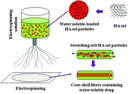
Water-solubility facilitates drug transportation and distribution of drugs throughout the body and hence effectively promotes their absorption. While there have been a number of techniques for incorporating water-soluble drugs into electrospun fibers to realize sustained release of them, problems including burst and uncontrolled release still remain to be solved. In this study, we developed a microsol-electrospinning technique for fabricating core–shell microfibers to achieve incubated, controlled and sustainable release of water-soluble drugs such as chloroquine (CQ). In this approach, nanoparticles made of CQ-loaded hyaluronic acid (HA) sol were first prepared using the emulsification method. Next, the HA-sol nanoparticles were dispersed in poly(L-lactide) (PLLA) electrospinning solution to form a uniform suspension, which was used for fabricating composite microfibers through microsol-electrospinning. Judging from SEM and TEM, the composite microfibers had smooth, uniform morphology and core–shell structure. Further tests showed that the microsol-electrospun microfibers had similar physical, chemical, and mechanical properties as microfibers fabricated using a conventional electrospinning approach. In vitro drug release tests showed that compared to conventional electrospun microfibers, the burst release of CQ was significantly reduced in microsol-electrospun microfibers. Meanwhile, the release time of CQ was markedly extended, being as long as more than 40 days. Importantly, the drug release rate could be readily adjusted by changing the concentration of microsol particles and the amount of drug in the microfibers. Together, findings from this study have revealed that microsol-electrospinning is a facile technique for loading water-soluble drugs into electrospun microfibers and releasing them in a controlled fashion, which may expand the applications of water-soluble drugs.


1. Introduction

Water-soluble drugs are a major category of common drugs, which can be quickly and easily dissolved in water, and absorbed in vivo. Due to the high solubility, rapid dissolution, tachymetabolism and ready diffusion of water-soluble drugs in aqueous solution, it is generally difficult to achieve sustainable release of them without using appropriate loading systems. To improve the effectiveness of water-soluble drug therapies, many carriers, including liposomes,1 microcapsules,2,3 nanospheres,4,5 and hydrogels,6 have been developed and applied to lesions. However, such delivery approaches may be problematic from various aspects. For example, rapid drug release, non-three-dimensional scaffolds, difficulty to maintain appropriate local dosage at the lesion site and so on. Developing new drug loading and release systems, therefore, is appealing for achieving controlled local release of water-soluble drugs in lesions.

Electrospinning is an effective technique that has been extensively used to fabricate ultra-fine polymer fibers with diameters ranging from several microns down to less than 100 nm.7 Among the numerous applications of electrospun fibers in biomedicine, drug carrier is one of the most exciting one.8 To date, a couple of electrospinning techniques have been developed to fabricate polymer nanofibers for drug delivery, including mixing electrospinning,9 emulsion electrospinning,10 and coaxial electrospinning.11 In order to incorporate water-soluble drugs in polymer microfibers, emulsion electrospinning and coaxial electrospinning are most frequently used. Coaxial electrospinning results in microfibers with core–shell structure, in which the water-soluble drug is confined in the core and protected by a polymer shell.12 While such a core–shell structure of fibers markedly help reduce burst release and maintain stable release rate, achieving coaxial electrospinning is technically demanding due to the complexity of spinneret and potential instability of coaxial flow upon feeding.13,14 A much simpler approach, emulsion electrospinning is used for carrying water-soluble drugs by emulsifying aqueous solution in a polymer solution and then electrospinnning the emulsion.15 Nonetheless, it suffers low efficiency of drug loading. Hence, it remains challenging to develop a simple yet reliable technique for fabricating electrospun polymer microfibers loaded with water-soluble drugs.

In addition to controlled loading and release, preserving the activity of water-soluble drugs is also a critical issue affecting their applications. Hydrosols, a type of sol in which water is the dispersed phase and possesses high dispersion stability, have shown promise in drug delivery applications.16,17 Specifically, hydrosol nanoparticles may be obtained by ultrasonic dispersion and have been used as efficient drug carriers.18 In hydrosol nanoparticles, the drug is dissolved into the aqueous solution and can be readily released from them in a controlled fashion as a result of free diffusion of water through the wall of nanoparticles. The hydrosol nanoparticles could form stable water-in-oil (W/O) emulsions in organic solvents, which are beneficial for fabrication of hydrosol-loaded carriers. However, hydrosol nanoparticles resist diffusion of organic solvents and can effectively prevent organic solvent from contacting the encapsulated drug within them. As a result, hydrosol may function as an ideal isolated system that protects the activity of drug within it.

However, it is a constant challenge to realize the controlled and delayed release of water-soluble drugs from electrospun polyester fibers, because (1) water-soluble drugs fast dissolved in water solution; (2) water-soluble drugs could not dissolve into the organic solvent during the preparation of electrospun solution. Taking advantages of hydrosol nanoparticles, in this study we aimed to develop an efficient microsol-electrospinning technique for fabricating core–shell polymer microfibers for water-soluble drug loading and release. By incorporating water-soluble drug into lipid-soluble electrospun fibers, such an approach may achieve controlled loading and sustainable release of water-soluble drugs, and meanwhile protect the drug activity. Chloroquine phosphate salt (CQ), a common drug for treating falciparum malaria, vivax malaria, and other diseases,19,20 is used as a model drug in this study. CQ is typical water-soluble drug which can be easily dissolved in water but is hard to dissolve in organic solvents such as ethyl alcohol, chloroform, and benzene.21 As a natural polymer and exists richly in the extracellular matrix (ECM) of most tissues, hyaluronic acid (HA) has been widely used in drug delivery systems.22 In this study, we first prepared CQ-loaded HA hydrosol nanoparticles using ultrasonic emulsification method. We then uniformly dispersed the HA-sol nanoparticles in poly(L-lactide) solution and used the suspension to fabricate core–shell electrospun microfibers by a one-step electrospinning process, i.e., microsol-electrospinning. In addition to morphological characterizations, the physical, chemical, and mechanical properties of the electrospun microfibers were extensively studied. More importantly, the vitro loading and release behaviors of CQ-loaded microfibers fabricated using microsol-electrospinning were investigated.

2. Materials and methods

2.1 Materials

Poly(L-lactide) (PLLA, Mw = 50 kDa, Mw/Mn = 1.61) was purchased from Jinan Daigang Co. (Jinan, China). Fermentation-derived hyaluronan (HA, sodium salt, Mw = 0.5 MDa) was purchased from Yuancheng Technology Co. (Wuhan, China) and used without further purification. Chloroquine phosphate salt (CQ) was purchased from Sigma (United States). Dichloromethane (DCM) was purchased from Jiangsu Qiangsheng Functional Chemistry Co., Ltd. N,N-Dimethylformamide (DMF) was obtained from Shanghai Qiangshun Chemical Reagent Co., Ltd (Shanghai, China). Span-80 was purchased from Sinopharm Chemical Reagent Co., Ltd (Shanghai, China).

2.2 Preparation of microsol solutions

HA hydrosols (1 wt%, obtained by adding 0.1 g HA into 9.9 g distilled water and stirring till complete dissolution) were first prepared in triplicate. Each group contained 0.5, 1.0, 1.5 wt% CQ (with respect to PLLA), respectively. The drug-loaded HA-sol was added into a solution of DCM and 1% Span-80 (with respect to PLLA). In order to prepare uniform water-in-oil (W/O) emulsions containing micro-sol particles, the mixture was stirred severely for 20 minutes. Then 1.0 g PLLA and 2.0 g DMF were added into the emulsion successively and dissolved entirely to obtain homogeneous electrospinning solution. PLLA solution as control was prepared as the following protocol: 1.0 g PLLA was added into 4.0 g DCM and stirred completely, then 2.0 g DMF was added into the mixture to obtain the control electrospinning solution. The size of HA-microsol particles in the prepared emulsion were measured using dynamic light scattering (DLS, Zetasizer, Malvern, Nano-ZS90).

2.3 Electrospinning

The electrospinning process was carried out at room temperature through the experimental set-up which included a high voltage direct current power supply, purchased from Tianjin Dongwen high voltage power supply factory, and a digitally controlled and extremely accurate syringe pump, purchased from Baoding Longer Precision Pump Co., Ltd. The electrospinning solution was transferred to a 10 mL syringe. The applied voltage was 12 kV, and was attached to the tip of a syringe needle (inner diameter, 0.9 mm). The flow rate of the syringe pump was 0.8 mL h−1, and the electrospun microfibers were collected using a piece of electrically grounded aluminum foil which was placed at a distance of 15 cm from the tip of syringe needle. The fabricating compositions of electrospun microfiber samples used in this study are shown in Table 1.
Table 1 The compositions for fabricating electrospun microfiber samples
Sample name Amount of 1% HA (g) Percentage of CQ (with respect to PLLA)
PLLA-HA05-CQ05 0.05 0.5%
PLLA-HA05-CQ10 0.05 1.0%
PLLA-HA05-CQ15 0.05 1.5%
PLLA-HA10-CQ05 0.1 0.5%
PLLA-HA10-CQ10 0.1 1.0%
PLLA-HA10-CQ15 0.1 1.5%


2.4 Characterizations of electrospun microfibers

The morphologies of the prepared microfibers membranes were examined by field-emission scanning electron microscopy (FESEM) which was using a Hitachi 4800 system with an acceleration voltage of 3.0 kV. Before SEM observation, the surface of the fibers was sputter-coated with platinum.

Transmission electron microscopy (TEM) images of individual microfiber's inner structure were obtained by a Hitachi HT7700 at 120 kV.

Fourier transform infrared spectroscopy (FTIR, Nicolet 6700) was used for investigating the composites, pure CQ, and HA.

Wide angle X-ray diffraction (WAXRD) pattern was recorded by using an X-ray diffractometer (X' Pert-Pro MPD) using Cu Kα radiation with a Ni filter (1.542 Å). The samples were scanned from 5° to 90° at a scanning rate of 5° min−1.

The static water contact angles (WCA) of microfibrous membranes were measured using a contact angle analyzer (DSA25S, Data Physics Corporation).

Glass transition temperatures (Tg) were determined using a Perkin-Elmer differential scanning calorimeter (DSC, DIAMOND) apparatus. The samples were heated from 20 to 150 °C with a heating rate of 10 °C min−1 under nitrogen atmosphere.

For mechanical tests, the dry microfibrous membranes were punched into dumbbell shaped specimens (15.0 × 3.0 × 0.13 mm3). Uniaxial tensile tests were performed using a mechanical testing machine (Hengyi, 5 mm min−1, n = 5).

2.5 In vitro drug release test

To detect the drug content in the fibers, drug-loaded electrospun fibrous membranes (about 30 mg) were dissolved in 2.0 g DCM. Then 2.0 mL PBS was added into the mixture to extract the supernatant liquor. After 25 repeated extraction processes, all of the drugs in the organic solution were completely extracted. The drug loading efficiency (LE) of electrospun fibers was calculated as follows:
 
image file: c4ra07518h-t1.tif(1)

As measured, the LEs of all CQ-loaded electrospun membranes were about 80% in this study.

To identify the in vitro release behavior of CQ, small square samples, which had approximately identical size and thickness with a total mass of about 100 mg, were cut from the fibrous membranes and then immersed in 20 mL of 154 mM phosphate buffered saline (PBS, pH 7.4), The suspension was maintained in a constant-temperature shaking air bath (Taichang Huamei Biochemical Instruments, Jiangsu, China) with a shaking speed of 100 cycles per min at a temperature of 37 °C. At predetermined time intervals, 3.0 mL buffer was removed for analysis and 3.0 mL fresh PBS was added back for continuing incubation. The concentrations of CQ in the samples were measured by an UV-Vis spectrophotometer (WFZ UV-2102 Unique Technology Shanghai) at wavelength of 257 nm. The results were showed in terms of cumulative release as a function of release time:12

 
image file: c4ra07518h-t2.tif(2)
where Mt is the amount of CQ released at time t and M is the total amount of CQ in the fibrous membranes.

2.6 Statistical analysis

All data were obtained at least in triplicates and presented as mean ± standard deviation (SD). One way ANOVA together with the Tukey's post-hoc test were used to discern the statistical difference between groups. A probability value (p) of less than 0.05 is considered statistically significant.

3. Results

3.1 Preparation of microsol particles

The DLS measurement indicated the size distribution of HA-microsol particles in DCM (Fig. 1). It can be seen that the diameter of particles ranges from 100 nm through 2500 nm. Nonetheless, about 96.5% of particles are less than 1000 nm and the PDI is 0.235, indicating the relatively good uniformity of microsol particles. In addition, the particles size distribution remained unchanged within 2 h.
image file: c4ra07518h-f1.tif
Fig. 1 Diameter of micro-gel particles in organic solvents.

3.2 Morphology of electrospun fibers

The surface morphology of electrospun fiber membranes are shown in Fig. 2. The fibers appear uniform and no particles are seen on their surface. The diameters of fibers in PLLA, PLLA-HA05-CQ05, PLLA-HA05-CQ10, PLLA-HA05-CQ15, PLLA-HA10-CQ05, PLLA-HA10-CQ10, and PLLA-HA10-CQ15 membranes were 1.48 ± 0.44, 1.56 ± 0.26, 1.52 ± 0.30, 1.48 ± 0.11, 1.36 ± 0.17, 1.44 ± 0.17, and 1.33 ± 0.32 μm, respectively. Addition of different amounts of HA-microsol and CQ did not seem to apparently affect the diameter of electrospun fibers.
image file: c4ra07518h-f2.tif
Fig. 2 SEM images of PLLA (a), PLLA-HA05-CQ05 (b), PLLA-HA05-CQ10 (c), PLLA-HA05-CQ15 (d), PLLA-HA10-CQ05 (e), PLLA-HA10-CQ10 (f), and PLLA-HA10-CQ15 (g), respectively. The corresponding images in WCA tests are shown in the insets.

The inner structure of electron fibers was further examined using TEM (Fig. 3). Clearly, relatively uniform core–sheath structures were formed by microsol-electrospinning. The HA-microsol phase as the core was well packaged into the PLLA phase as the shell. The diameters of cores are 0.29, 0.42, 0.75, 0.33, 0.58, and 0.83 μm for PLLA-HA05-CQ05, PLLA-HA05-CQ10, PLLA-HA05-CQ15, PLLA-HA10-CQ05, PLLA-HA10-CQ10, and PLLA-HA10-CQ15 fibers, respectively. Apparently, increase of the content of HA-sol and CQ resulted in fibers with larger cores.


image file: c4ra07518h-f3.tif
Fig. 3 TEM images of core–shell structures of PLLA (a), PLLA-HA05-CQ05 (b), PLLA-HA05-CQ10 (c), PLLA-HA05-CQ15 (d), PLLA-HA10-CQ05 (e), PLLA-HA10-CQ10 (f), and PLLA-HA10-CQ15 (g) fibers, respectively.

3.3 Characterizations of the fibrous membranes

The surface chemistry of fibrous membranes was characterized by ATR-FTIR (Fig. 4). The peak at 3436 cm−1 indicates the C–N stretch of CQ, while the peak at 1613 cm−1 is due to phenyl and its conjugated structure. Pure HA shows absorption bands at 3408 (O–H, N–H) and 1616 (C–N) cm−1, which is in accordance with previously reported IR spectrum of HA.23 In the IR spectrum of PLLA, the peak at 1755 cm−1 indicates C[double bond, length as m-dash]O stretching vibration of ester linkage, and the peaks at 2924 cm−1 and 2853 cm−1 are from C–H stretch.24 The IR spectra of fibrous membranes incorporated with different amounts of HA and CQ appear similar as the spectrum of pure PLLA. No characteristic peaks of HA and CQ are seen in the IR spectra of fibrous membranes, implying that there were no HA and CQ presented at the surface of fibers. This again demonstrates that all the particles of HA-microsol were packaged into the fibers fabricated using microsol electrospinning.
image file: c4ra07518h-f4.tif
Fig. 4 FTIR spectra of CQ and HA and various electrospun fibrous membranes.

In addition, WCA tests were performed to characterize the surface wettability of electrospun membranes. The water contact angles are 130.17° ± 2.12°, 131.15° ± 2.92°, 130.12° ± 3.10°, 130.70° ± 1.13°, 131.95° ± 2.67°, 131.14° ± 2.31°, 132.10° ± 1.92° for PLLA, PLLA-HA05-CQ05, PLLA-HA05-CQ10, PLLA-HA05-CQ15, PLLA-HA10-CQ05, PLLA-HA10-CQ10, and PLLA-HA10-CQ15 membranes, respectively (insets in Fig. 2). The wettability of all electrospun membranes did not apparently differ from each other, suggesting that all the particles of HA-microsol were incorporated in the PLLA fibers as a result of the hydrophilicity of HA and CQ. The results from WCA tests well echo the ATR-FTIR results.

Further, the XRD profiles of HA, CQ, and electrospun fibrous membranes were examined (Fig. 5). In contrast to the amorphous PLLA and HA which show no characteristic peaks in XRD curves, pure CQ was crystallized with a set of major peaks in the range of 5–30°. However, there are no CQ peaks at all in all the drug-loaded fibrous membranes. Therefore, addition of HA-microsol and CQ did not apparently affect the crystalline state of the electrospun fibers.


image file: c4ra07518h-f5.tif
Fig. 5 XRD images of PLLA (a), PLLA-HA05-CQ05 (b), PLLA-HA05-CQ10 (c), PLLA-HA05-CQ15 (d), PLLA-HA10-CQ05 (e), PLLA-HA10-CQ10 (f), PLLA-HA10-CQ15 (g) electrospun fibrous membranes, HA (h) and CQ (i), respectively.

To investigate the effect of HA on the thermodynamic behavior of electrospun fibrous membranes, DSC analysis was performed for the membranes with and without HA-microsol entrapment.25,26 As measured, the glass transition temperature (Tg) are 61.50, 62.47, 62.36, 63.41, 62.79, 62.82, 62.68 °C for PLLA, PLLA-HA05-CQ05, PLLA-HA05-CQ10, PLLA-HA05-CQ15, PLLA-HA10-CQ05, PLLA-HA10-CQ10, and PLLA-HA10-CQ15, respectively. Apparently, addition of HA and CQ resulted in little change in the Tg of electrospun membranes, meaning that microsol-electrospinning did not affect the thermodynamic behavior of PLLA.

The stress–strain profiles of electrospun fibrous membranes measured from tensile tests are shown in Fig. 6. The tensile strengths of the fibrous membranes of PLLA, PLLA-HA05-CQ05, PLLA-HA05-CQ10, PLLA-HA05-CQ15, PLLA-HA10-CQ05, PLLA-HA10-CQ10, and PLLA-HA10-CQ15 membranes are 4.12 ± 0.25, 3.91 ± 0.16, 3.81 ± 0.95, 3.49 ± 0.91, 3.39 ± 0.83, 3.39 ± 0.71, and 3.38 ± 0.85 MPa, respectively. The breaking elongations are 78.89 ± 0.23, 71.17 ± 0.45, 63.48 ± 0.33, 70.25 ± 0.71, 73.15 ± 0.89, 74.77 ± 0.56, 75.47 ± 0.43%, respectively. Therefore, it appears that the tensile strength and breaking elongation of PLLA were slightly decreased as a result of HA and CQ incorporation. However, the tensile moduli of all the fibrous membranes are similar, being approximately 44.8 MPa.


image file: c4ra07518h-f6.tif
Fig. 6 Stress–strain curves of electrospun fibrous membranes, respectively.

3.4 In vitro release study

The release behaviors of CQ from core phase HA-sol in the fibrous membranes were examined in vitro (Fig. 7). With the increase of CQ amount in HA-sol, the final accumulated release percentage of it increased. For example, in the membrane prepared from 0.05 g HA-sol with 0.5% CQ, the final accumulated release percentage of CQ was 43%, while they were 59% and 67% with 1.0% and 1.5% CQ loading, respectively (Fig. 7a). Similarly, the final accumulated release percentages of CQ were 65%, 88%, and 100% in membranes prepared from 0.1 g-HA-sol with 0.5%, 1.0%, 1.5% CQ loading, respectively (Fig. 7b).
image file: c4ra07518h-f7.tif
Fig. 7 Cumulative in vitro release profiles of fibrous membranes fabricated using microsol-electrospinning. (a) PLLA-HA05-CQ05, PLLA-HA05-CQ10, PLLA-HA05-CQ15; (b) PLLA-HA10-CQ05, PLLA-HA10-CQ10, and PLLA-HA10-CQ15, respectively.

As can be clearly seen from the release curves, the CQ-loaded microsol-electrospun fibrous membranes can achieve stable and long release of CQ. For instance, in the membrane prepared from 0.10 g HA-sol with 1.0% CQ (PLLA-HA10-CQ10), consistent drug release lasted as long as 6 weeks (Fig. 7b). Interestingly, the release time was highly dependent on the loading amounts of HA and CQ. Fibrous membranes with lower levels of HA and CQ released CQ more slowly and the total release time could be significantly longer than 6 weeks. However, there seems to be an upper limit of drug loading, above which the release of CQ was significantly sped up. This is clearly shown in the case of PLLA-HA10-CQ15, from which almost 100% drug release was achieved within as short as 2 weeks (Fig. 7b). Depending on the HA and CQ amounts, initial burst release might also be reduced in microsol-electrospun fibrous membranes. With the same content of HA, more CQ will cause more burst release and shorter time sustained release. Membranes with more CQ will release more at the same time. With the same concentration of CQ, more HA loaded will cause more initial burst release, shorter time sustained release, and more release capacity at the same time.

4. Discussion

As an advanced version of emulsion electrospinning and coaxial electrospinning for controlled release of water-soluble drugs from polymer microfibers, in this study, a facile and efficient microsol-electrospinning technology has been developed to incorporate water-soluble drugs into fibrous membranes for sustained drug release. A stable and evenly dispersed microsol particle solution was first prepared for electrospinning solution through ultrasonic emulsification method. With such a structure, the microsol particles were uniformly dispersed in the organic phase to form a more stable emulsion compare to emulsion electrospinning. The latter functioned as a protective layer and isolated the water-soluble drug molecules from the organic solvent which might cause activity loss of the drugs. The soft particles were uniformly dispersed in DCM and most of them were smaller than 1000 nm in diameter. These particles remained stable in the solution for at least 3 h without apparent aggregation, which assured stable fabrication of drug-loaded electrospun fibrous membranes. Upon electrospinning, the viscosity of outer PLLA phase increased more rapidly than that of inner microsol particles due to the faster evaporation of chloroform compared to water. The viscosity of fiber gradually changed from the outer layer to the inner layer, resulting in the movement and stretch of inner HA microsol. Eventually, the HA-sol particles merged under the electrostatic field force and were incorporated into PLLA fibers as the core phase, forming a typical core–shell structure (Fig. 8). While the HA-sol particles were exclusively parceled in the fibers (Fig. 3), incorporation of HA-sol particles did not affect the chemical and physical properties of PLLA fibers, including the surface chemistry (Fig. 4), crystalline state (Fig. 5), Tg, and mechanical properties (Fig. 6).
image file: c4ra07518h-f8.tif
Fig. 8 Schematic illustration of the formation of composite fibers with core–shell structure during microsol-electrospinning.

The microsol-electrospinning and emulsion electrospinning share the same mechanism of preparation of electrospun solution and the process of electrospining. However, compared to the emulsion electrospinning and coaxial electrospinning techniques that have been commonly used for loading hydrophilic drugs, microsol-electrospinning offers an alternate approach which does not require complicated setup, yet achieves more stable electrospinning process. Meanwhile, it can achieve an ideal drug loading efficiency (about 80%) with this technology. The total release time of drugs from micro-electrospun fibrous membranes was also increased, being as long as 6 weeks (Fig. 7). In addition, depending on the amount of HA and drug, initial burst release could also be minimized in microsol-electrospun fibrous membranes (Fig. 7). Therefore, the microsol-electrospinning technique not only enables stable and continuous electrospinning and markedly improves drug loading efficiency, but also reduces the initial burst release and realizes long controlled release. Such a method can be effectively used to package and consistently release a number of water-soluble macromolecules, especially the molecules whose activity is highly sensitive to organic solvents, such as proteins and growth factors. Further improvement of this study is undergoing, including increasing loading efficiency to minimize drug loss and optimizing the conditions for HA-sol formation.

5. Conclusion

A novel, convenient, and effective technology has been developed through microsol-electrospinning to incorporate water-soluble drugs into fibrous composite membranes. The microsol-electrospun membranes were composed of smooth and uniform fibers with a core–shell structure, with CQ-loaded HA being the core and PLLA being the shell. The composite membrane showed typical physical and chemical properties of PLLA. The loading efficiency of CQ in microsol-electrospun membranes was about 80%. During release, burst release of CQ from microsol-electrospun membranes happened at the initial stage, following with steady and slow release for as long as 42 days. The drug release rate could be readily adjusted by changing either the content of microsol particles or the amount of drug in the fibers. Taken together, microsol-electrospinning holds good potential as a facile and versatile technology for high-capacity loading and controlled long-term release of water-soluble drugs.

Acknowledgements

This work was supported by the National Natural Science Foundation of China (81171479, 51373112, 51303120 and 81471790), Scientific Research Foundation for Returned Overseas Chinese Scholars, State Education Ministry (K524703513), and Jiangsu Provincial Special Program of Medical Science (BL2012004).

References

  1. V. P. Torchilin, Nat. Rev. Drug Discovery, 2005, 4, 145–160 CrossRef CAS PubMed.
  2. W. Song, Q. He, H. Möhwald, Y. Yang and J. Li, J. Controlled Release, 2009, 139, 160–166 CrossRef CAS PubMed.
  3. J. Ye, C. Wang, P. Su, H. Wu, Y. Zhou, K. Yao, J. Yang and Z. Gou, J. Mater. Chem., 2010, 20, 9025–9028 RSC.
  4. G. Pan, Q. Guo, C. Cao, H. Yang and B. Li, Soft Matter, 2013, 9, 3840–3850 RSC.
  5. C. Hu and W. Cui, Adv. Healthcare Mater., 2012, 1, 809–814 CrossRef CAS PubMed.
  6. N. Han, J. Johnson, J. J. Lannutti and J. O. Winter, J. Controlled Release, 2012, 158, 165–170 CrossRef CAS PubMed.
  7. Q. P. Pham, U. Sharma and A. G. Mikos, Tissue Eng., 2006, 12, 1197–1211 CrossRef CAS PubMed.
  8. X. Hu, S. Liu, G. Zhou, Y. Huang, Z. Xie and X. Jing, J. Controlled Release, 2014, 185C, 12–21 CrossRef PubMed.
  9. E.-R. Kenawy, G. L. Bowlin, K. Mansfield, J. Layman, D. G. Simpson, E. H. Sanders and G. E. Wnek, J. Controlled Release, 2002, 81, 57–64 CrossRef CAS.
  10. X. Xu, X. Zhuang, X. Chen, X. Wang, L. Yang and X. Jing, Macromol. Rapid Commun., 2006, 27, 1637–1642 CrossRef CAS PubMed.
  11. A. Yarin, Polym. Adv. Technol., 2011, 22, 310–317 CrossRef CAS PubMed.
  12. Y. Zhang, X. Wang, Y. Feng, J. Li, C. Lim and S. Ramakrishna, Biomacromolecules, 2006, 7, 1049–1057 CrossRef CAS PubMed.
  13. C. L. He, Z. M. Huang, X. J. Han, L. Liu, H. S. Zhang and L. S. Chen, J. Macromol. Sci., Part B: Phys., 2006, 45, 515–524 CrossRef CAS.
  14. W. Cui, Y. Zhou and J. Chang, Sci. Technol. Adv. Mater., 2010, 11, 014108 CrossRef.
  15. X. Xu, L. Yang, X. Xu, X. Wang, X. Chen, Q. Liang, J. Zeng and X. Jing, J. Controlled Release, 2005, 108, 33–42 CrossRef CAS PubMed.
  16. Y. Matsuura, M. Morimoto, A. Masui and R. Uda, Polym. J., 2012, 44, 730–732 CrossRef CAS.
  17. Z. Li, X. Chen, Y. Yang, X. Yang, L. Zhang, G. Yang, R. Tang and Z. Gou, Chem.–Asian J., 2013, 8, 2729–2738 CrossRef CAS PubMed.
  18. H.-K. Chan and P. C. L. Kwok, Adv. Drug Delivery Rev., 2011, 63, 406–416 CrossRef CAS PubMed.
  19. S. Gemma, V. Travagli, L. Savini, E. Novellino, G. Campiani and S. Butini, Recent Pat. Anti-Infect. Drug Discovery, 2010, 5, 195–225 CrossRef CAS.
  20. A. Bajpai and J. Choubey, J. Mater. Sci.: Mater. Med., 2006, 17, 345–358 CrossRef CAS PubMed.
  21. H. Vogelpoel, J. Welink, G. Amidon, H. Junginger, K. Midha, H. Möller, M. Olling, V. Shah and D. Barends, J. Pharm. Sci., 2004, 93, 1945–1956 CrossRef CAS PubMed.
  22. B. P. Purcell, I. L. Kim, V. Chuo, T. Guenin, S. M. Dorsey and J. A. Burdick, Biomater. Sci., 2014, 2, 693–702 RSC.
  23. H. Tan, C. R. Chu, K. A. Payne and K. G. Marra, Biomaterials, 2009, 30, 2499–2506 CrossRef CAS PubMed.
  24. A. Gao, F. Liu and L. Xue, J. Membr. Sci., 2014, 452, 390–399 CrossRef CAS PubMed.
  25. G. Ozkoc and S. Kemaloglu, J. Appl. Polym. Sci., 2009, 114, 2481–2487 CrossRef CAS PubMed.
  26. C. Xie, X. Li, X. Luo, Y. Yang, W. Cui, J. Zou and S. Zhou, Int. J. Pharm., 2010, 391, 55–64 CrossRef CAS PubMed.

This journal is © The Royal Society of Chemistry 2014
Click here to see how this site uses Cookies. View our privacy policy here.