Ultra metal ions and pH sensing characteristics of thermoresponsive luminescent electrospun nanofibers prepared from poly(HPBO-co-NIPAAm-co-SA)

Liang-Nien Chena, Chi-Ching Kuo*b, Yu-Cheng Chiuc and Wen-Chang Chen*ac
aInstitute of Polymer Science and Engineering, National Taiwan University, Taipei 106, Taiwan. E-mail: chenwc@ntu.edu.tw; Fax: +886-2-23623040; Tel: +886-2-23628398
bInstitute of Organic and Polymeric Materials, National Taipei University of Technology, 10608 Taipei, Taiwan. E-mail: kuocc@mail.ntut.edu.tw
cDepartment of Chemical Engineering, National Taiwan University, Taipei 106, Taiwan

Received 22nd July 2014 , Accepted 4th September 2014

First published on 4th September 2014


Abstract

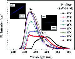
Novel multifunctional fluorescent electrospun (ES) nanofibers were prepared from random copolymers of poly{2-{2-hydroxyl-4-[5-(acryloxy)hexyloxy]phenyl}benzoxazole}-co-(N-isopropylacrylamide)-co-(stearyl acrylate)} (poly(HPBO-co-NIPAAm-co-SA)) using free-radical polymerization, followed by electrospinning. The moieties of HPBO, NIPAAm, and SA were designed to exhibit zinc ion (Zn2+) and pH sensing, thermoresponsiveness, and physical cross-linking, respectively. The ES nanofibers prepared from the P4 copolymer (1[thin space (1/6-em)]:[thin space (1/6-em)]93[thin space (1/6-em)]:[thin space (1/6-em)]6 composition ratio for HPBO/NIPAAm/SA), showed ultrasensitivity to Zn2+ (as low as 10−8 M) because of the large blue-shifting of 75 nm of the emission maximum and the 2.5-fold enhancement of the emission intensity. Furthermore, the nanofibers exhibited a substantial volume (or hydrophilic–hydrophobic) change during the heating and cooling cycle between 10 °C and 40 °C, attributed to the low critical solution temperature of the thermoresponsive NIPAAm moiety. Such temperature-dependent variation of the prepared nanofibers under the presence of Zn2+ or basic conditions led to a distinct on-off switching of photoluminescence. The high surface-to-volume ratio of the prepared ES nanofibers significantly enhanced their sensitivity compared to that of thin films. These results indicated that the prepared multifunctional ES nanofibers could be potentially used in metal ion, pH, and temperature sensing devices.


Introduction

Developing multifunctional copolymers with environmental-stimulus-responsive fluorescent probes that can be used as metal-ion, temperature, pH, DNA, sugar, and gas sensors is crucial for sensory applications in biotechnology and environmental sciences.1–6 Metal ion detection is especially vital for applications in gene transcription, cell apoptosis, cell transport, and the investigations related to the metabolism of bacteria or its notorious biological membranes.7–10 Zn2+ plays an indispensable role in biological processes and it is the second most abundant transition metal ion found in the body; a Zn2+ deficiency may lead to prostate cancer, diabetes, and neuron disorders.10–13

Several fluorescent Zn2+ probes have been developed based on small molecules,14–16 copolymers,10,17 peptides,18 and nanoparticles.19 2-(2-Hydroxyphenyl) benzoxazole (HPBO)-based functional materials can be used to sense Zn2+ or pH because they undergo an excited-state intramolecular proton transfer (ESIPT) through structural transformations, resulting in a large Stokes shift in fluorescence emission on sensing Zn2+ or change in pH.20–22 We demonstrated that multifunctional sensing material, HPBO-containing poly(N-isopropylacrylamide) (PNIPAAm), used a selective Zn2+ sensor in an aqueous solution and considerably affected the fluorescence properties with temperature or pH variation.23–25 Note that PNIPAAm26,27 and poly(2-(dimethylamino)ethyl methacrylate) (PDMAEMA)28,29 are widely studied thermoresponsive polymers. Furthermore, Liu et al. demonstrated that responsive polymer-based dual fluorescent chemosensors, micelles, could enter into living cells and sensitively respond in vitro to Zn2+ ions and change of temperatures in aqueous media.10 However, most of the aforementioned studies11–25 were based on solutions or thin films, rather than nanofibers. The high surface-to-volume ratio of the nanofibers facilitates rapid responses for multifunctional sensory materials.

The electrospinning technique has been used widely in recent studies because it is inexpensive and facile, and enables nanometer-scaled fibers to be fine tuned.30–32 Multifunctional stimulus-responsive electrospun (ES) nanofibers could alter one or more properties, including morphology, photoluminescence, and wettability, upon exposure to external signals such as pH, temperature, and the presence of metal ions or volatile organic compounds.33–36 Thus, the combination of ES nanofibers with multifunctional polymers could lead to the production of highly sensitive materials or devices.

In this study, we developed sensory ES nanofibers that comprise multifunctional poly(HPBO-co-NIPAAm-co-SA) containing a metal ion/pH sensing moiety (HPBO), a hydrophilic thermo-responsive material (PNIPAAm), and a physically cross-linkable segment (PSA), as shown in Scheme 1. The nanofibers prepared from the three-component random copolymers were first synthesized by free-radical polymerization, followed by electrospinning. Our results showed that the prepared ES nanofibers exhibited the multifunctional sensing characteristics of Zn2+, temperature, and pH, which were substantially more sensitive than the spin-coated films.


image file: c4ra07422j-s1.tif
Scheme 1 (a) Synthesis of poly(HPBO-co-NIPAAm-co-SA) random copolymers, the ESIPT mechanism, and the structure of its zinc complex under aqueous Zn2+ solution or basic condition. (b) The SEM image of ES nanofibers and the confocal image of them in the presence of Zn2+.

Experimental part

Materials

6-Bromohexanol, 2,4-dihydroxybenzaldehyde, N-ethyl-diisopropylamine, chloromethyl ether, 2-aminophenol, p-toluene sulfonic acid, acrylic acid, N-(3-dimethylaminopropyl)-N′-ethylcarbodiamidehydrochloride (EDC), 4-dimethylaminopyridine, and benzyltriethyl ammonium chloride (BTEAC) were purchased from Aldrich and used as received. NIPAAm, AIBN, and SA were purified through recrystallization from hexane. ZnCl2, CaCl2, NaOMe, KF, MgBr2, LiClO4, FeCl3, CuCl2, and NiCl2 salts, methylene chloride, tetrahyrdofuran, and ethanol were purchased from Acros and used as received.

Synthesis of 2-(2-hydroxyphenyl)benzoxazole monomer

Precursors of the metal-ion-sensing HPBO moiety were synthesized according to Scheme S1 (ESI) by using a method similar to that described in our previous report.17 The chemical structure of HPBO is shown in the 1H NMR spectra of Fig. S1 (ESI).

Synthesis of poly(HPBO-co-NIPAAm-co-SA) terpolymers

Terpolymers were synthesized by conducting free-radical copolymerization of three monomers, HPBO, NIPAAm, and SA, as shown in Scheme 1(a). Polymers with different monomer feed molar ratios are denoted P1–P5 (Table 1). All of the polymers were placed in absolute ethanol under a nitrogen atmosphere, and then stirred and refluxed at 90 °C for 2–3 days. The mixture was poured into hexane and precipitated for 3 days, and then dried in vacuum to obtain the polymer. The synthesis and characterization of P4 are described in the following sections, and those of the other four polymers are in the ESI. The number-averaged molecular weight (Mn) and poly-dispersity index (PDI) estimated from GPC (DMF eluent) are listed in Table 1.
Table 1 Composition, molecular weights, and properties of poly(HPBO-co-NIPAAm-co-SA) random copolymers
Polymer Feed molar ratio HPBO–NIPAAm–SA Experimental ratioa Mnb PDI Td (°C) LCST (°C)
a Molar ratio (%) estimated from the 1H NMR spectra.b Mn and PDI determined by GPC with DMF eluent.c LCST measured by DSC.
P1 0.1[thin space (1/6-em)]:[thin space (1/6-em)]96.9[thin space (1/6-em)]:[thin space (1/6-em)]3 0.08[thin space (1/6-em)]:[thin space (1/6-em)]96.5[thin space (1/6-em)]:[thin space (1/6-em)]2.7 43[thin space (1/6-em)]130 1.85 355 29
P2 3[thin space (1/6-em)]:[thin space (1/6-em)]94[thin space (1/6-em)]:[thin space (1/6-em)]3 2.8[thin space (1/6-em)]:[thin space (1/6-em)]94.5[thin space (1/6-em)]:[thin space (1/6-em)]2.7 35[thin space (1/6-em)]490 1.89 350 28.5
P3 0[thin space (1/6-em)]:[thin space (1/6-em)]94[thin space (1/6-em)]:[thin space (1/6-em)]6 0[thin space (1/6-em)]:[thin space (1/6-em)]94.6[thin space (1/6-em)]:[thin space (1/6-em)]5.4 52[thin space (1/6-em)]010 1.97 356 28.2c
P4 1[thin space (1/6-em)]:[thin space (1/6-em)]93[thin space (1/6-em)]:[thin space (1/6-em)]6 0.8[thin space (1/6-em)]:[thin space (1/6-em)]93.7[thin space (1/6-em)]:[thin space (1/6-em)]5.5 48[thin space (1/6-em)]010 1.92 352 28.1c
P5 1[thin space (1/6-em)]:[thin space (1/6-em)]89[thin space (1/6-em)]:[thin space (1/6-em)]10 0.9[thin space (1/6-em)]:[thin space (1/6-em)]89.8[thin space (1/6-em)]:[thin space (1/6-em)]9.3 42[thin space (1/6-em)]110 1.98 358 27.7c


Synthesis of P4

82 mg of HPBO (0.21 mmol), 2.26 g of NIPAAm (20 mmol), 0.418 g of SA (1.29 mmol), 8.2 mg of AIBN (0.05 mmol) were dissolved in 7 mL ethanol and reacted for 3 days to obtain a white solid in 62% yield.1HNMR (d-CD2Cl2, 400 MHz): δ(ppm) = 0.8–2.5 [m, H58, –O–CH2 (CH2)16–CH3, –NH–CH–(CH3)2, –CH2–CH–CH2–CH–CH2–CH–, –OCH2–(C4H8)–CH2O–], 4.0 [m, H7, –OCH2–(C4H8)–CH2O–, –NH–CH–(CH3)2, –O–CH2−(CH2)16–CH3], 6.2–7.1 [m, H1, –NH–CH–(CH3)2], 7.1–7.9 (m, H7, phenyl). Elemental anal. Calcd for P4: C, 66.49; H, 9.36; N, 10.33. Found: C, 66.13; H, 9.42; N, 10.86.

Processing of electrospun fibers and drop-cast film

A single-capillary spinneret ES device was employed to produce ES nanofibers by using a method similar to that described in our previous report.33–36 The concentrations of P3, P4, and P5 in methanol were all at 25 wt%. To increase the conductivity of the polymer solution, 5 wt% of BTEAC (with respect to the polymer) was added to the solution. The polymer solution was fed into a metallic needle by using syringe pumps (KD Scientific Model 100, USA) at a feed rate of 0.8–1.0 mL h−1. The tip of the metallic needle was connected to a high-voltage power supply (Chargemaster CH30P SIMCO, USA), and a piece of aluminum foil or quartz was placed 10 cm below the tip of the needle for 6 h to collect the ES nanofibers. The ES nanofibers were fabricated at 25 °C with humidity of approximately 50%, and spinning voltage of 15 kV.

Polymer films corresponding to the ES nanofibers were prepared by drop-casting on a quartz substrate from the same blending solutions and dried in an air-flow hood, and the properties of the films were compared with those of the ES nanofibers.

Characterization

1H nuclear magnetic resonance of the prepared copolymers was measured by a Bruker AV 400 MHz spectrometer using deuterated methylene chloride and deuterated chloroform as solvents. All GPC analyses were performed using a Lab Alliance RI2000 instrument and a polymer–DMF solution, and the instrument was calibrated using a polystyrene curve. The thermal decomposition temperature was determined using a Perkin-Elmer thermal gravimetric analyzer, which heated the samples from 100 °C to 800 °C at a rate of 10 °C min−1 under a nitrogen atmosphere. UV-visible (UV-Vis) absorption spectra were recorded using a Shimadzu UV-Vis spectrophotometer. The morphologies of the ES nanofibers were analyzed using a JEOL JSM-6330F FE-SEM. FE-SEM samples were sputtered with platinum prior to the characterization of images and analyzed by accelerating at 10 kV. Photoluminescence (PL) experimental data were recorded using a Fluorolog-3 spectrofluorometer (Horiba Jobin Yvon).

The variation in the optical absorption and photoluminescence of the prepared ES nanofibers or films with different temperatures can be described as follows: to ensure that the beam excites at the same point on the prepared samples at each measurement, the ES nanofibers or drop-cast films were fixed in cuvettes by an adhesive tape and filled with aqueous metal ion solution at the concentration of 10−10 to 10−4M or under acidic and basic aqueous solution. Each measurement was carried out for 15 min to ensure that equilibrium of the chelating reaction was reached. Then, the ES nanofibers or films were heated and cooled using the variable temperature device for the measurement of photophyscial property. All the temperatures were maintained for 30 min to ensure that a steady state was reached.

Results and discussion

Characterization of poly(HPBO-co-NIPAAm-co-SA)

The synthetic scheme of multifunctional poly{2-{2-hydroxyl-4-[5-(acryloxy) hexyloxy]phenyl}benzoxazole}-co-(N-isopropylacrylamide)-co-(steary lacrylate)}, poly (HPBO-co-NIPAAm-co-SA) copolymers is shown in Scheme 1 (a).Fig. 1 shows the 1H NMR spectrum of P4 in CD2Cl2, with the mole ratio of 1[thin space (1/6-em)]:[thin space (1/6-em)]93[thin space (1/6-em)]:[thin space (1/6-em)]6 for the HPBO, NIPAAm, and SA moieties. Proton peak X in Fig. 1 represents D-dichloromethane. A proton signal for the phenyl group and aromatic ring of the HPBO moiety is observed at 7.1–7.9 ppm (Peak g). The proton peak at 6.2–7.1 ppm (Peak f) represents the secondary amine moiety of NIPAAm. The peak at 4.0 ppm (Peak e) is attributed to the methylene moiety neighbor of the oxygen in HPBO and SA. Peaks at 0.8–2.5 ppm (Peaks a, b, c, and d) represent the alkyl chains on the polymer. The HPBO/NIPAAm/SA composition of the copolymer P4 estimated based on the ratio obtained through the NMR peak area integration is 0.8[thin space (1/6-em)]:[thin space (1/6-em)]93.7[thin space (1/6-em)]:[thin space (1/6-em)]5.5, which is in a fair agreement with the element analysis result. The chemical structures of the other four copolymers, P1–P3 and P5, are shown in the 1H NMR spectra, Fig. S2 (ESI). The copolymer ratios of HPBO/NIPAAm/SA estimated from NMR spectra and EA results are 0.08[thin space (1/6-em)]:[thin space (1/6-em)]96.5[thin space (1/6-em)]:[thin space (1/6-em)]2.7, 2.8[thin space (1/6-em)]:[thin space (1/6-em)]94.5[thin space (1/6-em)]:[thin space (1/6-em)]2.7, 0[thin space (1/6-em)]:[thin space (1/6-em)]94.6[thin space (1/6-em)]:[thin space (1/6-em)]5.4, and 0.9[thin space (1/6-em)]:[thin space (1/6-em)]89.8[thin space (1/6-em)]:[thin space (1/6-em)]9.3 for the copolymers P1, P2, P3, and P5, respectively. The good agreement between the feeding ratio and experimental composition suggests the successful preparation of the target copolymers.
image file: c4ra07422j-f1.tif
Fig. 1 1H NMR spectra of P4 in d-CD2Cl2.

The molecular weights, thermal properties, and low critical solution temperature (LCST) of the five copolymers, P1–P5, are listed in Table 1. The number-average molecular weight and polydispersity index (Mn, PDI) of P1–P5 are (43[thin space (1/6-em)]130, 1.85), (35[thin space (1/6-em)]490 and 1.89), (52[thin space (1/6-em)]010 and 1.97), (48[thin space (1/6-em)]010 and 1.92), and (42[thin space (1/6-em)]110 and 1.98), respectively. The thermal decomposition curves of the prepared polymers are shown in Fig. S3 (ESI) with a similar thermal decomposition temperatures (Td) ranging from 350 to 358 °C because of the similar NIPAAm composition. Fig. S4 in the ESI shows the typical optical transmittance (at 520 nm) versus temperature curves of P1 and P2, which exhibits a thermoreversible soluble-to-insoluble phase transition in an aqueous medium at approximately 28–29 °C (LCST). The LCST of P3, P4, and P5 is around 28 °C, as identified by DSC in Fig. 2.37,34 P1 and P2, with a low SA molecular ratio, are soluble in water below its LCST, but P3–P5, with a high SA composition, are not soluble. Note that SA is hydrophobic and physical cross-linking moiety, and this explains the above results. The LCSTs of P1–P5 range from 27 to 29 °C and are slightly lower than that of PNIPAAm at 32 °C38 because of the hydrophobic characteristic of the SA moiety.34 Furthermore, the increased hydrophobic SA moiety facilitates chain aggregation in an aqueous solution and lowers the LCSTs of P3 (28.2 °C), P4 (28.1 °C), and P5 (27.7 °C) compared with those of P1 (29 °C) and P2 (28.5 °C). P1 and P2 are soluble in aqueous solution and thus are not suitable for preparing ES nanofibers for sensing metal ions. Thus, only P3–P5 ES nanofibers were prepared and used for the sensory applications.


image file: c4ra07422j-f2.tif
Fig. 2 LCST of P3, P4 and P5 determined by DSC.

Morphologies of poly(HPBO-co-NIPAAm-co-SA) electrospun fibers

Fig. 3 shows the FE-SEM images of the electrospun (ES) nanofibers prepared from P3, P4, and P5. The inset figure shows the enlarged FE-SEM image of the ES nanofibers. As shown in Fig. 3, the P3, P4, and P5 ES nanofibers in a dry state have average diameters of 386 ± 87, 395 ± 103, and 402 ± 92 nm, respectively. Note that the average diameter value was the statistical average of the diameters of fifty fibers from each sample.33–36 Moreover, note that all the ES nanofibers fabricated using methanol as the processing solvent exhibited a smooth, nonporous surface.39 The diameters of the P3–P5 ES nanofibers are similar in a dry state because the concentration the ES solution is fixed, and the molar ratios of the HPBO and SA moieties in the copolymers are lower than that of NIPAAm.
image file: c4ra07422j-f3.tif
Fig. 3 FE-SEM images of P3, P4, and P5 ES nanofibers at the dry state and after treatment with water when the temperature is varied from 10 to 40 °C (all scale bars represent 5 μm). The inset FE-SEM images are the enlarged view of the aforementioned fibers (scale bars represent 100 nm).

To observe the morphologies of the ES nanofibers in pure water at various temperatures, the P3–P5 ES nanofibers were collected on a small piece of aluminum foil and immersed in water at 10 or 40 °C. After 10 min, the nanofibers were placed in liquid nitrogen to fix their morphologies. The residual water was immediately removed, and the nanofibers were kept in vacuum for 30 min to ensure that they retained their original morphologies. The SEM images of the ES nanofibers prepared using these procedures, as the temperature varied from the dry state to 10 or 40 °C are shown in Fig. 3. P3–P5 ES nanofibers are not soluble in water and could maintain their fiber morphologies irrespective of whether the temperature is under or over the LCST because of the sufficient composition of physically cross-linking SA. However, both P1 and P2 ES nanofibers dissolve in water below 32 °C because of their low SA molecular ratios, as shown in Fig. S5 (ESI). The diameters of the P3, P4, and P5 ES nanofibers at 10 °C are 1721 ± 487, 1623 ± 413, and 1382 ± 321 nm, respectively, while those at 40 °C are 1125 ± 289, 1032 ± 327, and 1074 ± 321 nm. All dry-state ES nanofibers immersed in pure water at 10 °C and 40 °C swell considerably in water because of the hydrophilic NIPAAm chain. However, these swollen nanofibers still maintain their cylindrical shape and do not dissolve in water at 10 °C because of the physically cross-linked SA moiety. Furthermore, the fiber diameters at 10 °C are greater than those at 40 °C, due to the hydrophilic NIPAAm chain below the LCST. Conversely, the nanofiber diameters are reduced from approximately 1.5 to 1.0 μm as the temperature increased from 10 to 40 °C, as shown in Fig. 3. Such volume variation is because of the reduced water content as the temperature exceeds the LCST. Although the P3 ES nanofibers exhibit a structural variation with respect to the temperature, they do not exhibit fluorescent emission after sensing Zn2+ because they do not contain the HPBO moiety. Therefore, only P4–P5 ES nanofibers with the HPBO moieties were used to chelate Zn2+, as discussed in the following section.

Metal ion sensing of poly(HPBO-co-NIPAAm-co-SA)

Fig. 4 shows the variations in the photoluminescence (PL) spectra of P4 in a THF solution containing various types of metal ions at a concentration of 10−4 M. Under the initial condition without metal ions, the PL spectra of P4 shows a main emission peak with the maximum (λPLmax) wavelength around 470 nm, which is attributed to the HPBO moiety.40 As shown in Fig. 4, an enhanced fluorescence intensity and a clear blue shift in the emission peak from 470 to 410 nm are observed when Zn2+, Ca2+, Mg2+, Na+, K+, and Li+ are added. Compared to the initial condition, the fluorescence intensity is considerably higher on chelating Zn2+. Furthermore, the green-blue fluorescence changes considerably to sky-blue, as shown in the inset figure, attributed to the interruption of the ESIPT process due to zinc complexation (Scheme 1(a)).40 The ESIPT process is shown in detail in Scheme S2 (ESI). A complete quench of fluorescence is observed, when the probe is chelated by heavy metal ions, such as Fe3+, Ni2+, and Cu2+; similar to those reported by Wu et al.6 and Iwata et al.41
image file: c4ra07422j-f4.tif
Fig. 4 PL spectra of P4 in presence of different metal ions in THF solutions (10−4 M). The two inset figures show the color changes in P4 in presence of 10−4 M zinc ions under 254 nm UV light.

The variation in UV–visible absorption of P4 in THF solutions using various concentrations (10−10 to 10−4 M) of Zn2+ is shown in Fig. S6 (ESI). As the concentration of zinc ions is increased, the absorption peak maxima (λabsmax) at both 325 nm and 341 nm decrease, whereas the intensity of a broad band from 350 to 400 nm increases. The absorption band at the longer wavelength indicates the formation of a zinc complex, as shown in Scheme 1(a). An isosbestic point is observed at 350 nm, suggesting that a simple complex formation process occurs.22,42 Fig. 5(a) and (b) shows the variation in the PL spectra of P4 and P2 in THF solutions with different zinc ion concentrations. The PL intensity of P1 is considerably lower than that of P2, and thus the sensing of P2 is used to compare with that of P4. As shown in the figure, the concentration of Zn2+ increasing from 10−10 to 10−8 M results in a clear blue shift in the emission peak from 470 to 410 nm. Also, the concentration of Zn2+ increasing from 10−8 to 10−4 M enhances the PL intensities of P4 and P2. Furthermore, when the zinc ion concentration is at an extremely dilute level of 10−8 M, the emission maximum shift (Δλmax) was observed to be as high as 60 nm, and it resulted in the change of color that can be easily observed by the naked eye. In contrast, the higher sensing composition (HPBO) in the polymer (P2, 2.8%; P4, 0.8%) at 10−4 M of Zn2+ leads to the larger PL intensity enhancement of 3.5 and 2.5 times compared to that under the initial condition, respectively. However, the P2 could not be used as an ES nanofiber for sensing Zn2+ aqueous solution because of its solubility in water.


image file: c4ra07422j-f5.tif
Fig. 5 Variation of the PL spectra of (a) P4 and (b) P2 in THF solutions with various concentrations of Zn2+ metal ions (10−10 to 10−4 M).

Fig. 6(a) and (b), and the inset figures show the variation in the PL spectra of P4/P5 ES nanofibers and thin films using the various concentrations of Zn2+ aqueous solutions, respectively. As shown in Fig. 6, the high Zn2+ concentration increases the PL intensity and causes a blue shift from 500 to 425 nm in the emission spectra. The lowest detectable Zn2+ concentration for the thin film is 10−7 M, but 10−8 M for the ES nanofibers because the latter has a high surface/volume ratio. The PL intensity If/I0 of the P4 ES nanofibers (I0 denotes the original PL intensity at 500 nm of λPLmax, and If denotes the PL intensity at 425 nm of λPLmax at 10−4 M of Zn2+) is 2.5, higher than that of the P4 thin film, which is 2.0. This difference is due to the different surface-to-volume ratio of the nanofibers.43,44 Furthermore, the sensitivity on Zn2+ based on the If/I0 ratio can be observed in the following order: P4 ES nanofibers (2.5) > P5 ES nanofibers (2.0) > P4 thin films (1.5) > P5 thin films (1.0), as shown in Fig. 6(a) and (b). The substantially improved Zn2+ sensing performance is attributed to the higher hydrophilicity of P4 compared to P5 because of the higher SA composition.


image file: c4ra07422j-f6.tif
Fig. 6 Variation of the PL spectra of (a) P4 and (b) P5 ES nanofibers with various concentrations of Zn2+ aqueous solutions (10−10 to 10−4 M). The PL spectra of the corresponding thin films are shown in the inset figures.

Temperature and pH sensing of poly(HPBO-co-NIPAAm-co-SA)

The effect of pH on fluorescence spectra is shown in Fig. 7. As shown in the figure, the PL intensity of the P4 ES nanofibers in the acidic (pH value: 4–6) or neutral (pH value: 7) aqueous solution is almost the same, but it is sensitive to the basic condition, especially in the pH range of 9–12. The fluorescence intensity of the P4 ES nanofibers at pH 12 is two times greater than that at pH 7, and the λPLmax is blue shifted from 500 nm (pH 7) to 425 nm (pH 12). In a basic aqueous solution, the phenolate anions produced through intermolecular proton transfer to the hydroxyl anions disrupts the ESIPT process.17,45 This explains the aforementioned variation in fluorescence, which is similar to the mechanism shown in the bottom figure of Scheme 1(a).
image file: c4ra07422j-f7.tif
Fig. 7 Variation of the PL spectra of P4 ES nanofibers with pH values under (a) acidic condition and (b) basic condition.

The variation of the temperature on the fluorescence of the P4 ES nanofibers at pH values of 4 and 10 under air atmosphere are shown in Fig. 8(a) and (b), respectively. Fig. 8(a) shows that the ES nanofibers exhibit a negligible change in PL intensity at pH 4 as the temperature is varied. However, the clear fluorescence spectra and intensity changes are observed in the ES nanofibers at pH value of 10 (Fig. 8(b)), when the temperature increases. As shown in Fig. 8(b), as the temperature increases from 10 °C to 40 °C, the emission peak at 425 nm decreases, and the emission peak at 500 nm increases possibly due to the aggregation of the free phenolated HPBO under the basic condition. When the temperature is higher than the LCST, the PNIPAAm chain becomes hydrophobic, causing the hydrophilic phenolated HPBO to aggregate in the aqueous solution and leads to a red shift in emission. It indicates that the HPBO (under the acidic condition) is not sensitive to the change from hydrophilic to hydrophobic environments at the LCST (Fig. 8(a)), but this is not the case for the phenolated HPBO (under the basic condition, Fig. 8(b)).


image file: c4ra07422j-f8.tif
Fig. 8 Variation of the PL spectra of P4 ES nanofibers with temperature in aqueous solutions with (a) pH = 4 and (b) pH = 10.

Fig. 9 shows the variation in fluorescence of the P4 ES nanofibers in a 10−4 M Zn2+ environment as the temperature is varied from 40 to 10 °C. The fluorescence plots exhibit an apparent “off-on switching” characteristic, when the temperature increases from 40 to 10 °C. When the temperature is 40 °C, which is higher than the LCST of P4, the P4 ES nanofibers exhibit a hydrophobic characteristic and are not sensitive to Zn2+. When the temperature gradually decreases from 40 to 10 °C, the fluorescence becomes sensitive to Zn2+, and the λPLmax is blue shifted from 500 nm (40 °C) to 425 nm (10 °C). This suggests that the hydrophobic P4 ES nanofibers became hydrophilic, resulting in the absorption due to Zn2+. The emission intensity of λPLmax from 500 to 425 nm increases when the temperature is reduced because of the hydrophilic and hydrophobic switching of the LCST. Therefore, the fluorescence spectra show the off state when Zn2+ is not detected at 40 °C and the on state when Zn2+ is detected at 10 °C. The confocal images (the inset image of Fig. 9) show that the P4 ES nanofibers with larger diameters comparatively swell more in water at 10 °C than at 40 °C because of the hydrophilic PNIPAAm moiety. In addition, the P4 ES nanofibers emit a green-blue light at 40 °C and a lighter pure-blue light at 10 °C. It indicates that the off state is switched to the on state because of the formation of a HPBO–zinc complex within the P4 ES nanofibers at temperatures below the LCST. However, the PL intensity and λPLmax do not change when the temperature increases to 40 °C. This suggests that the P4 ES nanofibers is chelated with Zn2+, forming the HPBO–zinc complex, and are no longer sensitive to the change from a hydrophilic to a hydrophobic environment. These results indicate that the as-prepared ES nanofibers have potential application in multifunctional sensing devices.


image file: c4ra07422j-f9.tif
Fig. 9 Variation of the PL spectra of P4 ES nanofibers with temperature using a Zn2+ concentration of 10−4 M. The two inset figures show confocal images of the nanofibers at 40 °C and 10 °C.

Conclusion

New multifunctional ES nanofibers featuring high sensitivity for Zn2+ and pH were successfully prepared from poly(HPBO-co-NIPAAm-co-SA) using a single-capillary spinneret. The moieties of HPBO, NIPAAm, and SA were designed to exhibit the functionalities of zinc ion (Zn2+) and pH sensing, thermoresponsive, and physical cross-linking, respectively. The ES nanofibers prepared from P4 and P5 maintained their fiber structure in water and sensitivity for Zn2+ and pH because of the sufficient SA composition. In addition, the SA content considerably affected the Zn2+ detection ability because of the change in the hydrophilic–hydrophobic characteristics. Therefore, the Zn2+ sensing performance of the P4 nanofibers was superior to that of the P5 nanofibers. The P4 ES nanofibers exhibited considerable blue shifts in photoluminescence spectra and had enhanced emission intensity for detecting an extremely dilute concentration of Zn2+ (10−8 M and pH 9). Furthermore, the LCST of NIPAAm moiety in the P4 nanofibers showed a significantly temperature-dependent variation in volume (or hydrophilic–hydrophobic change). It led to distinct on-off switching of photoluminescence when the nanofibers were under Zn2+ or basic condition. Furthermore, the P4 ES nanofibers showed a higher surface-to-volume ratio than the corresponding thin films, thus yielding an enhanced performance. The present study demonstrated that the prepared multifunctional ES nanofibers could be used as advanced devices for sensing metal ions, pH, and temperature.

Acknowledgements

The financial support from the Ministry of Science and Technology of Taiwan and Textile Research institute are highly appreciated.

Notes and references

  1. E. M. Nolan and S. J. Lippard, Chem. Rev., 2008, 108, 3443–3480 CrossRef CAS PubMed.
  2. C. G. Wu, H. C. Lu, L. N. Chen and Y. C. Lin, J. Polym. Sci., Part A: Polym. Chem., 2008, 46, 1586–1597 CrossRef CAS.
  3. S. S. Balamurugan, G. B. Bantchev, Y. Yang and R. L. McCarley, Angew. Chem., Int. Ed., 2005, 44, 4872–4876 CrossRef CAS PubMed.
  4. K. Iwai, Y. Matsumura, S. Uchiyama and A. P. de Silva, J. Mater. Chem., 2005, 15, 2796–2800 RSC.
  5. A. Munoz-Bonilla, M. Fernandez-Garcia and D. M. Haddleton, Soft Matter, 2007, 3, 725–731 RSC.
  6. W. C. Wu, Y. Tian, C. Y. Chen, C. S. Lee, Y. J. Sheng, W. C. Chen and A. K.-Y. Jen, Langmuir, 2007, 23, 2805–2814 CrossRef CAS PubMed.
  7. J. M. Hu, L. Dai and S. Y. Liu, Macromolecules, 2011, 44, 4699–4710 CrossRef CAS.
  8. X. Q. Chen, T. Pradhan, F. Wang, J. S. Kim and J. Y. Yoon, Chem. Rev., 2012, 112, 1910–1956 CrossRef CAS PubMed.
  9. C. H. Li and S. Y. Liu, J. Mater. Chem., 2010, 20, 10716–10723 RSC.
  10. T. Liu and S. Y. Liu, Anal. Chem., 2011, 83, 2775–2785 CrossRef CAS PubMed.
  11. T. V. Ohalloran, Science, 1993, 261, 715–725 CAS.
  12. T. Koike, T. Watanabe, S. Aoki, E. Kimura and M. Shiro, J. Am. Chem. Soc., 1996, 118, 12696–12703 CrossRef CAS.
  13. K. R. Gee, Z. L. Zhou, W. J. Qian and R. Kennedy, J. Am. Chem. Soc., 2002, 124, 776–778 CrossRef CAS PubMed.
  14. E. Tomat, E. M. Nolan, J. Jaworski and S. J. Lippard, J. Am. Chem. Soc., 2008, 130, 15776–15777 CrossRef CAS PubMed.
  15. Z. C. Xu, K. H. Baek, H. N. Kim, J. N. Cui, X. H. Qian, D. R. Spring, I. Shin and J. Yoon, J. Am. Chem. Soc., 2010, 132, 601–610 CrossRef CAS PubMed.
  16. C. S. He, W. P. Zhu, Y. F. Xu, Y. Zhong, J. A. Zhou and X. H. Qian, J. Mater. Chem., 2010, 20, 10755–10764 RSC.
  17. C. C. Yang, Y. Tian, C. Y. Chen, W. C. Chen and A. K.-Y. Jen, Macromol. Rapid Commun., 2007, 28, 894–899 CrossRef CAS.
  18. B. P. Joshi, W. M. Cho, J. Kim, J. Yoon and K. H. Lee, Bioorg. Med. Chem. Lett., 2007, 17, 6425–6429 CrossRef CAS PubMed.
  19. M. J. Ruedas-Rama and E. A. H. Hall, Anal. Chem., 2008, 80, 8260–8268 CrossRef CAS PubMed.
  20. J. K. Lee, H. J. Kim, T. H. Kim, C. H. Lee, W. H. Park, J. Kim and T. S. Lee, Macromolecules, 2005, 38, 9427–9433 CrossRef CAS.
  21. R. Tangirala, E. Baer, A. Hitner and C. Weder, Adv. Funct. Mater., 2004, 14, 595–604 CrossRef CAS.
  22. M. Taki, J. L. Wolford and T. V. O'Halloran, J. Am. Chem. Soc., 2004, 126, 712–713 CrossRef CAS PubMed.
  23. G. Masci, L. Giacomelli and V. Crescenzi, Macromol. Rapid Commun., 2004, 25, 559–564 CrossRef CAS.
  24. C. Zheng, W. D. He, W. J. Liu, J. Li and J. F. Li, Macromol. Rapid Commun., 2006, 27, 1229–1232 CrossRef CAS.
  25. C. C. Yang, Y. Q. Tian, A. K.-Y. Jen and W. C. Chen, J. Polym. Sci., Part A: Polym. Chem., 2006, 44, 5495–5504 CrossRef CAS.
  26. A. Muñoz-Bonilla, M. Fernández-García and D. M. Haddleton, Soft Matter, 2007, 3, 725–731 RSC.
  27. F. Bougard, M. Jeusette, L. Mespouille, P. Dubois and R. Lazzaroni, Langmuir, 2007, 23, 2339–2345 CrossRef CAS PubMed.
  28. J. Li, W. D. He, S. Han, X. Sun, L. Li and B. Zhang, J. Polym. Sci., Part A: Polym. Chem., 2009, 47, 786–796 CrossRef CAS.
  29. M. Nakayama and T. Okano, Macromolecules, 2008, 41, 504–507 CrossRef CAS.
  30. D. H. Reneker and I. Chun, Nanotechnology, 1996, 3, 216–223 CrossRef.
  31. A. G. MacDiarmid, W. E. Jones Jr, I. D. Norris, J. Gao, A. T. Johnson Jr, N. J. Pinto, J. Hone, B. Han, F. K. Ko, H. Okusaki and M. Llaguno, Synth. Met., 2001, 119, 27–30 CrossRef CAS.
  32. H. Okusaki, T. Takahashi, N. Miyajima, Y. Suzuki and T. Kuwabara, Macromolecules, 2006, 39, 4276–4278 CrossRef.
  33. C. C. Kuo, Y. C. Tung and W. C. Chen, Macromol. Rapid Commun., 2010, 31, 65–70 CrossRef CAS PubMed.
  34. Y. C. Chiu, C. C. Kuo, J. C. Hsu and W. C. Chen, ACS Appl. Mater. Interfaces, 2010, 2, 3340–3347 CAS.
  35. Y. C. Chiu, G. Chen, C. C. Kuo, S. H. Tung, T. Kakuchi and W. C. Chen, ACS Appl. Mater. Interfaces, 2012, 4, 3387–3395 CAS.
  36. L. N. Chen, Y. C. Chiu, J. J. Hung, C. C. Kuo and W. C. Chen, Macromol. Chem. Phys., 2014, 215, 286–294 CrossRef CAS.
  37. K. Kobayashi, H. Yan and H. Okusaki, Macromolecules, 2009, 42, 5916–5918 CrossRef.
  38. M. Nakayama, T. Okano and F. M. Winnik, Mater. Matters, 2010, 5, 56–62 CAS.
  39. F. H. Anka and K. J. Balkus Jr, Ind. Eng. Chem. Res., 2013, 52, 3473–3480 CrossRef CAS.
  40. J. K. Lee, H. J. Kim, T. H. Kim, C. H. Lee, W. H. Park, J. Kim and T. S. Lee, Macromolecules, 2005, 38, 9427–9433 CrossRef CAS.
  41. K. Kanata, T. Kumagi, H. Aoki, M. Deguchi and S. Iwata, J. Org. Chem., 2001, 66, 7328–7333 CrossRef PubMed.
  42. M. M. Henary and F. C. J. ahrni, J. Phys. Chem. A, 2002, 106, 5210–5220 CrossRef CAS.
  43. X. J. Peng, J. J. Du, J. L. Fan, J. Y. Wang, Y. K. Wu, J. Z. Zhao, S. G. Sun and T. Xu, J. Am. Chem. Soc., 2007, 129, 1500–1501 CrossRef CAS PubMed.
  44. X. Wang, C. Drew, S. H. Lee, K. J. Senecal, J. Kumar and L. A. Samuelson, Nano Lett., 2002, 2, 1273–1275 CrossRef CAS.
  45. K. Das, N. Sarkar, A. K. Ghosh, D. Majumdar, D. N. Nath and K. Bhattacharyya, J. Phys. Chem., 1994, 98, 9216–9221 CrossRef.

Footnote

Electronic supplementary information (ESI) available: Synthesis and 1H NMR spectra of HPBO monomer. Synthesis and 1H NMR spectra of HPBO, P1, P2, P3 and P5. TGA of P1–P5. Variation in optical transmittance of P1 and P2 at temperatures of 15–50 °C. SEM images of P1 and P2 ES nanofibers. The UV-Vis absorption spectra for P4 in THF solution in presence of Zn2+. ESIPT process and energy level diagram. See DOI: 10.1039/c4ra07422j

This journal is © The Royal Society of Chemistry 2014
Click here to see how this site uses Cookies. View our privacy policy here.