Immobilization of titania nanoparticles on the surface of cellulose fibres by a facile single step hydrothermal method and study of their photocatalytic and antibacterial activities

Indu Chauhan and Paritosh Mohanty*
Indian Institute of Technology Roorkee, Department of Applied Science and Engineering, Saharanpur Campus, Saharanpur-247 001, U.P., India. E-mail: paritosh75@gmail.com; Fax: +91-1322714311; Tel: +91-1322714338

Received 21st July 2014 , Accepted 17th October 2014

First published on 17th October 2014


Abstract

Cellulose fibers immobilized with 2.5 to 21.0 wt% titania (TiO2) nanoparticles of diameter 10–20 nm were prepared by a facile single step hydrothermal method. Paper matrices were prepared by a standard handsheet making procedure using these TiO2 immobilized cellulose fibers. These paper matrices successfully degraded ∼82% of formaldehyde and completely degraded methyl orange in 180 min, when irradiated with sunlight. Moreover, the results improved on increasing either the irradiation time or TiO2 content. Furthermore, these paper matrices showed promising antibacterial activity against Escherichia coli in visible light. The successful immobilization of TiO2 nanoparticles on paper matrices was characterized by field emission scanning electron microscopy (FESEM) and X-ray diffraction studies (XRD).

Introduction

With the alarming increase in the pollutant level in the environment, attributed to industrial and other sources, there is an utmost need for the development of low-cost and efficient systems for water and air purification through the degradation of pollutants.1 Recently, titania (TiO2) has attracted significant attention as a solar light driven photocatalyst to degrade non-biodegradable dyes and volatile organic compounds (VOCs).1,2 It is economical and chemically stable with a high oxidizing power when illuminated with light and generates hydroxyl radicals and superoxide ions that can oxidize VOCs.2 Photocatalysis, being a surface and interface phenomenon, increases on increasing the surface to volume ratio of the material; this can be made possible by synthesizing nanoparticles.3 However, stabilizing individual nanoparticles is always a challenge because agglomeration is a major problem. One way to resolve this issue is using a surfactant or stabilizing agent to chemically stabilize the nanoparticles. Another more efficient way is to immobilize the nanoparticles on the surface of substrates. This in turn is very helpful in recovering the highly dispersed photocatalyst from the treated solution.1,4 Recently, various research groups have reported the immobilization of TiO2 over various substrates, such as polymers, glass, and natural fibers.1,2,4–6 Abidi et al., Yuranova et al. and Bedford et al. have demonstrated the self cleaning and UV-protection properties of textiles made from TiO2 modified cotton fibers.1,2,6 Mellott et al. have studied the formation of TiO2 thin film on the glass slide, which could degrade stearic acid, and thus have a potential to be used as self cleaning glasses for windows.7 Similarly, Lee and co-workers, Xu et al. and Paz et al. have studied the photocatalytic activity of TiO2 nanoparticle coated polymer matrices for water remediation and VOCs degradation.8–11 However, most of the studied substrates are expensive, non-renewable and are non-biodegradable. Therefore, there was a need to develop a TiO2 modified substrate, which could circumvent these issues and add environmental and economical benefits.5 Cellulose, a natural biopolymer, being one of the most economical, easy to access and biodegradable materials has the potential to be used as a substrate for TiO2 immobilization. Construction materials, such as paints and organic binders, liberate VOCs and exposure to these poses a serious health problem called sick building syndrome.12–14 Recently, indoor products derived from TiO2 are being marketed as the solution for this problem. Thus, TiO2 based paper matrices can be a potential solution because paper products are very common in living areas.15–18 After the pioneering work of Matsubara et al., TiO2 modified cellulose fibers or paper assisted photocatalytic degradation of VOCs have been extensively studied.15–18 In addition, the antimicrobial activity of TiO2 based products was recently demonstrated.4,19 Matsunaga et al., for the very first time reported the microbiocidal effect of platinised TiO2.19 Recently, Chawengkijwanich et al., studied the antibacterial activity of TiO2 coated packaging film.20 Similarly, Li et al., have reported the fabrication of TiO2 modified paper using layer-by-layer deposition, and then studied the photocatalytic and antibacterial activity.18 Moreover, the synthesis and photocatalytic and antibacterial applications of TiO2–cellulose composites have also been studied by Luo et al. and Li et al.21,22 Thus, TiO2 modified paper products can not only degrade VOCs but can also inhibit the growth of pathogens.16–18

In all these reports the TiO2 incorporation/immobilization in the paper matrices is a multistep process, where the first step involves the synthesis of TiO2 nanoparticles and the incorporation of these synthesized nanoparticles into the paper is executed in the second step.16,17,23 Uniform distribution, retention and agglomeration of the nanoparticles are big challenges in these processes.16,23 Here, we report a facile method, where the synthesis and the immobilization of the nanoparticles on the surface of the cellulose fibers are carried out in a single step. These nanoparticle immobilized cellulose fibers were used to make paper matrices by a standard handsheet making procedure. These paper matrices were then used for the degradation of methyl orange (a non-biodegradable dye) and formaldehyde (a VOC) in the sunlight and also the antibacterial activity was demonstrated against E. coli bacteria in the visible light.

Experimental

Materials

Titanium isopropoxide (Avra Synthesis Pvt. Ltd., India), ethanol (99.9% pure, Merk), methyl orange (Qualigen fine chemicals, India), formaldehyde (Himedia laboratories Pvt. Ltd., India), bleached softwood cellulose fibers (Star Paper Mill Ltd., India), peptone (Rankem, RFCL Ltd., India), beef extract (Himedia laboratories Pvt. Ltd., India), agar powder (Himedia laboratories Pvt. Ltd., India) and E. coli (MTCC no. 1698, Microbial Type Culture Collection and Gene Bank, India) were used. All the reagents were used as received without any further purification.

Immobilization of TiO2 nanoparticles on cellulose fibers

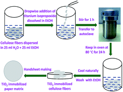
Cellulose fibers immobilized with different amounts of TiO2 nanoparticles were prepared by hydrothermally treating the cellulose fibers with titanium isopropoxide Ti{OCH(CH3)2}4 in ethanol and water solution at the temperature of 80 °C for 24 h (Scheme 1).
image file: c4ra07372j-s1.tif
Scheme 1 Schematic representation of the immobilization of TiO2 nanoparticles on the surface of cellulose fiber of paper.

In a typical synthesis, x mL (x = 0.21, 0.42, 0.64 and 1.3 mL, corresponding to the 5, 10, 15 and 30 wt% of TiO2 in 1.2 g of paper matrix) of titanium isopropoxide was dissolved in 25 mL of ethanol and added dropwise to the cellulose fiber suspension (cellulose fibers dispersed in a solution of 25 mL H2O and 25 mL EtOH) with continuous stirring. The whole suspension was stirred for 1 h at RT. The suspension was then transferred into a Teflon-lined stainless steel autoclave and kept in an oven at 80 °C for 24 h. The suspension was naturally cooled and washed several times with ethanol. An identical hydrothermal reaction of titanium isopropoxide was carried out to make TiO2 in the absence of cellulose fibers.24

Preparation of the paper matrices from the TiO2 nanoparticle immobilized cellulose fibers

The obtained TiO2 immobilized cellulose fibers were then used for the fabrication of paper matrices by the standard handsheet making procedure using the British sheet former (Pulp Evaluation Apparatus, manufactured by Mavis Engg. Co. Ltd., application no. 1092).16,23 Typically, the TiO2 immobilized cellulose fibres were dispersed in 500 mL of H2O and then poured into the sheet former filled with 8 L of water. It was agitated with the help of a perforated stirrer, which was moved up and down in 6 ± 1 s to uniformly disperse the cellulose fibers in the suspension. The wet paper matrices were then recovered with the help of blotting paper after draining the water under suction. These wet paper matrices were dried using a machine glazed (MG) drier (Aktiebolaaget. c.j. wennbergs, Mek. verkstad, karlstad, Sweden) at 80 °C for 90–120 min.23

Photocatalytic degradation of methyl orange (MO) and formaldehyde

The efficacy of the paper matrices immobilized with TiO2 nanoparticles for the degradation of MO and formaldehyde was probed in the presence of sunlight. Typically one fourth of the circular paper matrix with an area of 200 cm2 was dipped in 20 mL of methyl orange (0.3 mM) and 20 mL of formaldehyde (145 mM) solutions and kept in the dark to reach the equilibrium of dye adsorption. Then, the solutions were exposed to sunlight for various time intervals. At certain time intervals, the solutions were collected and the change in the absorbance was analyzed by a UV-visible spectrophotometer (UV1800 Shimadzu UV spectrophotometer) in the range of 200–700 nm. For MO and formaldehyde, the λmax was observed at 464 and 298 nm, respectively.25–27 The photocatalytic decomposition efficiency of MO and formaldehyde was calculated using the following equation:
image file: c4ra07372j-t1.tif
where η1 is the decomposition efficiency of MO or formaldehyde and AB and AT represent the absorbance values of blank and tested samples, respectively.

Antibacterial activity of TiO2 nanoparticles immobilized paper matrices

The antibacterial activity of the TiO2 immobilized paper matrices was tested against E. coli under the illumination of an incandescent lamp (1.933 J cm−2). Before testing, the paper strips (8 cm × 1 cm) were sterilized in an autoclave. The sterilized paper strips were then dipped into 10 mL of bacterial suspension with an initial bacterial concentration of 468 × 105 CFU mL−1. The bacterial suspensions were incubated at 36 °C for 72 h. During the process, after every 4 h, the treated bacterial suspensions were exposed to an incandescent lamp for 30 min. The serial dilutions of the treated bacterial samples were prepared, and then 100 μL of each dilution was spread on nutrient agar media (NAM) in petri-plates. The plates were then incubated at 36 °C for 24 h and the number of colonies was counted by the plate count method.18

Characterization

The microstructure of the paper matrices immobilized with TiO2 nanoparticles was studied by field emission scanning electron microscopy (FESEM) (MIRA3 TESCAN) operated at a voltage of 3.0 kV. The crystal structure of the paper matrices was investigated by X-ray diffraction (XRD) (Bruker D8 FOCUS) with CuKα radiation (λ = 0.15405 nm) at a scanning speed of 2° min−1. The retention of TiO2 nanoparticles on paper matrices was analyzed by the combustion method. The TiO2 content was calculated by subtracting the ash content of the blank paper matrix from the TiO2 immobilized paper matrix.

Results and discussion

To determine the amount (wt%) of the TiO2 nanoparticles immobilized in the paper matrices, different paper matrices of the same dimensions of 4 × 4 cm were heated at 525 °C in air for 4 h in a muffle furnace. This is a very common method to determine the contents of inorganic filler in the paper products.16 Heating the paper matrices above 500 °C in air would decompose or oxidize the organic components (cellulose and hemicellulose) leaving behind the inorganic fillers, if any, such as TiO2. A complete mass loss (ash wt% ∼0.009) was observed in the case of the paper matrix without the immobilized TiO2 nanoparticles (sample a, w/o TP). However, in samples b, c, d and e, the mass remaining after the combustion was around 2.5, 9.0, 13.0, and 21.0 wt%, respectively. Based on the conc. of the TiO2 nanoparticles retained in the paper matrices, the sample paper matrices were designated as 2.5TP, 9.0TP, 13.0TP and 21.0TP for the paper matrices immobilized with 2.5, 9.0, 13.0, and 21.0 wt%, of TiO2 nanoparticles, respectively. The microstructure of the cellulose fibers immobilized with TiO2 nanoparticles of the paper matrices was analyzed by FESEM. The representative FESEM images of the specimens are shown in Fig. 1. It can clearly be seen from the FESEM images that the surface of the cellulose fibers of the blank paper matrix (w/o TP) (Fig. 1a) is smooth and clean, in contrast with the cellulose fibers immobilized with the different concentrations of the TiO2 nanoparticles (Fig. 1b–e). As expected, the content of the nanoparticles in the paper matrices increases with increasing TiO2 to cellulose ratio, as can be seen with the high magnification FESEM images shown in the insets (Fig. 1b–e). Furthermore, it can be seen that the nanoparticles are uniformly distributed throughout the fiber surfaces.
image file: c4ra07372j-f1.tif
Fig. 1 FE-SEM images of paper sheets (a) without TiO2, and immobilized with (b) 2.5, (c) 9.0, (d) 13.0, and (e) 21.0 wt% of TiO2 nanoparticles. (f) The FESEM image of the TiO2 sample synthesized in the absence of cellulose fibers. The high magnification FESEM images are shown in the inset of (b) to (e) to demonstrate the increase in the content of the nanoparticles in the paper matrices with increasing TiO2 concentration.

The spherical nanoparticles were observed to have diameters ranging between 10 to 20 nm, irrespective of the TiO2 content. Even the TiO2 nanoparticles synthesized under an identical reaction condition in the absence of the cellulose fibers had similar shape and size in the range of 10 to 20 nm (Fig. 1f).

It can be conceived that the hydroxyl groups present on the surface of the cellulose fibers interact with the Ti–O through H-bonding (Fig. 2).28,29 This further helps in the generation of the TiO2 nuclei on the surface of the cellulose fibers. As the reaction proceeds, the nuclei grow and form nanoparticles of TiO2 on the surface of the cellulose fiber (Fig. 2). This was further confirmed because the nanoparticles were deposited only on the surface and in no other places. The good retention capacity in the range of 50–90% further supports the mechanism. Otherwise, the nanoparticles would have drained out during the paper fabrication process.


image file: c4ra07372j-f2.tif
Fig. 2 Mechanism for the nucleation and growth of the TiO2 nanoparticles on the surface of cellulose fibers.

The phase analysis of the TiO2 immobilized over the cellulose fiber of paper matrices was investigated by XRD (Fig. 3). Two characteristic overlapped weaker diffraction peaks at d-values of 0.5823 and 0.5335 and a sharp peak at 0.3896 nm corresponding to (101), (10[1 with combining macron]) and (002) reflections of cellulose-I, respectively, were observed in all these samples.30 Few additional peaks corresponding to CaCO3 can also be seen (Fig. 3b).31 The papers with TiO2 nanoparticles have peaks at d-values of 0.3503, 0.2371, 0.1897, 0.1699, 0.1480 and 0.1353 nm corresponding to the (101), (004), (200), (105), (204) and (116) reflections of the anatase phase of the TiO2, which fairly matched with the JCPDS card no. 78-2486 (Fig. 3c–f). A similar anatase phase was observed in the case of the nanoparticles synthesized under similar experimental conditions (Fig. 3a).24


image file: c4ra07372j-f3.tif
Fig. 3 XRD patterns of (a) as-synthesized TiO2 nanoparticles and (b) w/o TP (c) 2.5TP, (d) 9.0TP, (e) 13.0TP and (f) 21.0TP [*CaCO3 as filler in supplied raw cellulose fibers].

The non-biodegradable dye effluents released from the textile industries and VOCs, either in gaseous or liquid form, released into the environment from various sources have become a serious threat to the existence of living organisms.18 Most of the dyes used in textile industries are azo dyes.18 Thus, in the present investigation MO, an anionic azo dye, was selected as a model compound, and formaldehyde was chosen as a model VOC for studying their photo-degradation by the TiO2 nanoparticles immobilized in paper matrices under the irradiation of sunlight. Fig. 4a shows the photocatalytic degradation (%) of MO by the TiO2 nanoparticles immobilized in paper matrices. As expected, the concentration of the MO decreases either by increasing the exposure time or by using the paper matrices having higher TiO2 contents. It was observed that ∼6% of MO was degraded by 2.5TP under the sunlight irradiation of 90 min (Fig. 4a). On further increasing the irradiation time to 120 min, ∼10% decrease in the concentration of MO was observed. However, on further exposing the 2.5TP to sunlight for 180 min, only 11% of the MO was degraded. In addition, 9.0TP, 13.0TP and 21.0TP degraded around 64, 79 and 92% of MO, respectively, after 90 min irradiation. The 9.0TP showed around 97% MO degradation in 180 min. However, almost complete degradation of MO was observed in 180 min for the 13.0TP and 21TP specimens (Fig. 4a).


image file: c4ra07372j-f4.tif
Fig. 4 Photocatalytic degradation of (a) methyl orange and (b) formaldehyde by (■) 2.5TP, (●) 9.0TP, (▲) 13.0TP and (▼) 21.0TP as a function of sunlight irradiation time.

A similar degradation study was performed against formaldehyde using these paper matrices. It was observed that 2.5TP degraded ∼18% of formaldehyde in just 30 min (Fig. 4b). However, on further increasing the irradiation time to 120 min, only ∼22% formaldehyde degradation was observed. The total percent degradation of formaldehyde observed by 2.5TP in 180 min was ∼23%. The 9.0TP degraded ∼36% of formaldehyde in 90 min, and on further increasing the irradiation time to 180 min ∼67% of formaldehyde was degraded. However, a substantial decrease in the formaldehyde concentration was observed at higher concentration of TiO2. The paper matrices 13.0TP and 21.0TP in 90 min degraded around 60% and 51% of formaldehyde, respectively. On further increasing the irradiation time to 180 min, around 80% and 82% degradation of formaldehyde was observed for 13.0TP and 21.0TP samples, respectively (Fig. 4b). Thus, increase in the conc. of the TiO2 nanoparticles in the paper matrices increases the reaction active sites for the generation of hydroxyl and superoxide radicals under the sunlight illumination, which enhances the degradation of MO and formaldehyde.18,32

Furthermore, the antibacterial activity of these paper matrices was investigated by inhibiting the growth of E. coli (Fig. 5). It was observed that the paper matrices without TiO2 nanoparticles showed no antibacterial activity. However, the plate count method for 2.5TP, 9.0TP, 13.0TP and 21.0TP showed a reduction of log[thin space (1/6-em)]0.23, log[thin space (1/6-em)]0.43, log[thin space (1/6-em)]0.50 and log[thin space (1/6-em)]1.19, respectively, as compared to the initial concentration of bacteria (468 × 105 CFU mL−1). The initial concentration of the bacteria in the broth was very high and in the presence of weak visible light (incandescent lamp), the paper matrices showed moderate antibacterial activity (Table 1). The results support the fact that the photoactivation of the TiO2 nanoparticles causes the peroxidation of the polyunsaturated phospholipids of the cell membranes of the bacteria and also results in the loss of respiratory activity of the bacteria, which in turn kills the bacteria.33,34


image file: c4ra07372j-f5.tif
Fig. 5 Log reduction values of E. coli bacterial count by the paper matrices immobilized with different conc. of TiO2 nanoparticles.
Table 1 Antibacterial activity of test paper matrices against E. coli 1698. The viable bacteria were monitored by counting the number of colony-forming units (CFU)
S. no. Paper matrices Bacterial count (CFU mL−1)
1 w/o TP 468 × 105
2 2.5TP 276 × 105
3 9.0TP 174 × 105
4 13.0TP 146 × 105
5 21.0TP 030 × 105


Conclusions

A facile single step hydrothermal method was developed to immobilize TiO2 nanoparticles with diameter of 10–20 nm on the cellulose fibers of paper matrices. The paper matrices immobilized with different concentrations of the TiO2 nanoparticles showed promising high-end applications for the degradation of non-biodegradable dyes, such as methyl orange, and volatile organic compounds (VOCs), such as formaldehyde, in only 180 min. The paper matrices further showed antibacterial activity by inhibiting the growth of E. coli in the presence of visible light. Thus, the presently used hydrothermal method is an effective method to immobilize nanoparticles on cellulose fibers in a single step and with a high retention capacity. This method can also be extended for the immobilization of other metal/metal oxide nanoparticles on cellulose fibers. Furthermore, because of the photocatalytic and antibacterial activities showed by the paper matrices immobilized with nanoparticles, these could have potential usage as wall paper, window sheet or decontamination substrates for the degradation of VOCs and dyes present in the environment and in food packaging.

Acknowledgements

The work is supported by IIT Roorkee, India (Grant no. FIG-400131-DPT/12-13). IC acknowledges the Ministry of Human Resource Development (MHRD), Govt. of India for the financial support.

Notes and references

  1. N. M. Bedford and A. J. Steckl, ACS Appl. Mater. Interfaces, 2008, 2, 2448 CrossRef.
  2. N. Abidi, L. Cabrales and E. Hequet, ACS Appl. Mater. Interfaces, 2009, 10, 2141 Search PubMed.
  3. G. Guisbiers, G. Abudukelimu and D. Hourlier, Nanoscale Res. Lett., 2011, 6, 396 CrossRef PubMed.
  4. E. V. Skorb, L. I. Antonouskaya, N. A. Belyasova, D. G. Shchukin, H. Möhwald and D. V. Sviridov, Appl. Catal., B, 2008, 84, 94 CrossRef CAS PubMed.
  5. N. M. Bedford, M. Pelaez, C. Han, D. D. Dionysiou and A. Steckl, J. Mater. Chem., 2012, 22, 12666 RSC.
  6. T. Yuranova, D. Laub and J. Kiwi, Catal. Today, 2007, 122, 109 CrossRef CAS PubMed.
  7. N. P. Mellott, C. Durucan, C. G. Pantano and M. Guglielmi, Thin Solid Films, 2006, 502, 112 CrossRef CAS PubMed.
  8. J. A. Lee, K. C. Krogman, M. Ma, R. M. Hill, P. T. Hammond and G. C. Rutledge, Adv. Mater., 2009, 21, 1252 CrossRef CAS.
  9. J. A. Lee, Y. S. Nam, G. C. Rutledge and P. T. Hammond, Adv. Funct. Mater., 2010, 20, 2424 CrossRef CAS.
  10. Q. F. Xu, Y. Liu, F.-J. Lin, B. Mondal and A. M. Lyons, ACS Appl. Mater. Interfaces, 2013, 5, 8915 CAS.
  11. Y. Paz and Z. Luo, J. Mater. Res., 1995, 10, 2842 CrossRef CAS.
  12. M. Takeuchi, J. Deguchi, S. Sakai and M. Anpo, Appl. Catal., B, 2010, 96, 218 CrossRef CAS PubMed.
  13. W. J. Meggs, Environ. Health Perspect., 1993, 101, 234 CrossRef CAS.
  14. M. Hodgson, Environ. Health Perspect., 2002, 110, 663 CrossRef CAS.
  15. H. Matsubara, M. Takada, S. Koyama, K. Hashimoto and A. Fujishima, Chem. Lett., 1995, 24, 767 CrossRef.
  16. Y. Iguchi, H. Ichiura, T. Kitaokas and H. Tanaka, Chemosphere, 2003, 53, 1193 CrossRef CAS.
  17. H. Ichiura, T. Kitaoka and H. Tanaka, Chemosphere, 2003, 50, 79 CrossRef CAS.
  18. H. Li, S. Fu and L. Peng, Fibers Polym., 2013, 14, 1794 CrossRef CAS.
  19. T. Matsunaga, R. Tomoda, T. Nakajima and H. Wake, FEMS Microbiol. Lett., 1985, 29, 211 CrossRef CAS PubMed.
  20. C. Chawengkijwanich and Y. Hayata, Int. J. Food Microbiol., 2008, 123, 288 CrossRef CAS PubMed.
  21. Y. Luo, J. Xu and J. Huang, CrystEngComm, 2014, 16, 464 RSC.
  22. S.-M. Li, Y.-Y. Dong, M.-G. Ma, L.-H. Fu, R.-C. Sun and F. Xu, Carbohydr. Polym., 2013, 96, 15 CrossRef CAS PubMed.
  23. I. Chauhan, S. Chattopadhya and P. Mohanty, Mater. Express, 2013, 3, 343 CrossRef CAS PubMed.
  24. Z. Zhang, C.-C. Wang, R. Zakaria and J. Y. Ying, J. Phys. Chem. B, 1998, 102, 10871 CrossRef CAS.
  25. C. Chen, J. Liu, P. Liu and B. Yu, Adv. Chem. Eng. Sci., 2011, 1, 9 CrossRef CAS.
  26. M. Ge, C. Guo, X. Zhu, L. Ma, Z. Han, W. Hu and Y. Wang, Front. Environ. Sci. Eng. China, 2009, 3, 271 CrossRef CAS PubMed.
  27. U. Azimov, N. Kawahara and E. Tomita, Fuel, 2012, 98, 164 CrossRef CAS PubMed.
  28. Y. Suda, T. Morimoto and M. Nagao, Langmuir, 1987, 3, 99 CrossRef CAS.
  29. L.-M. Liu, P. Crawford and P. Hu, Prog. Surf. Sci., 2009, 84, 155 CrossRef CAS PubMed.
  30. Y. Liu and H. Hu, Fibers Polym., 2008, 9, 735 CrossRef CAS PubMed.
  31. K. Ghule, A. V. Ghule, B. J. Chen and Y. C. Ling, Green Chem., 2006, 8, 1034 RSC.
  32. A. Sarkar, A. Shchukarev, A.-R. Leino, K. Kordas, J.-P. Mikkola, P. O. Petrov, E. S. Tuchina, A. P. Popov, M. E. Darvin, M. C. Meinke, J. Lademann and V. V. Tuchin, Nanotechnology, 2012, 23, 475711 CrossRef PubMed.
  33. B. A. Jucker, H. Harms, S. J. Hug and A. J. B. Zehnder, Colloids Surf., B, 1997, 9, 331 CrossRef CAS.
  34. M. J. Hajipour, K. M. Fromm, A. A. Ashkarran, D. J. Aberasturi, I. R. Larramendi, T. Rojo, V. Serpooshan, W. J. Parak and M. Mahmoudi, Trends Biotechnol., 2012, 30, 499 CrossRef CAS PubMed.

This journal is © The Royal Society of Chemistry 2014
Click here to see how this site uses Cookies. View our privacy policy here.