One-step fabrication of a superhydrophobic polymer surface from an acrylic copolymer containing POSS by spraying

Hui Li*a, Xiaoyun Zhaoa, Guohong Chua, Shuxiang Zhang*a and Xiaoyan Yuanb
aSchool of Chemistry and Chemical Engineering, and Shandong Key Laboratory of Fluorine Chemistry and Chemical Engineering Materials, University of Jinan, Jinan 250022, China. E-mail: chm_lih1@ujn.edu.cn; fhx_zhangsx@163.com; Fax: +86 531 82767096; Tel: +86 531 82767096
bSchool of Materials Science and Engineering, and Tianjin Key Laboratory of Composite and Functional Materials, Tianjin University, Tianjin 300072, China

Received 15th July 2014 , Accepted 10th November 2014

First published on 14th November 2014


Abstract

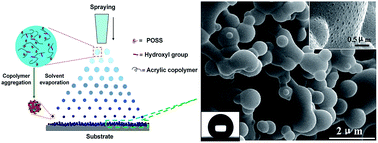
A superhydrophobic polymer surface with hierarchical structures is prepared by a one-step spraying process with a POSS-acrylic random copolymer solution. The morphology of the superhydrophobic surface consists of micron/nano-scale spherical protrusions. The possible formation mechanism of the superhydrophobic surface is proposed. The as-prepared surface has good superhydrophobicity and blood compatibility.


In the past decade, superhydrophobic surfaces have received considerable attention in the field of materials and surface sciences, due to their outstanding performance, such as in oil–water separation, and anti-icing applications, as well as in blood compatibility.1–4 Serving as the best engineer and designer, nature forms unartificial superhydrophobic surfaces in many animals and plants such as water strider's legs and lotus leaves.5,6 Inspired by nature, a myriad of approaches, such as template synthesis, electrospinning, sol–gel method, casting and plasma etching, have been developed to fabricate superhydrophobic surfaces with water contact angle higher than 150° and low contact angle hysteresis.7–9 In general, to achieve superhydrophobic surfaces, a combination of low surface free energy materials and surface roughness is required. Thus far, a wide variety of materials including inorganic, organic and composite materials have been utilised to prepare superhydrophobic surfaces. Among them, polymeric materials have been considered to be appropriate substrates because of their unique and versatile properties.10 For example, Yuan et al. prepared superhydrophobic poly(vinyl chloride) (PVC) surfaces by the template and casting method,6 and Levkin et al. obtained a superhydrophobic porous poly(octyl cyanoacrylate) coating by immersing a substrate coated with cyanoacrylate in an aqueous solution.11 However, most of the techniques for the fabrication of superhydrophobic surfaces either involve multi-step processes and sometimes harsh conditions or are unsuitable for large-sized flat surfaces, which limit their practical applications. A larger-scale fabrication of superhydrophobic surfaces on substrates by using polydivinylbenzene chalk was reported, but the surfaces of these substrates required a certain microstructure with rough mophology.12

Spray coating is a simple and well established technique in the modern surface-coating industry, that generates superhydrophobic surfaces by spraying a low concentration suspension on flat substrates.13–16 Ogihara et al. reported, for example, a superhydrophobic surface by spraying an ethanol suspension of SiO2 nanoparticles on a paper substrate.17 However, based on this method, polymeric materials have rarely been used in the preparation of superhydrophobic surface by themselves. In a previous report, our group successfully fabricated superhydrophobic surfaces through spraying a micelle solution of a fluorinated acrylic copolymer.4 In this communication, we present a one-step spraying-coating process for the fabrication of a superhydrophobic surface with a polyhedral oligomeric silsesquioxane (POSS)-acrylic random copolymer solution. Compared with the above-mentioned approaches, the copolymer solution used herein is relatively low-priced, can be easily prepared, is translucent, and is applicable to multiple substrates. Of note, the superhydrophobic surface exhibits regular morphology and consists of micron- and nano-scale spherical protrusions or particles, leading to the formation of hierarchical micro-nano structures. The as-prepared surface shows excellent superhydrophobicity and good blood compatibility. The possible formation mechanism of the superhydrophobic polymer surface is also proposed.

At first, a POSS-acrylic copolymer (PAC) was synthesized by free radical solution polymerization. The structure of the PAC is illustrated in Scheme 1. Experiments are described in detail in the ESI and the Fourier transform infrared (FT-IR) and 1H nuclear magnetic resonance (NMR) spectra of the PAC are shown in Fig. S1 and S2, respectively. As confirmed by gel permeation chromatography (GPC), the copolymer had a number-average molecular weight of 35[thin space (1/6-em)]000 with a polydispersity index of 1.91. Moreover, only one peak appeared at the elution volume for GPC (Fig. S3), indicating copolymer formation.


image file: c4ra07113a-s1.tif
Scheme 1 The structure of the POSS-acrylic copolymer (PAC).

Then the resultant PAC was dissolved in ethyl acetate to form a translucent solution containing 10 wt% of the copolymer. The diluted copolymer solution was deposited onto a copper net and dried at room temperature. The morphology of the PAC self-assembly aggregates was characterised by transmission electron microscope (TEM) (Fig. 1). The copolymer was prone to aggregation, forming many nano-scale spherical particles with diameters of 20–40 nm after solvent evaporation. The self-assembly of the copolymer and surface mobility of the POSS moieties in the copolymer possibly contributed to sphere formation.18–20


image file: c4ra07113a-f1.tif
Fig. 1 TEM micrograph of the PAC self-assembly aggregates.

After spraying the above-mentioned solution on a glass substrate and subsequent drying process, the copolymer formed a superhydrophobic POSS-acrylic copolymer (SPAC) surface. Meanwhile, the hydrophobic POSS-acrylic copolymer (HPAC) surface was also fabricated by spin coating for comparison (the preparation method of the HPAC surface is seen in the ESI). The morphologies of the HPAC and SPAC surfaces were examined by scanning electron microscopy (SEM). The HPAC surface was smooth (Fig. S4), and the static water contact angle (CA) on the HPAC surface was 106 ± 2.3° (Fig. S4, inset). However, the SPAC surface consisted of many connected micron-scale spherical protrusions, similar to the previous reported morphology,21 forming a rough structure (Fig. 2a). The high-magnification SEM image (Fig. 2a, inset) clearly showed numerous nano-scale outshoots on the surface of each micron-scale spherical protrusion. The XPS spectrum of the SPAC is shown in Fig. 2b, and the carbon, oxygen and silicone atomic percentages were estimated to be approximately 65.5%, 24.7% and 9.8%, respectively. There is a higher ratio of silicone to carbon in the XPS data (9.8%[thin space (1/6-em)]:[thin space (1/6-em)]65.5% ≈ 0.149[thin space (1/6-em)]:[thin space (1/6-em)]1) than the theoretical value of the PAC molecule (≈0.036[thin space (1/6-em)]:[thin space (1/6-em)]1), suggesting that the migration of the POSS groups containing silicone to the coating surface, which is essential to obtain superhydrophobic surfaces with low surface energy. As shown in the insets of Fig. 2c and d, the water CA on the SPAC surface could reach 158 ± 3.5° and the sliding angle (SA) was less than 3°, exhibiting outstanding superhydrophobicity. Moreover, these large-scale superhydrophobic surfaces were easily formed on other substrates including paper, PVC film, steel mesh and aluminum plate (Fig. S5a–d). Although the coated film exhibited a decrease in transmittance compared with the PVC film (Fig. S5e), caused by the light scattering and diffuse reflection of the surface roughness,12 the samples were visible after spray-coating the PAC solution (Fig. S5a and S5b).


image file: c4ra07113a-f2.tif
Fig. 2 SEM image (a) and XPS survey spectrum (b) and optical images of the water CA (c) and SA (d) of SPAC surface.

The possible formation mechanism for the superhydrophobic polymer surface prepared by spraying can be described as follows (Fig. 3a). During spray coating, the polymer solution is atomised to large numbers of tiny liquid droplets with different particle sizes by the pressure at the spray gun nozzle. Fig. 3b shows the cumulative particle size distribution curve of liquid droplets (the test method is seen in the ESI). The results indicate that approximately 50% of liquid droplets have sizes at or below 19.78 μm, including approximately 10% of liquid droplets less than 9.38 μm, and 40% of those have sizes between 19.78 and 34.26 μm. Before these tiny liquid droplets impact the substrate, the droplets gradually become smaller owing to the rapid evaporation of the solvent in air. Simultaneously, this process is accompanied by the self-assembly aggregation of the POSS-acrylic copolymer. The POSS functional groups can easily transfer to the air–copolymer solution interface (outside of the droplets) due to their self-aggregation behaviour and surface mobility,19,20 while the hydrophilic hydroxyl groups attached to the copolymers can migrate to the inside of droplets. This leads to the formation of submicron-sized polymeric particles. As a result, these particles stack and adhere together irregularly on the substrate when these particles hit the substrate surface, finally bringing about the formation of different-sized spherical protrusions of 500 nm to 3 μm (Fig. 2a). The surfaces of these micron-scale spherical protrusions appear a significant amount of nano-scale outshoots because of the self-aggregates behaviour of POSS functional groups (Fig. 2a, inset), ultimately leading to the formation of hierarchical micro-nanostructures. As indicated above, the possible morphologies of the relatively rough surface may be affected by a number of factors, such as the particle size distribution of liquid droplets, concentration of the polymer solution, evaporation rate of solvent, and self-assembly aggregates behaviour.


image file: c4ra07113a-f3.tif
Fig. 3 Schematic representation of forming SPAC surface (a) and the cumulative particle size distribution curve of “liquid droplets” prepared by spraying (b), D10 = 9.38 μm, D50 = 19.78 μm, D90 = 34.26 μm.

In theory, superhydrophobic surfaces have been speculated to possess good hemocompatibility.22 A widely accepted postulate in biomaterials science is that protein adsorption (especially, fibrinogen) is the first step in the blood–material interaction, followed by platelet adhesion and activation, leading to thrombus formation.23 Fig. 4 shows the amounts of bovine serum albumin (BSA) and bovine fibrinogen (BFG) adsorbed on the HPAC and SPAC surfaces from their respective PBS solutions (1 mg ml−1). The SPAC surface had lower amounts of adsorbed BSA and BFG as compared with the HPAC surface. This result indicates that the SPAC surface could resist protein adsorption to some extent in accordance with the superhydrophobic surfaces reported by Shirtcliffe and Advincula.24,25 Furthermore, the amount of BSA adsorbed on the SPAC surface decreased by approximately 27% as compared with that adsorbed on the HPAC surface; however, more importantly, the amount of adsorbed BFG decreased by approximately 64%, which is deemed to be one of the major factors affecting platelet adhesion.26,27


image file: c4ra07113a-f4.tif
Fig. 4 BSA and BFG adsorbed amounts on the surfaces of HPAC (a) and SPAC (b).

It is well known that platelet adhesion and activation are considered as key events in blood coagulation.27 In order to further evaluate the blood compatibility of the superhydrophobic surface, the platelet adhesion experiments on the HPAC and SPAC surfaces were also carried out. For comparison, similar experiments were conducted on PVC surfaces. As shown in Fig. S6, numerous adhered platelets were spread out on the PVC surface, and pseudopodia were clearly observed, implying that they have been extremely activated. However, for HPAC surface, the number of adhered platelets dramatically decreased (∼79%) as compared with that on the PVC surface (Fig. S7), and their morphology was maintained with almost no pseudopodia (Fig. 5a). More exhilaratingly, the adhering platelets nearly disappeared (Fig. S7), and few pseudopodia as well as deformation were observed on the SPAC surface (Fig. 5b). This should be attributed to the superhydrophobicity and the relative low protein adsorption on the SPAC surface.22,24,28


image file: c4ra07113a-f5.tif
Fig. 5 SEM images of the platelets adhesion on the surfaces of HPAC (a) and SPAC (b).

In conclusion, we successfully developed a simple one-step procedure to fabricate superhydrophobic polymer surfaces through spraying a POSS-acrylic copolymer solution. The hierarchical structure of the superhydrophobic surface was constructed by taking advantage of the self-assembly of the POSS-acrylic copolymer in the spraying process. The as-prepared surface by the spraying method presents excellent superhydrophobicity, outstanding anti-adhesion to platelet and protein adsorption resistance. Although the formed superhydrophobic polymer surface is not completely transparent, this study describes a facile method for fabricating large-scale superhydrophobic surfaces with potential applications in blood compatibility, anti-icing, antifouling, and water repellency.

Acknowledgements

This work is financially supported by the National Natural Science Foundation of China (51103061).

Notes and references

  1. C. R. Crick, J. A. Gibbins and I. P. Parki, J. Mater. Chem. A, 2013, 1, 5943–5948 CAS .
  2. Z. Shi, I. Wyman, G. Liu, H. Hu, H. Zou and J. Hu, Polymer, 2013, 54, 6406–6414 CrossRef CAS PubMed .
  3. B. J. Privett, J. Youn, S. A. Hong, J. Lee, J. Han, J. H. Shin and M. H. Schoenfisch, Langmuir, 2011, 27(15), 9597–9601 CrossRef CAS PubMed .
  4. H. Li, Y. Zhao and X. Yuan, Soft Matter, 2013, 9, 1005–1009 RSC .
  5. X. Gao and L. Jiang, Nature, 2004, 432, 36 CrossRef CAS PubMed .
  6. Z. Yuan, H. Chen and J. Zhang, Appl. Surf. Sci., 2008, 254, 1593–1598 CrossRef CAS PubMed .
  7. K. Acatay, E. Simsek, C. O. Yang and Y. Z. Menceloglu, Angew. Chem., Int. Ed., 2004, 43, 5210–5213 CrossRef CAS PubMed .
  8. M. Hikita, K. Tanaka, T. Nakamura, T. Kajiyama and A. Takahara, Langmuir, 2005, 21, 7299–7301 CrossRef CAS PubMed .
  9. X. Du and J. H. He, ACS Appl. Mater. Interfaces, 2011, 3, 1269–1277 CAS .
  10. N. J. Shirtcliffe, G. McHale and M. I. Newton, J. Polym. Sci., Part B: Polym. Phys., 2011, 49, 1203–1217 CrossRef CAS .
  11. X. Du, J. S. Li, L. X. Li and P. A. Levkin, J. Mater. Chem. A, 2013, 1, 1026–1029 CAS .
  12. Y. L. Zhang, J. N. Wang, Y. He, Y. He, B. B. Xu, S. Wei and F. S. Xiao, Langmuir, 2011, 27, 12585–12590 CrossRef CAS PubMed .
  13. P. A. Levkin, F. Svec and J. M. J. Fréchet, Adv. Funct. Mater., 2009, 19, 1993–1998 CrossRef CAS PubMed .
  14. W. Wu, X. Wang, X. Liu and F. Zhou, ACS Appl. Mater. Interfaces, 2009, 1, 1656–1661 CAS .
  15. M. Stepien, J. J. Saarinen, H. Teisala, M. Tuominen, M. Aromaa, J. Kuusipalo, J. M. Mäkelä and M. Toivakka, Appl. Surf. Sci., 2011, 257, 1911–1917 CrossRef CAS PubMed .
  16. H. S. Hwang, N. H. Kim, S. G. Lee, D. Y. Lee, K. Cho and I. Park, ACS Appl. Mater. Interfaces, 2011, 3, 2179–2183 CAS .
  17. H. Ogihara, J. Xie, J. Okagaki and T. Saji, Langmuir, 2012, 28, 4605–4608 CrossRef CAS PubMed .
  18. N. C. Escudé and E. Y. X. Chen, Chem. Mater., 2009, 21, 5743–5753 CrossRef .
  19. R. Y. Kannan, H. J. Salacinski, P. E. Butler and A. M. Seifalian, Acc. Chem. Res., 2005, 38, 879–884 CrossRef CAS PubMed .
  20. S. T. Iacono, S. M. Budy, J. M. Mabry and D. W. Smith, Macromolecules, 2007, 40, 9517–9522 CrossRef CAS .
  21. X. Chen, P. Fei, K. A. Cavicchi, W. Yang and N. Ayres, Colloid Polym. Sci., 2014, 292, 477–484 CAS .
  22. T. L. Sun, H. Tan, D. Han, Q. Fu and L. Jiang, Small, 2005, 1, 959–963 CrossRef CAS PubMed .
  23. Z. Zhang, M. Zhang, S. Chen, T. A. Horbett, B. D. Ratner and S. Jiang, Biomaterials, 2008, 29, 4285–4291 CrossRef CAS PubMed .
  24. R. B. Pernites, C. M. Santos, M. Maldonado, R. R. Ponnapati, D. F. Rodrigues and R. C. Advincula, Chem. Mater., 2012, 24, 870–880 CrossRef CAS .
  25. Y. Koc, A. J. Mello, G. McHale, M. I. Newton, P. Roacha and N. J. Shirtcliffe, Lab Chip, 2008, 8, 582–586 RSC .
  26. A. Wörz, B. Berchtold, K. Moosmann, O. Prucker and J. Rühe, J. Mater. Chem., 2012, 22, 19547–19561 RSC .
  27. L. R. Wang, H. Qin, S. Q. Nie, S. D. Sun, F. Ran and C. S. Zhao, Acta Biomater., 2013, 9, 8851–8863 CrossRef CAS PubMed .
  28. C. Mao, C. Liang, W. Luo, J. Bao, J. Shen, X. Hou and W. Zhao, J. Mater. Chem., 2009, 19, 9025–9029 RSC .

Footnote

Electronic supplementary information (ESI) available: Experimental section, characterization methods, FT-IR and 1H NMR spectra, GPC curve of PAC, SEM image and the water contact angle of the surface of HPAC, photographs of water droplets on the spray-coated different substrates, SEM image of the platelets adhesion on PVC surface. Number of the platelets adhesion on the different surfaces. See DOI: 10.1039/c4ra07113a

This journal is © The Royal Society of Chemistry 2014
Click here to see how this site uses Cookies. View our privacy policy here.