Improvement of boehmite nanoparticles' aqueous dispersability by controlling their size, shape and crystallinity

Pablo Pardo, José Miguel Calatayud and Javier Alarcón*
University of Valencia, Department of Inorganic Chemistry, Calle Doctor Moliner 50, 46100-Burjasot, Valencia, Spain. E-mail: javier.alarcon@uv.es

Received 27th June 2014 , Accepted 15th September 2014

First published on 15th September 2014


Abstract

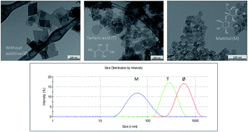
In this work, results on the control of size, shape, crystallinity and aqueous dispersability of boehmite prepared by a hydrothermal process are reported. The two step synthetic procedure entailed the precipitation of a xerogel by adding NaOH until pH 10 to a solution of aluminum nitrate at 100 °C without or with additives such as tartaric acid and maltitol, and a subsequent hydrothermal treatment at 150 and 200 °C for different periods of time. The final materials were characterized by transmission electron microscopy (TEM), X-ray diffraction (XRD) and elemental analysis. The stability and other characteristics of the boehmite nanoparticle aqueous dispersions were determined by measuring the zeta potential (ζ), and the particle size distribution (PSD) by dynamic light scattering (DLS). Additive-free boehmite hydrothermally processed at 150 °C for 24 h displayed average lengths of around 20 nm. On increasing the temperature to 200 °C and the holding time up to 168 h, both the particle length increased (ca. 95 nm) and the particle size distribution widened. Comparatively, the growth of the boehmite nanoparticles in the samples prepared with additives was severely restricted up to 20% of the length of additive-free obtained particles, leading to narrower particle size distributions. Modifications in their shape were also allowed. Interestingly, improved aqueous dispersion stabilities were observed for samples prepared in the presence of an additive. A higher dispersability over a wide pH window was found in maltitol-grafted boehmite nanoparticle dispersions with hydrodynamic sizes closer to the single-particle sizes, as observed by TEM. The different formation and stabilization mechanisms of the boehmite nanoparticles obtained in the presence of additives through the synthetic procedure are also discussed.


Introduction

Nanocrystals with different shapes have been extensively studied during the last two decades, because they have allowed for the development of materials with new properties and the potential for new applications in fields such as catalysis, biomedicine or ceramics.1–3 One type of nanoparticle of great interest is based on aluminum oxohydroxide or, more specifically, on the γ-AlOOH polymorph known as boehmite because of its role as a precursor to γ-Al2O3, a catalyzer support often used in the petrochemical industry and as a support to molecules and/or biomolecules with specific functionalities in different media.4–6 The structure of boehmite consists of Al(O,OH)6 units that form octahedral layers by sharing edges. Layers are stacked in the [010] direction and are linked by hydrogen bonds. The most widely accepted space group is Cmcm, with cell parameters of a = 2.86, b = 12.21 and c = 3.69.7 Depending on the preparation conditions, different nanoparticle morphologies are obtained7 with four types of exposed surfaces, namely (100), (010), (001) and (101), whose differences in prevalence determine the physical and chemical properties of the material. Recently, we reported the functionalization of boehmite nanoparticles with different molecules to develop potential sensors for cations and anions in aqueous solutions by fluorescent spectroscopy as well as potential contrast agents for NMR imaging.8,9 The good performance of these supporting molecules on the boehmite surface is caused by the non-aggregation of nanoparticles over a wide pH range. The stability of the aqueous dispersions allowed in situ emission measurements in solution, because the colloidal particles did not produce any scattering. It is also well known that boehmite has long been used as an adjuvant in practically all high-volume vaccines, and has been safe to use for this purpose for many years.10 All these contrasting abilities of boehmite nanoparticles allow forecasting their potential use after functionalization. However, the preparation of stable dispersions of well-shaped, monosized boehmite nanocrystals in a wide pH window is necessary for improving their applicability.

There are three preparation methods usually used in the synthesis of boehmite nanoparticles in aqueous solution. Precipitation from aqueous aluminum solutions has been widely used, but it requires the strict control of many reaction parameters.11 The hydrolysis and condensation of aluminum alkoxides followed by peptization in an acidic medium has also produced boehmite.12 However, the dispersion stabilities of the obtained boehmites in aqueous solution decrease with increasing pH, i.e. at pH > 9, the sol solution transforms into a gel. Hydrothermal techniques have also been extensively utilized in the preparation of boehmites in aqueous solution.13–16 Some additives (or ligands) such as polyols, polycarboxylic acids, anions, and different surfactants have been used with the goal of controlling the size, morphology and even the dispersability of boehmite nanoparticles in aqueous solutions.17–23 It has been proved that the efficiency of acyclic polyols as complexing agents depends on both the number of OH groups bonded to the carbon chain and their stereochemistry.17 This ability causes a modification in the usual morphologies observed for boehmite nanoparticles, specifically resulting in an increase in the (101) face areas. In order to check the effect of a large-volume polyol in which the stabilization of the boehmite colloidal dispersions would have a steric component, maltitol was chosen as an additive in the hydrothermal preparation of boehmite in this work. The influence of some organic acids on the synthesis of boehmite has also been reported.20 It has been proposed that these additives completely inhibit gibbsite formation and facilitate boehmite nucleation through different amounts of complex formation by multidentate ligands. In our view, a comparison of the effect of a carboxylic acid such as tartaric acid with that of a large-volume polyol on the size, morphology and aqueous dispersability of boehmites prepared by the hydrothermal method is worthy.

According to the reported data, two distinct boehmite morphologies can be obtained after hydrothermal processing of the precipitated solid (xerogel) depending on the pH of the starting solution. Although boehmite is the main crystalline phase detected in these precipitates, bayerite and/or gibbsite have also been found.24 Boehmite nanocrystals are fibers in acidic conditions (pH = 2–5), whereas they form plates under basic media (pH = 10–11).25–28 Results have also showed that the final sizes of boehmite nanocrystals are mainly dependent on the temperature and time of the hydrothermal process. The higher the temperature and/or longer processing time, the larger is the size of boehmite nanocrystals. However, no quantitative evaluation on the size variation has been reported so far.

In order to avoid the presence of any aluminum hydroxide in the precipitate and to favor the precipitation of boehmite as the only crystalline phase in the final solid, some authors have stated that the precipitation should be carried out in a hot aqueous solution.29,30 Thus, the first goal of this paper is to evaluate the effects of the temperature and the hydrothermal processing time of xerogels obtained at 100 °C on the morphologies, sizes and aqueous dispersion stabilities of the final boehmites. Moreover, we intend to evaluate the effects of the two additives, a large-volume polyol as maltitol and a carboxylic acid as tartaric acid, present in the aqueous solution before the precipitation of boehmite in the first step, on the final size and morphology of boehmite as a function of the temperature and time of the hydrothermal processing. In addition, the aqueous dispersabilities of the boehmites prepared with and without additives will be compared.

Experimental section

Preparation procedure

The preparation of the boehmite nanoparticles proceeded in an aqueous medium through a gel precipitation by the alkalization of a solution of aluminum nitrate and its subsequent hydrothermal aging. The synthetic methodology is based on a general procedure previously used by several authors.17 Besides extending the temperature range of the hydrothermal aging, the procedure applied in this study had a higher temperature precipitation step, i.e. at 100 °C, which was reported to favor the formation of boehmite among other aluminum hydroxide phases in non-aqueous routes.29 The effect of the two additives, tartaric acid and maltitol, on the final morphologies, microstructures, and aqueous dispersion stabilities of the boehmites was tested versus the materials obtained without additives.

An initial 0.1 M Al(NO3)3·9H2O (Panreac) solution (pH ca. 2) was heated to 100 °C with stirring in a reflux system. In the samples with an additive, 0.01 mol L−1 of tartaric acid (C4H6O6, Sigma-Aldrich) or maltitol (C12H24O11, Sigma-Aldrich) were added to the initial solution. Once the desired temperature was reached, NaOH (5 M) was added dropwise until the pH was adjusted to 10. A white, dense precipitate of boehmite was formed during the addition. The as-obtained precipitate was then hydrothermally aged for 24, 72 and 168 h at two different temperatures. For the treatments at 150 °C, 100 mL PTFE vessels were used. Moreover, the aging at 200 °C was carried out in PTFE lined stainless steel autoclaves. After the hydrothermal aging, which occurred without pH variation, samples were centrifuged and washed three times with water. The hydrogels were finally dried overnight in a furnace at 100 °C. Assuming quantitative precipitation of boehmite from the aluminum nitrate initial solution, yields of ca. 65% were achieved after the complete procedure. Materials were labeled X_YYY_ZZZ, where X indicates the absence of an additive (0) or the use of tartaric acid (T) or maltitol (M), and YYY and ZZZ correspond to the temperature (in °C) and time (in hours) of the hydrothermal treatment, respectively.

Material characterization

Characterization was mainly focused on the morphological and microstructural features of the boehmite nanoparticles and on the properties of their aqueous suspensions.

The sizes and shapes of the final nanoparticles were observed by transmission electron microscopy (TEM) at 100 kV (Model 1010, Jeol, Tokyo, Japan). Samples were prepared by dispersing the as-produced powders in water and setting a drop of the suspensions on copper grids that had previously been coated with a porous thin carbon film. The average size of the boehmite nanoparticles was determined by randomly selecting between 225 and 50 nanoparticles from the TEM images and manually measuring the major diagonal of their bases using the Image J software. Likewise, the particle thicknesses were measured by the occurrence of edgewise lying particles. In a typical measurement, a ratio of 4 pixels per nm was set as the resolution to magnification ratio of the image. From the set of measurements, histograms were generated to determine the full width at half maximum (FWHM) of the particle length (size) distributions. The aspect ratio of the nanoparticles was calculated by dividing the average diagonal length by the corresponding average thickness.

HRTEM images were collected using a Tecnai G2 F20 field emission electron microscope operating at 200 kV and equipped with a Gatan CCD camera. Samples were also prepared by the evaporation of very dilute aqueous suspensions onto carbon-coated grids. Data treatment was performed with Digital Micrograph software.

The XRD analyses of the powder samples were performed in a Bruker D-8 Advance diffractometer, using Cu Kα radiation, with 1 and 3 mm divergence and antiscattering slits, respectively, and a 3° 2θ range Lynxeye linear detector. The X-ray powder diffraction patterns were run from 5 to 80° 2θ with a step size of 0.02° 2θ and an accumulated counting time of 0.2 s. EVA software was used for profile analysis and an application of Scherrer's equation (λ = 1.541874 Å; K = 1) was used for the determination of crystallite size in different crystallographic directions.

The elemental analysis was carried out in a CE Instruments EA1110 Elemental Analyzer, set for the detection of nitrogen, carbon and hydrogen, and using atropine as a standard.

The particle size distribution (PSD) of the suspensions prepared by dispersion of the obtained powders in water was measured by dynamic light scattering using Zetasizer Nano ZS (Malvern Instruments) equipment. Stable 0.3 g L−1 dispersions of boehmite that were suitable for measurement were obtained by sonication (10 s, 150 W) and a subsequent adjustment to pH = 10 with NaOH. The same equipment was used in the measurements of the ζ-potential in 0.1 g L−1 suspensions of boehmite nanoparticles adjusted to pH = 8.7.

Results and discussion

Electron microscopy observations

The TEM images in Fig. 1 summarize the most significant morphological features of the samples prepared in this study. The left column shows the micrographs of the additive-free samples, and the middle and right columns correspond to the samples with added tartaric acid and maltitol, respectively. In rows, different conditions of hydrothermal synthesis are displayed; from top to bottom, 150 °C (24 h and 168 h), 200 °C (24 h and 168 h). As can be seen in Fig. 1, the sizes and shapes of the boehmite nanoparticles were strongly influenced by the presence of the additives as well as by the length and temperature of the hydrothermal treatment over the range of experimental conditions used in this study. The scale bars in Fig. 1 are 100 nm for all the micrographs except for J, K, and L which are 200 nm long.
image file: c4ra06322h-f1.tif
Fig. 1 TEM micrographs of the samples prepared in this work: 0_150_24 (A), T_150_24 (B), M_150_24 (C), 0_150_168 (D), T_150_168 (E), M_150_168 (F), 0_200_24 (G), T_200_24 (H), M_200_24 (I), 0_200_168 (J), T_200_168 (K), and M_200_168 (L). Scale bar is 100 nm except for B (20 nm) and J, K and L (200 nm).

For the additive-free prepared boehmites, the particle size increased with an increase in the temperature and holding time of the treatment. This fact can be observed for samples 0_150_24, 0_150_168, 0_200_24 and 0_200_168 in the micrographs shown in Fig. 1A, D, G and J. The PSDs calculated from the length measurements of the TEM images shown in Table 1 and Fig. 1S shifted to greater size values and broadened over a wider range as the time and temperature increased. The relatively small boehmites obtained at 150 °C for 24 h showed sizes between ca. 14–26 nm. With a temperature increase up to 200 °C during 24 h, the size range increased to values between ca. 45–95 nm. After 168 h at 200 °C, the boehmite nanoparticles showed sizes in the range ca. 70–120 nm.

Table 1 Descriptive parameters of particle size distributions as measured on TEM images. L, average length of major diagonal of the platelets; sL, L standard deviation; ML, mode of L; FWHML, full width at half maximum of L distribution; nL, number of length measurements; e, average thickness of the particles; se, e standard deviation; ne, number of thickness measurements; r, aspect ratio (e/L)
Sample L (nm) sL (nm) ML (nm) FWHML (nm) nL e (nm) se (nm) ne r
0_150_24 21.1 10.6 14.43 13.3 115 5.2 3.0 68 4.0
T_150_24 21.2 11.3 12.12 17.4 142 4.9 1.0 95 4.3
M_150_24 15.4 5.3 9.0 11.8 224 4.7 1.1 108 3.3
0_150_168 32.8 13.9 22.1 26.5 90 4.7 1.7 65 7.0
T_150_168 28.7 11.3 19.3 16.5 93 5.8 1.4 38 4.9
M_150_168 10.7 3.7 7.9 9.5 48 3.7 1.1 38 2.9
0_200_24 69.6 24.0 63.1 53.9 96 15.9 5.7 20 4.4
T_200_24 41.9 13.3 28.2 22.9 136 9.6 1.9 44 4.4
M_200_24 15.8 6.2 14.2 16.7 194 4.8 4.8 85 3.3
0_200_168 94.7 30.5 76.0 51.34 120 25.2 6.0 19 3.8
T_200_168 59.4 24.2 36.4 39.33 169 14.0 2.8 40 4.2
M_200_168 17.6 10.3 6.3 10.42 121 7.5 1.9 120 2.3


The effect of the presence of an additive can be easily observed from the variations in the materials obtained at 200 °C, in which the differences between the samples are notably larger than in the preparations at 150 °C, although the trends were preserved. The particles in sample 0_200_24 (Fig. 1G) consisted of well-defined diamond-shaped platelets with sharp edges forming angles of 104° and 76°. The same morphology was described for commercial31 and boehmite nanoparticles obtained from basic-media.17 The particles obtained in the presence of an additive (1H, 1I) exhibited rather square morphologies, with angles closer to 90°. These differences were not so clear in the samples obtained at 150 °C for 24 h (Fig. 1, upper row, 1A vs. 1B and 1C) but they were even more pronounced for materials prepared at 200 °C for 168 h (Fig. 1 bottom row, 1J vs. 1K and 1L). Based on the measurements of the major diagonal of the particles performed on the TEM images, the average length of the particles decreased in the sequence 0 > T > M. Table 1 summarizes the main descriptive parameters of the size distributions calculated from the TEM measurements represented in Fig. 1S. The distribution maxima also shifted towards lower length values while their respective widths (FWHM) narrowed in the sequence 0-T-M in all of the series analysed. From these values and from the direct observations of the distributions in Fig. 1S, it can be stated that the presence of the additives resulted in a ca. 40% size reduction of the particles with the addition of tartaric acid and an ca. 80% reduction with maltitol. The presence of some edgewise lying particles in the micrographs of samples obtained at 200 °C allowed for the observation and measurement of the thicknesses. The average values obtained and the total numbers of measurements are also shown in Table 1. These measurements allowed for the calculation of the aspect ratio (r) of the particles as the ratio of their major diagonal to their thickness.

As can be observed in Table 1, the studied additives each had a different effect on the final shape of the nanoparticles. The tartaric acid route yielded particles with a similar aspect ratio of those obtained without an additive (ca. 4, Table 1). The particles obtained with maltitol showed lower aspect ratio values which decreased further with the hydrothermal treatment time (from 3.3 to 2.3). In agreement with this, previous works found in the literature observed that the presence of polyol type additives in the preparation of boehmite led to an increase in the amount of particle lateral surfaces (i.e. (100), (001) and (101)) relative to basal planes as a result of either a lateral surface stabilization induced by the presence of xylitol through adsorption or by lowering the crystal growth kinetics.18 The thermodynamic adsorption of xylitol described in the aforementioned study and of other acyclic polyols with backbones of 2 to 6 carbon atoms included in a later work17 was reported to occur preferentially at the lateral faces, where cavities containing –OH groups in the structure of boehmite stabilized the interaction with the polyols through hydrogen bonds. This so-called “nest effect” favoured the stabilization of lateral faces leading to their higher morphological expression in the final particles. The extent of this effect also depended on the complexing strength of the polyol, which was significantly affected by its stereochemistry. In the present work, maltitol was carefully chosen from among other polyols in order to study the effect of its voluminous structure (12 carbon atoms including a pyran cycle) on the final particle size and on the stability of the aqueous suspensions of nanoparticles. Previous results on other polyols were obtained in soft hydrothermal experiments at 95 °C,17 so a direct comparison could be misleading. Still, the presence of maltitol restrained the size of the nanoparticles when higher hydrothermal temperatures were used, and thus helped to achieve well-crystallized nanomaterials.

Tartaric acid seems to present a different role in the synthesis of boehmite nanoparticles. The tartrate anion present in the solution in alkaline conditions is comprised of two planar halves with all four carbon atoms lying in a plane and each half containing a carboxyl group, a tetrahedral carbon and a hydroxyl oxygen atom.31 With this configuration, the tartrate anion can coordinate Al3+ to form different tartrate–aluminum complex anions depending on the pH.32 At high pH values such as the ones used in this work, the formation of the binuclear species [Al2(C4O6H2)2]2− (log[thin space (1/6-em)]β = −18.95) competed with tetrahydroxyl-aluminium(III) [Al(OH)4] (log[thin space (1/6-em)]β = −23.46)32 among the other aquohydroxo complexes [Al(OH)h(OH2)6−h](3−h)+ involved in condensation through the olation/oxolation mechanism.33 This hindered the Al3+ polymerization and consequently slow down the boehmite formation, which was reported in the literature to increase over pH 5 in the hydrothermal aging process due to the enhancement of the dissolution–reprecipitation reaction. Due to their smaller size, particles obtained with the tartaric acid addition had higher specific surface area, thus obtaining a higher proportion of lateral faces than additive-free nanoparticles with the same aspect ratio and a reported effect of the lowering of crystal growth kinetics.18

As expected, the increased temperature of the hydrothermal aging increased the particle size of the materials. Remarkably developed shapes and sizes were achieved when working at 200 °C compared to the results of the procedure at 150 °C. In contrast, the main effect of a prolonged treatment time on the samples was not an increased nanoparticles growth, but is rather the enhancement of their stability in an aqueous dispersion, as we will see below. The better particle shape definition achieved with an increasing holding time favoured the electrostatic repulsion and/or steric hindrance, and thus stabilized the suspensions. In similar hydrothermal procedures with sodium polyacrylate as an additive,22 an evolution of the particle shape from platelets to fibres with time (within 168 h) has been reported. Unlike the observations in the present work, the authors noticed a decrease in the pH values during the hydrothermal aging that may be the cause of the morphological transformation. As has been established in the literature,34,35 the pH strongly affects the morphology of the boehmite particles, with elongated shapes being favoured as the pH value decreases.

X-ray diffraction and HRTEM microstructural analysis

XRD patterns of the precipitated boehmite solid (xerogels) obtained with and without additives are shown in Fig. 2S. Broader peaks with lower intensities were observed for M and T xerogels compared to the additive-free sample. Fig. 3S–5S display the set of XRD patterns prior to and after the hydrothermal processing at different temperatures and time periods for the gels without an additive, with tartaric acid and with maltitol, respectively. It can be seen that even the diffractograms corresponding to the boehmite xerogels that had been hydrothermally processed for a short time showed sharper and more intense diffraction peaks than the starting materials. This fact can be associated with the improvement of the crystallinity and/or the presence of much larger crystallites. A preferred orientation effect is clearly observed in the diffraction patterns based on the high relative intensity of the diffraction peak (020). This indicates the presence of a certain texture in the material that is a consequence of a particle shape with high aspect ratio, as was previously determined by TEM measurements. The effect is more pronounced in the additive-free hydrothermally treated material due to its more developed particle growth. It is notable that the differences observed in the XRD patterns of the xerogels both with and without additives remained after hydrothermal treatment. Thus, the additive-free samples always showed narrower profiles with higher peak intensities than materials prepared in the presence of tartaric acid or maltitol, which showed an increased broadening in their diffraction profiles in that order. An example of these differences is represented in Fig. 2, in which the xerogels were processed at 200 °C for 168 h.
image file: c4ra06322h-f2.tif
Fig. 2 Powder X-ray diffraction patterns of the samples prepared at 200 °C for 168 h without an additive, with tartaric acid and with maltitol. Bars indicate the position and relative intensity of boehmite reflections (Ref. code: 00-005-0190).

Assuming that the prepared boehmites reached high crystallinity, it would be possible to evaluate their microstructures from the performed XRD experiments. A simpler methodology is the application of the Scherrer method, with an estimation of the instrumental broadening by means of a standard (LaB6). The main purpose of the crystallite size determination was to compare the trend in the variation of these estimated crystallite sizes to the trend of the nanoparticle sizes measured from the TEM micrographs.

Regarding these microstructural modifications, the analysis of the samples prepared at 200 °C clearly showed the effects of the hydrothermal treatment and of the presence of additives in the final materials. As a representative comparison, the three materials obtained at 200 °C for 168 h displayed in Fig. 2 were considered. Table 2 summarizes the measured position of the basal reflection (020), its full width at half maximum and the crystallite size (〈Dv〉) calculated using the Scherrer equation in the [020] crystallographic direction for all samples prepared at 200 °C. The largest crystallite sizes were observed in samples prepared without an additive. In samples prepared in the presence of tartaric acid, reductions of as much as 60% were observed in the crystallite size in the [020] direction relative to the additive-free materials for 168 h. The decrease in the [020] crystallite size was even greater in the case of the samples prepared with maltitol, showing reduction percentages of up to 80% for 168 h reaction time. The calculated [020] crystallite sizes matched the corresponding particle thicknesses values (e, Table 1) obtained from the TEM measurements of the analysed samples. This agreement supported the methodology employed in the size determinations from the TEM images, confirming its suitability.

Table 2 Position (Pos.), Full Width at Half Maximum (FWHM) and crystallite size (〈Dv〉) obtained from the (020) diffraction profile of materials prepared at 200 °C
Sample Pos. (°2θ) FWHM (°2θ) 〈Dv〉 (nm)
0_200_24 14.424 0.366 13.8
0_200_72 14.420 0.260 23.4
0_200_168 14.436 0.238 33.1
T_200_24 14.292 0.653 10.5
T_200_72 14.317 0.585 12.4
T_200_168 14.328 0.541 13.1
M_200_24 14.346 1.053 5.0
M_200_72 14.380 1.011 6.5
M_200_168 14.340 0.950 7.2


A small amount of evidence of well-developed crystalline structures was found in the well-defined morphologies of the particles observed by TEM as well as in their X-ray diffraction profiles. The observed broadening in these profiles was mainly caused by the reduction of the size of the coherent diffraction domains as a consequence of the reduction in particle size, which reached a factor of 6 when comparing the extreme size values in Table 2. An important factor related to the crystallinity of the boehmite that has been widely discussed in the literature is the increase in the 020 interplanar distance. A consequence of this increase is the shift of the 020 reflection position to lower angles in the diffraction profile, sometimes in the range of units of °2θ,36 that has aroused some controversy in the literature.37 The shift was previously attributed to the presence of interlayer water38,39 and then to a relaxation of the attractive forces between the layers due to the effect of the peripheral absorption of water.40 Subsequent studies established that the small number of octahedral layers in the finely crystalline boehmite structure are the main reason for this shift, although both the d(020) displacement and its profile broadening were enhanced by the interlayer water molecules.36 Taking this into consideration, the occurrence of fine crystalline boehmite in the presence of an additive is also supported by the observed slight d(020) increase (in the order of tenths of °2θ, as summarized in Table 2)14 when compared with the 2θ positions obtained in the preparation without an additive. The relatively small d(020) shifts suggests the presence of a well-ordered crystalline structure in small coherent diffraction domains.

The HRTEM observations confirmed the well-developed crystallinity of the nanoparticles that were formed by single crystals with clearly visible interplanar distances. As a representative example, Fig. 6S shows a set of boehmite nanoparticles with different orientations that are constituted of single crystalline domains.

Elemental analysis

In order to assess the presence of additives in the final powder, elemental analyses were carried out for samples 0_200_168, T_200_168 and M_200_168. Table 3 summarizes the weight percentages of N, C and H that were determined for the three materials. A significant amount of carbon was detected in the sample prepared with maltitol, whereas negligible C contents were found in the samples without an additive or with tartaric acid. This result allowed us to establish that none of the carbon from the tartaric acid is present in the dry powder obtained by preparation with this additive. Thus, unlike the proposed interactions with maltitol, surface interactions between tartaric acid and the boehmite nanoparticles are discarded. As was mentioned above, the particles obtained in the presence of tartaric acid showed a smaller size but had similar aspect ratio to those obtained without an additive. This may point to a slower, surface interaction-free growth of the boehmite nanoparticles that is equivalent to that of samples prepared without an additive.
Table 3 Elemental analysis of the final powders
Sample N (wt%) C (wt%) H (wt%)
0_200_168 0.11 0.05 1.35
T_200_168 0.05 0.04 1.91
M_200_168 0.11 0.95 1.72


Characterization of the aqueous suspensions of boehmite nanoparticles

The stability of the boehmite nanoparticles in an aqueous dispersion is an important feature for many of their applications. Measurements of the PSDs and ζ-potentials of the aqueous suspensions of the nanoparticles obtained in this study help in the characterization of their stability.

Aqueous suspensions of materials prepared at 150 and 200 °C for 24 h, which are illustrative of the behaviour of rest of the materials, exhibited the PSDs plotted in Fig. 3. Although the dispersion achieved was enough to perform the DLS measurements properly, the suspensions presented some degree of particle aggregation. This led to a disagreement between the TEM and DLS size estimations.


image file: c4ra06322h-f3.tif
Fig. 3 Particle size distributions of the samples treated at 150 °C for 24 h (A) and at 200 °C for 24 h (B) determined by DLS.

A consideration of the effects of the additives showed that, in all cases, the dispersion efficiency was higher in samples prepared with maltitol, which also had smaller particle distributions. Increasing the treatment temperature enhanced the stabilization effect of maltitol, as can be observed in Fig. 3. Fundamental particles, i.e. particles with sizes corresponding to the TEM observations, were only detected in M-containing samples. Materials prepared with tartaric acid or without an additive contained a larger proportion of aggregates22 that shifted the distribution towards larger particle sizes. The nanoparticles prepared with tartaric acid had an improved dispersion with respect to the additive-free materials obtained when the hydrothermal treatment was carried out at 200 °C. The nanoparticles obtained at this temperature in the presence of an additive showed a remarkable enhancement in their dispersion ability in an aqueous suspension that can be clearly noticed in Fig. 3B. The hydrothermal treatment time also influenced the behaviour of boehmite nanoparticles in aqueous suspensions. Fig. 7SA and B compare the PSDs of 24 and 168 h additive-free materials treated at 150 and 200 °C, respectively. For both temperatures, the samples treated for 168 h showed better dispersions with lower PSD values.

Measurements of the ζ-potentials of the aqueous suspensions of the as-prepared boehmite nanoparticles (pH = 8.7) (Fig. 4) also showed differences in the behaviour of samples as a result of their preparation procedures. While no noticeable differences were observed in the ζ-potentials when working at 150 °C (Fig. 4B), samples prepared at 200 °C exhibited increasing absolute values in the sequence 0_200_24 < T_200_24 < M_200_24 (Fig. 4A). This pointed to an incremental change in the negative surface charge in materials prepared with additives, which was in good agreement with the enhanced dispersion and with the consequent improved stabilization of their aqueous suspensions observed in the DLS analysis.


image file: c4ra06322h-f4.tif
Fig. 4 Determination of the ζ-potential at pH 8.7 for samples 0, T and M prepared at (A) 200 °C for 168 h and (B) at 150 °C for 24 h.

It is notable that the addition of maltitol molecules in the preparation of boehmite presented a double effect on the aqueous suspension stabilization. On one side, the higher proportion of negatively charged lateral faces increased the surface charge of the particles, as was clearly established by the ζ-potential measurements (Fig. 4). On the other side, the detection of a significant carbon content through the elemental analysis of the final product obtained with the maltitol addition (Table 3) pointed to the presence of relatively large maltitol molecules anchored to the lateral surfaces of the particles which induced a steric hindrance effect. Both these consequences of the use of maltitol helped with the repulsion between the boehmite nanoparticles, allowing for a better dispersion that resulted in an increase in the stability of their aqueous suspensions, as supported by the DLS and ζ-potential analyses. Indeed, the balanced combination of the electrostatic repulsion with steric stabilization was reported to be optimal for the stabilization of highly concentrated suspensions of alumina.41

As the lateral faces are responsible for the surface charge, nanoparticles prepared in the presence of tartaric acid at 200 °C showed a slight increase in their ζ-potential absolute values, as presented in Fig. 4. This effect is not as pronounced as in the case of maltitol, where an effective change in the shape of the nanoparticles (the aspect ratio) was achieved. Still, the effect of tartaric acid in the preparation of the boehmite nanoparticles was enough to enhance the dispersion of the materials (Fig. 3B) and the stability of the aqueous suspensions.

Remarks on the roles of the additives

From the results obtained in this study, it can be inferred that the tartaric acid and maltitol additives used each play quite different roles in both the precipitation step and in the subsequent hydrothermal aging, leading to variations in the final properties of the prepared boehmites. For the sake of clarity, Table 4 summarizes the main morphological characteristics of the boehmite nanoparticles prepared, as well as their crystallinity and the stability of their aqueous dispersions.
Table 4 Summary of the main features of the prepared boehmites
Sample Shape L (nm) e (nm) 〈Dv〉020 (nm) Aqueous stability ζ (mV)
0_150_24 Square 21.1 5.2 Low −21.6
T_150_24 Square 21.2 4.9 Low −18.4
M_150_24 Square 15.4 4.7 High −17.4
0_150_168 Rhombic 32.8 4.7 Low
T_150_168 Square 28.7 5.8 Low
M_150_168 Square 10.7 3.7 High
0_200_24 Rhombic 69.6 15.9 13.8 Low
T_200_24 Square 41.9 9.6 10.5 Medium
M_200_24 Square 15.8 4.8 5.0 High
0_200_168 Rhombic 94.7 25.2 33.1 Low −12.4
T_200_168 Rhombic 59.4 14.0 13.1 Medium −17.8
M_200_168 Square 17.6 7.5 7.2 High −24.4


It is proposed that tartaric acid slows down the boehmite formation due to the coordination of Al3+ by tartrate anions which form tartrate–aluminium complexes. On the contrary, the ability of maltitol to coordinate Al3+ is negligible and its effect is based on its interaction with the lateral surfaces of the solid. Thus, the action of tartaric acid is related to the rate of nucleation and growth of the nanoparticles, while maltitol affects their surface chemistry, hindering the condensation of nuclei and modifying their morphology development. Both additives thus extend their effects to the whole preparation process.

In general, hydrothermal aging involves different mechanisms in the transformations of nanoparticles. When the nanoparticles are partially soluble, the concentration of the chemical species in solution may be high enough to nucleate a more stable crystalline phase. The formation of well crystallized particles then takes place by a slow dissolution–recrystallization process. Such a process leads to a new crystalline phase, which was not observed in the present study.

The solubility of the boehmite nanoparticles is very low, so the crystallization of the early amorphous or poorly crystallized solid can only proceed by an in situ solid state transformation. This transformation involves the diffusion of ions within the solid as well as a partial dehydration.

The main morphological differences between the three types of boehmite were primarily induced in the precipitation process; in the case of tartarte-containing samples, controlling the nucleation and growth rate, and in the maltitol system, by the stabilization by adsorption of maltitol molecules on the lateral faces of the early developed nuclei. The hydrothermal aging, on the other hand, increased the crystallinity of the nanoparticles as assessed by XRD, enhancing properties such as the aqueous dispersion stability.

Conclusions

A procedure to control the size and shape of boehmite nanoparticles synthesized by a hydrothermal process and to improve their dispersability in aqueous media is reported. Additive-free and tartaric- and maltitol-containing xerogels previously obtained from a solution of aluminum nitrate at pH 10 were hydrothermally treated at 150 and 200 °C holding times between 24 h and 168 h. A higher hydrothermal treatment temperature and longer treatment time resulted in an increase in the average particle size and in the broadening of the size distributions.

On the contrary, the presence of additives in the hydrothermal process led to both a size reduction between 40% and 70% and narrower particle size distributions. In addition, some shape changes also occurred.

With respect to the aqueous dispersion stability, it was found that longer stabilities were observed for samples prepared in the presence of an additive, with the longest being the maltitol-grafted boehmite nanoparticle dispersions which have hydrodynamic sizes of around 50–60 nm. The hydrothermal aging time was found to increase the water dispersability of the materials.

Finally, a quantitative evaluation of the size and particle size distribution was reported in this work from the analysis of the TEM images.

Acknowledgements

Financial support from the Spanish Ministry of Economy and Competitiveness through the project CONSOLIDER INGENIO 2010 CSD2010-00065 is acknowledged. Technical support from the Servei Central de Suport a l'Investigació Experimental (SCSIE) of the University of Valencia in the application of TEM and XRD techniques is also acknowledged.

References

  1. M. Li, B. Lebeau and S. Mann, Adv. Mater., 2003, 15, 2032 CrossRef CAS.
  2. Y. Xia, P. Yang, Y. Sun, Y. Wu, B. Mayers, B. Gates, Y. Yin, F. Kim and H. Yan, Adv. Mater., 2003, 15, 353 CrossRef CAS.
  3. J. Yang, Z. Quan, D. Kong, X. Liu and J. Lin, Cryst. Growth Des., 2007, 7, 730 CAS.
  4. M. Digne, P. Sautet, P. Raybaud, P. Euzen and H. Toulhoat, J. Catal., 2002, 211, 1 CrossRef CAS.
  5. M. Digne, P. Sautet, P. Raybaud, P. Euzen and H. Toulhoat, J. Catal., 2004, 226, 54 CrossRef CAS PubMed.
  6. M. Amoura, N. Nassif, C. Roux, J. Livage and T. Coradin, Chem. Commun., 2007, 4015 RSC.
  7. R. Aucejo, J. Alarcón, C. Soriano, M. C. Guillem, E. García-España and F. Torres, J. Mater. Chem., 2005, 15, 2920 RSC.
  8. E. Delgado-Pinar, J. C. Frías, L. J. Jiménez-Borreguero, M. T. Albelda, J. Alarcón and E. García-España, Chem. Commun., 2007, 3392 RSC.
  9. A. Rutenberg, V. V. Vinogradov and D. Avnir, Chem. Commun., 2013, 49, 5636 RSC.
  10. S. Musić, Đ. Dragčević and S. Popović, Mater. Lett., 1999, 40, 269 CrossRef.
  11. M. Nguefack, A. F. Popa, S. Rossignol and C. Kappenstein, Phys. Chem. Chem. Phys., 2003, 5, 4279 RSC.
  12. X. Bokhimi, J. A. Toledo-Antonio, M. L. Guzmán-Castillo and F. Hernández-Beltrán, J. Solid State Chem., 2001, 159, 32 CrossRef CAS.
  13. K. Okada, T. Nagashima, Y. Kameshima, A. Yasumori and T. Tsukada, J. Colloid Interface Sci., 2002, 253, 308 CrossRef CAS PubMed.
  14. J. Sánchez-Valente, X. Bokhimi and F. Hernández, Langmuir, 2003, 19, 3583 CrossRef.
  15. T. Tsukada, H. Segawa, A. Yasumori and K. Okada, J. Mater. Chem., 1999, 9, 549 RSC.
  16. D. Chiche, C. Chanéac, R. Revel and J. P. Jolivet, Phys. Chem. Chem. Phys., 2011, 13, 6241 RSC.
  17. D. Chiche, C. Chizallet, O. Durupthy, C. Chanéac, R. Revel, P. Raybaud and J. P. Jolivet, Phys. Chem. Chem. Phys., 2009, 11, 11310 RSC.
  18. T. He, L. Xiang and S. Zhu, Langmuir, 2008, 24, 8284 CrossRef CAS PubMed.
  19. B. Dash, B. C. Tripathy, I. N. Bhattacharya and B. K. Mishra, Dalton Trans., 2010, 39, 9108 RSC.
  20. H. Y. Zhu, J. D. Riches and J. C. Barry, Chem. Mater., 2002, 14, 2086 CrossRef CAS.
  21. Y. Mathieu, B. Lebeau and V. Valtchev, Langmuir, 2007, 23, 9435 CrossRef CAS PubMed.
  22. Z. Zhang and T. J. Pinnavaia, J. Am. Chem. Soc., 2002, 124, 12294 CrossRef CAS PubMed.
  23. S. Musić, Đ. Dragčević and S. Popović, Mater. Lett., 1995, 24, 59 CrossRef.
  24. P. A. Buining, C. Pathmamanoharan, M. Bosboom, J. B. H. Jansen and H. N. W. Lekkerkerker, J. Am. Ceram. Soc., 1990, 73, 2385 CrossRef CAS PubMed.
  25. T. Tsuchida, J. Eur. Ceram. Soc., 2000, 20, 1759 CrossRef CAS.
  26. C. Kaya, J. Y. He, X. Gu and E. G. Butler, Microporous Mesoporous Mater., 2002, 54, 37 CrossRef CAS.
  27. S. Musić, Đ. Dragčević, S. Popović and A. Turković, Mater. Lett., 1994, 18, 309 CrossRef.
  28. B. E. Yoldas, J. Appl. Chem. Biotechnol., 1973, 23, 803 CAS.
  29. J. P. Jolivet, S. Cassaignon, C. Chanéac, D. Chiche and E. Tronc, J. Sol-Gel Sci. Technol., 2008, 46, 299 CrossRef CAS PubMed.
  30. M. Moreaud, D. Jeulin, V. Morard and R. Revel, J. Microsc., 2012, 245, 186 CrossRef CAS PubMed.
  31. G. K. Ambady and G. Kartha, Acta Crystallogr., Sect. B: Struct. Crystallogr. Cryst. Chem., 1968, 24, 1540 CrossRef CAS.
  32. S. Desroches, S. Daydé and G. Berthon, J. Inorg. Biochem., 2000, 81, 301 CrossRef CAS.
  33. E. Marklund and L. O. Öhman, J. Chem. Soc., Dalton Trans., 1990, 755 RSC.
  34. J. P. Jolivet, C. Chanéac, D. Chiche, S. Cassaignon, O. Durupthy and J. Hernandez, C. R. Geosci., 2011, 343, 113 CrossRef CAS PubMed.
  35. X. Y. Chen, H. S. Huh and S. W. Lee, Nanotechnology, 2007, 18, 285608 CrossRef.
  36. X. Y. Chen, Z. J. Zhang, X. L. Li and S. W. Lee, Solid State Commun., 2008, 145, 368 CrossRef CAS PubMed.
  37. R. Tettenhorst and D. A. Hofmann, Clays Clay Miner., 1980, 28, 373 CAS.
  38. P. Alphonse and M. Courty, Thermochim. Acta, 2005, 425, 75 CrossRef CAS PubMed.
  39. D. Papee, R. Tertian and R. Biais, Bull. Soc. Chim. Fr., 1958, 1301 CAS.
  40. B. R. Baker and R. M. Pearson, J. Catal., 1974, 33, 265 CrossRef CAS.
  41. L. Palmqvist, O. Lyckfeldt, E. Carlström, P. Davoust, A. Kauppi and K. Holmberg, Colloids Surf., A, 2006, 274, 100 CrossRef CAS PubMed.

Footnote

Electronic supplementary information (ESI) available. See DOI: 10.1039/c4ra06322h

This journal is © The Royal Society of Chemistry 2014
Click here to see how this site uses Cookies. View our privacy policy here.