Unparalleled sensitivity of photonic structures in butterfly wings

Zhiwu Han*a, Shichao Niua, Meng Yanga, Zhengzhi Mua, Bo Lia, Junqiu Zhanga, Junfeng Yeb and Luquan Rena
aKey Laboratory of Bionic Engineering (Ministry of Education, China), Jilin University, Changchun 130022, P. R. China. E-mail: zwhan@jlu.edu.cn
bFirst Hospital of Jilin University, Changchun 130022, P. R. China

Received 23rd June 2014 , Accepted 21st August 2014

First published on 28th August 2014


Abstract

Butterflies are famous for their brilliant iridescent colors, which arise from the unparalleled photonic nanostructures of the scales on their wings. In this paper, the sensitivity characteristics of the photonic structures in butterfly wings to surrounding media were found. First, it was shown that the iridescent scales of Morpho menelaus butterfly give a different optical response to surrounding vapours of water, ether and ethanol. Then, the ultra-depth three-dimensional microscope and FESEM were used to observe the morphology and nanostructures of butterfly wing scales. The high spectral response characteristics were identified by using an Ocean Optics spectrometer USB4000. It was found that the reflectance spectra of the Morpho menelaus butterfly scales could provide information about the nature of the surrounding vapours. Afterwards, the theory of multilayer-thin-film interference was used to analyse the mechanism of this sensitivity. It was determined that the multilayer-thin-film interference structure constituted by alternating films with high and low refractive indexes, leading to the sensitivity of butterfly wings. The refractive indexes of surrounding media play an important role in gas sensitivity. These characteristics dramatically outperform those of existing nano-engineered photonic sensors and may have potential in the design of efficient and high sensitivity optical gas sensors.

Introduction

Over the past ten years, there has been increasing interests and efforts working in the photonic crystals in the visible wavelength range research of Morpho butterflies,1–4 spectroscopes, or display screens.5,6 Although photonic structures of a biological origin have been studied for their optical and morphological properties,7 methods for the potential applications in selective gas sensors,8 functional coatings,9,10 and structural color devices11,12 that help make large, cheap and operable photonic crystals working in the visible spectrums are highly required.13

Butterfly is one of the insects that show brilliant iridescences. The multi-functional characteristics of Morpho butterfly wing scales, including lightweight, visual effects, hydrophobicity, mechanically strong and thermal regulations, are revealed and have a relationship with the photonic nanostructures in the ridges of the scales.14 The optical properties due to the multi-functional characteristics of the butterfly wing scales are associated with a photonic crystal, and such optical properties depend strongly on the wavelength and incidence angle of the incident light and the viewing angle.15 The colors of butterfly wings could change with the viewing angle, which is referred to as structural color.16,17 It was found that some butterflies are famous for their structural color18 and multifunctions.19–21 Morpho aega and Eryphanis reevesi exhibit a strong iridescence, which is attributed to the discrete “Christmas-tree” structures in butterfly wings.22 Photonic crystal structures of Morpho butterfly wings can lead to diversity of colors and patterns.23 The photonic structures of butterfly wings are called as photonic band gap materials (PBG).24 The revealed “Christmas-tree” nanostructures in the ground and cover scales of Morpho menelaus butterfly are responsible for the observed iridescent blue color and the diffraction pattern of the wings.25

Although the materials replicated from photonic structures in butterfly wings may have numerous applications ranging from optical computing26 through tunable photonic circuits,27 more efficient lasers28 and fibers for optical communications,29 and less harmful pigments30 to photonic paper,31 its gas sensitivity was mentioned only a few. These nanostructures of butterfly wings have been found to be sensitive to surrounding medium.7 In fact, the nanostructures that caused the brilliant iridescent colors are formed by discrete multilayers of cuticle and air, which is also the basic reason of butterfly's sensitivity to environmental surrounding media.32–34 The brightness and color can be changed with the alteration of surrounding media.35 This special property could be implemented into the engineering designs for highly sensitive detecting optical gas sensors.36

In this work, the optical sensitivity of Morpho menelaus butterfly wings to surrounding vapours, such as water, ether and ethanol, were investigated, which is mainly based on several elementary optical processes, including multilayer interference, diffraction grating,37 and light scattering.38 It was found that the sensitivity of Morpho menelaus butterfly wings to gaseous ether and ethanol is more complicated than water vapor, and the optical sensitivity of Morpho menelaus butterfly wings to the surrounding vapours of water, ether and ethanol was very significant. Moreover, both the different sensitivities and the color changes accompanied with the different sensitivities were identified. The theory of multilayer-thin-film interference was used to analyse the sensitive mechanism of Morpho menelaus butterfly wings to ether and ethanol. This unparalleled sensitivity of photonic structures in Morpho menelaus butterfly wings could be applied to highly sensitive and selective bio-inspired sensors by designing and fabricating similar artificial nanostructures.

Experimental

In this work, the scales of Morpho menelaus butterfly were taken as the biological experimental sample. Due to the back wing's surface of Morpho menelaus butterfly being covered by mysteriously brilliant blue scales, only one part of Morpho menelaus butterfly wing was chosen as the experimental area of study.

Analytical grade reagents NaCl, ether and absolute alcohol were provided for experimental pre-treatment. The macroscopic morphology of the butterfly wings was characterized using a camera. The spatial arrangements of the scales were characterized using an ultra-depth three-dimensional microscope (VHX-2000). With the help of a scanning electron microscope (FESEM: JSM 6700-F), the dimensions and characteristics of the cross-sectional ultra-structure were obtained. A Spectrometer USB4000 was provided by the Ocean Optics for determining reflectance spectra of the scales during the process of interaction with vapours of water, ether and ethanol.

In these experiments, specimens of butterfly wings were rinsed three times by 0.65% NaCl solution, and then soaked in ether for 10 min to get rid of the dust, fat, and proteins on the wing surface.39 Afterwards, a series of dehydration pretreatments in graded ethanol solution were conducted. At last, in order to demonstrate that the Morpho menelaus butterfly wings have highly sensitivity response to vapours of water, ether and ethanol, the specimens were placed into a sealed box in a dark room. The aim of this operation is to exclude the interference of other light. The water was heated by an alcohol lamp. The ether and ethanol were heated by a hot water bath to make water, ether and ethanol evaporate fully. Then, the vapours of water, ether and ethanol were individually introduced into the equipment gradually, and the reflectance spectra was recorded by Spectrometer USB4000 during the whole experimental process.

Results and discussion

The beautiful colors of butterfly wings usually arise from two sources: chitin periodical structures and pigments, which are also referred as “physical” colors and “chemical” colors, respectively.24 Notably, the spatial structure arrangement of the scales plays a key role in structural (physical) color.17,40 For the purpose of verifying the high selectivity of butterfly wing's surface structure to surrounding mediums, the “physical” color of its wings is the prerequisite condition for choosing the right type of butterfly to be studied. The Morpho menelaus butterfly has a bright blue wing scale, and it is well known by its iridescent blue color, as shown in Fig. 1. The wingspan is about 12.0 cm. A simple alcohol discoloration experiment proved that its iridescent color is structure-based and it has an angle-dependent discoloration effect. It can be distinguished by varying the observation angles with the naked eye. The Morpho menelaus butterfly lives in tropical forests, in particular in Central and South America, including Brazil, Koda, and Venezuela. Brazil is a kingdom of butterfly. The fore and hind wings all exhibit brilliant blue with the edges having a black color. However, only the dorsal surface exhibits the blue coloration, which is shown in Fig. 1a. The ventral surface is brown in appearance, which is shown in Fig. 1b.
image file: c4ra06117a-f1.tif
Fig. 1 Macroscopic appearance of the original Morpho menelaus butterfly wings: (a) the dorsal wing exhibits blue coloration and (b) the ventral surface is brown in appearance.

The “Christmas-tree” nanostructures in the scales of the Morpho menelaus butterfly wing are responsible for the observed iridescent blue color.41 In fact, this kind of “Christmas-tree” nanostructure is an alternative multilayer structure of chitin and air. The cuticle of chitin is transparent with little pigment. The reflection and absorption of the chitin/air multilayer system can provide not only the most pure structural colors but also the stability of color against variations in the multilayer structure.42 The iridescent blue color of the Morpho menelaus butterfly dorsal wing is shown in Fig. 2. It has two layers of scales: cover scales without color and ground scales with structural color. Along with the light intensity weakening, the blue color of ground scales was changed from blue to colorless. However, there is no color change with the cover scales. It is also indicated that the color of the ground scales is structural color. The reason is that the color would not be changed if it is a pigment (chemical) based color. Therefore, the gas response characteristics of its wings must have a relationship with the surface microstructures. Hence, the surface ultrastructures of butterfly scales need to be meticulously researched. The cover scales lie above the ground scales and exhibit high levels of transparency as shown in Fig. 3a. Generally, the iridescent blue color of Morpho menelaus butterfly wings is mainly caused by strong constructive interference within the nanostructures of each delicately sculpted scale. Thus, the cross-section images was used to reveal the “Christmas-tree” nanostructure of Morpho menelaus butterfly's ground scales, which is the fundamental reason for the iridescent blue color shown in Fig. 3. The tower structures are composed of a thin-film multilayer nanostructure, as shown in Fig. 3b. It can be observed that the Morpho menelaus butterfly scales have tower structures, which are composed of alternating layers of chitin and air. This multilayer system has some multilayered exhibition arms of chitin interspersed into air gap layers of approximately 80 and 110 nm thickness, respectively. The multilayer structure has a size of 1.8 μm in height with a 0.5 μm width. The distance between adjacent ridges is approximately 0.8 μm. It is obvious that each ridge has 8–9 pairs of exhibition arms (chitin layers) and the lamellae distributes on both sides of the ridge axis symmetrically in width. The branches of the tree-like structure run parallel or near-parallel to the base of the scale. These dimensions are very important because such a structure will cause the bright blue color and interact with the vapors.


image file: c4ra06117a-f2.tif
Fig. 2 The color change of Morpho menelaus butterfly scales under different light intensities. This process was characterized using a ultra-depth three-dimensional microscope. It was magnified 200 times. With the order of light intensity being (a) > (b) > (c) > (d), the scales changed from blue to colorless. This indicates that the blue color of the ground scales is structure-based.

image file: c4ra06117a-f3.tif
Fig. 3 (a) High-magnification FESEM images of the cross-section microstructures of Morpho menelaus butterfly wing scales. Its configured multilayer structures are revealed clearly. Arrow 1 and arrow 2 are ground scales with blue color and arrow 3 is cover scale without color. (b) The multilayer structure of the ground scales is arrow 4. A shelf-like multilayer structure and its dimensions are obtained from analyzing the FESEM images.

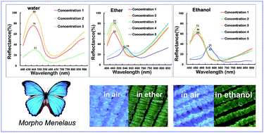
The reflectance spectrum was measured to detect the sensitivity of the Morpho menelaus butterfly to vapours of water, ether and ethanol. The incident light was emitted from a LS-1 tungsten halogen light source and transported by an optical fiber. Then, the light was irradiated on the scale samples. The reflected light was captured and inputted into Ocean Optics USB4000 spectrometer. The experimental device and its working principle are provided in the ESI. The reflective spectra of the Morpho menelaus butterfly scales in the presence of vapours of water, ether and ethanol were acquired and are shown in Fig. 4. The real concentrations of the vapours are provided in the ESI. Although the intensity of reflectance spectra of Morpho menelaus butterfly scales could be changed during the vapour concentration of water, ether and ethanol increase, the change quantity of the intensity of reflectance spectra is not same. It was shown that the structure of butterfly scales has highly spectrum selective response to different vapors.7,43 Molecules of water vapor enhanced the reflection. When the concentration of pneumatolytic water is added up to a certain level, a fog droplet is formed. Then, the structure displays hydrophobic behavior44 and the fog droplet acts to reduce the absorption of the probe light. The reflectivity is gradually reduced, as shown in the green curve in Fig. 4a. Vapor of ether and ethanol can not only make the reflection intensity increase, but also change the color variation of Morpho menelaus scales as shown in Fig. 4b and c. From the reflective spectra of the pneumatolytic ether and ethanol, it can be found that the pneumatolytic ether and ethanol can make reflection intensity changes increase. When the vapor concentration increases up to the point of forming a liquid, the reflective peak was red shifted (blue and purple curves in Fig. 4b and c) and the color of Morpho menelaus scales changed from blue to green (inset in Fig. 4b and c). It is indicated that the structure demonstrates high selectivity to pneumatolytic ether and ethanol before the two gases condense into a liquid.


image file: c4ra06117a-f4.tif
Fig. 4 The reflectance spectrum of Morpho menelaus butterfly wings with different vapours of water, ether and ethanol. Along with the increase in vapour concentration of pneumatolytic water, ether and ethanol, the butterfly scales show different spectral responses. In the reflection spectrum image of pneumatolytic (a) water, it can be found that along with the increase of concentration of water vapor, the reflection spectrum intensity increased firstly and then decreased; however, the peak position does not shift. As the vapour concentration increased for (b) ether and (c) ethanol, the reflection spectrum intensity increased at first and then it red shifted. At last, when the pneumatolytic ether and ethanol concentration increased and condensed to form liquids, the peak wavelength of reflection efficiency was at 548.45 nm and 547.19 nm, respectively, and the color of the scales turned green, as indicated by the arrow in the figure.

Models

2D optical models simplified from Morpho menelaus scales were designed to analyse the sensitivity mechanism of the nanostructure and are shown in Fig. 5a. Here, the models with three periods in vertical direction were used. The dimensions along the x-axis are infinite in this example.45,46 The lamellae of the model are symmetrically distributed on both sides of the trunk and are parallel to the base. The theory of thin-film multilayer interference of optical models was used to analyse the sensitivity mechanism of Morpho menelaus butterfly scales to ether and ethanol. Considering that the light is incident on the scale where interference occurred, the reflected light beams on the interfaces of the chitin and air may interfere with each other. If the constructive interference occurs, the light intensity will increase under conditions that the wavelength of the interference light is multiple of the half wavelength; however, if this is not the case, then it will reduce. It was viewed that the thin-film multilayer interference occurred in the periodic multilayer structure. The chitin and air layers were designated as x and y with thicknesses h1 and h2 and the refractive indices were set to n1 and n2, respectively, as shown in Fig. 5b. It is assumed that n1 > n2, in the present study.36 Here, the x layer is the material of chitin and the y layer is the surrounding medium. Thus, if the refractive index of the y layers was changed by surrounding media, the wavelength of reflective light will also change. It is the mechanism of the unparalleled sensitivity of photonic structures in Morpho menelaus butterfly wings to surrounding media. Considering the multilayer horizontal model layers, the phases of the reflected light are both at the upper and lower interfaces between chitin and air. The thin-film multilayer interference can be applied as follows:36
 
λ = 2(n1h1[thin space (1/6-em)]cos[thin space (1/6-em)]r1 + n2h2[thin space (1/6-em)]cos[thin space (1/6-em)]r2) (1)
where the angles of refraction in layers of chitin and air are r1 and r2, respectively. The wavelength of reflective light changes with the thickness “h”, refractive indices “n”, and the reflection angle “r”. Next, considering the visible spectrum effect, the lamellae height h1 and the height between lamellae of the nanostructure h2 of Morpho menelaus butterfly are measured. The calculated values and experimental results are shown in Table 1. It is obvious that the peak wavelength of the reflectance and transmittance spectra undergoes major changes when only the refractive indexes of the surrounding mediums parameter changes and all other parameters are unchanged. Therefore, this result proved the conclusion again that it is the change of refractive indexes of surrounding media, making butterfly wings have excellent sensitivity properties to surrounding media.

image file: c4ra06117a-f5.tif
Fig. 5 (a) The horizontal model simplified from Morpho menelaus butterfly and (b) configuration of multilayer-thin-film interference.
Table 1 Parameters and results of the calculationa
Butterfly Surrounding medium Incident angle r1 r2 h1 h2 n1 n2 λT λE
a Data used in eqn (1). The experimental reflective spectra of Morpho menelaus in air, ether and ethanol are obtained, respectively, as shown in Fig. 4. The wavelength of diffraction efficiency peak was named λE. λT = 2(n1h1[thin space (1/6-em)]cos[thin space (1/6-em)]r1 + n2h2[thin space (1/6-em)]cos[thin space (1/6-em)]r2), λE is diffraction efficiency peak of experimental data.
Morpho menelaus Air 0 0 0 80 110 1.555 1 468.80 463.77
Ether 0 0 0 80 110 1.555 1.349 546.90 548.45
Ethanol 0 0 0 80 110 1.555 1.362 548.44 547.19


Conclusions

The sensitive photonic crystal type nanostructures in Morpho menelaus butterfly wings were investigated by electron microscopy and reflectance spectroscopy in this study. This functional structures were composed of alternating layers of chitin and air. It was found that the blue color of ground scales is structure-based, and it was also found that its iridescent color has an angle-dependent discoloration effect. An unparalleled sensitivity of photonic structures in Morpho menelaus butterfly wings to different surrounding vapours was found. From the reflectance spectra it can be found that the wavelength and efficiency of the reflectance peak could change by altering the concentration and type of surrounding media. It was the surrounding medium that caused changes in the refractive index of the air layer in the thin-film multilayer nanostructure. The structural color of the scales would also change accordingly. In addition, the refractive indices of surrounding media in the thin-film multilayer nanostructure can affect the interference phenomenon. The refractive indexes of surrounding media plays an important role in gas sensitivity. More importantly, the calculation results from the theory of the thin-film multilayer nanostructure were consistent with the experimental results, which provides strong evidence for analyzing the sensitivity mechanism of butterfly scales to surrounding media. The sensitivity of the nanostructures of Morpho menelaus butterfly scales is meaningful. It bodes well for the discovery of new gas sensitive structural materials. Its structural parameters may provide the basis for various artificial manufacturing practical applications. This work paves a new pathway for extensive exploration of designing new optical gas sensors.

Acknowledgements

This work is supported by the National Natural Science Foundation of China (no. 51175220, 51325501, and 51290292), Science and Technology Development Project of Jilin Province (no. 20111808), and the Graduate Innovation Fund of Jilin University (no. 20121085).

Notes and references

  1. H. T. Ghiradella and M. W. Butler, J. R. Soc., Interface, 2009, 6, S243–S251 CrossRef PubMed.
  2. A. R. Parker, V. L. Welch, D. Driver and N. Martini, Nature, 2003, 426, 786–787 CrossRef CAS PubMed.
  3. S. Berthier, E. Charron and A. D. Silva, Opt. Commun., 2003, 228, 349–356 CrossRef CAS PubMed.
  4. Y. Ding, S. Xu and Z. L. Wang, J. Appl. Phys., 2009, 106, 074702 CrossRef PubMed.
  5. G. Subramania, Y. J. Lee, I. Brener, T. S. Luk and P. G. Clem, Opt. Express, 2007, 15, 13049–13057 CrossRef CAS.
  6. A. C. Arsenault, D. P. Puzzo, I. Manners and G. A. Ozin, Nat. Photonics, 2007, 1, 468–472 CrossRef CAS.
  7. A. H. G. Allogho, Opt. Commun., 2011, 284, 1656–1660 CrossRef PubMed.
  8. R. A. Potyrailo, H. Ghiradella, A. Vertiatchikh, K. Dovidenko, J. R. Cournoyer and E. Olson, Nat. Photonics, 2007, 1, 123–128 CrossRef CAS.
  9. W. Zhang, D. Zhang, T. X. Fan, J. J. Gu, J. Ding, H. Wang, Q. X. Guo and H. Ogawa, Chem. Mater., 2009, 21, 33–40 CrossRef.
  10. O. Sato, S. Kubu and Z. Z. Gu, Acc. Chem. Res., 2009, 42, 1–10 CrossRef CAS PubMed.
  11. M. Kolle, P. M. S. Cunha, M. R. J. Scherer, F. Huang, P. Vukusic, S. Mahajan, J. J. Baumberg and U. Steiner, Nat. Nanotechnol., 2010, 5, 511–515 CrossRef CAS PubMed.
  12. Y. Ding, S. Xu and Z. L. Wang, J. Appl. Phys., 2009, 106, 074702 CrossRef PubMed.
  13. Y. Chen, J. J. Gu, S. M. Zhu, T. X. Fan, D. Zhang and Q. X. Guo, Appl. Phys. Lett., 2009, 94, 053901 CrossRef PubMed.
  14. F. Liu, Y. P. Liu, L. Huang, X. H. Hu, B. Q. Dong, W. Z. Shi, Y. Q. Xie and X. Ye, Opt. Commun., 2011, 284, 2376–2381 CrossRef CAS PubMed.
  15. S. Banerjee, J. B. Cole and T. Yatagai, Micron, 2007, 38, 97–103 CrossRef PubMed.
  16. A. R. Parker, R. C. McPhedran, D. R. McKenzie, L. C. Botten and N. A. P. Nicorovici, Nature, 2001, 409, 36–37 CrossRef CAS PubMed.
  17. A. R. Parker, J. Opt. A: Pure Appl. Opt., 2000, 2, R15–R28 CrossRef.
  18. X. F. Yang, Z. C. Peng, H. B. Zuo, T. L. Shi and G. L. Liao, Sens. Actuators, A, 2011, 167, 367–373 CrossRef CAS PubMed.
  19. Z. W. Han, S. C. Niu, C. H. Shang, Z. N. Liu and L. Q. Ren, Nanoscale, 2012, 4, 2879–2883 RSC.
  20. Z. W. Han, S. C. Niu, M. Yang, J. Q. Zhang, W. Yin and L. Q. Ren, Nanoscale, 2013, 5, 8500–8506 RSC.
  21. Z. W. Han, S. C. Niu, W. Li and L. Q. Ren, Appl. Phys. Lett., 2013, 102, 233702 CrossRef PubMed.
  22. V. B. de Campos, Micron, 2011, 42, 801–807 CrossRef PubMed.
  23. J. Tang, S. M. Zhu, Z. X. Chen, C. L. Feng, Y. J. Shen, F. Yao, D. Zhang, W. J. Moon and D. M. Song, Mater. Chem. Phys., 2012, 131, 706–713 CrossRef CAS PubMed.
  24. L. P. Biró, K. Kertész, Z. Vértesy, G. I. Márk, Z. Bálint, V. Lousse and J. P. Vigneron, Mater. Sci. Eng., C, 2007, 27, 941–946 CrossRef PubMed.
  25. F. Liu, Y. P. Liu, L. Huang, X. H. Hu, B. Q. Dong, W. Z. Shi, Y. Q. Xie and X. Ye, Opt. Commun., 2011, 284, 2376–2381 CrossRef CAS PubMed.
  26. J. D. Joannopoulos, S. G. Johnson, J. N. Winn and R. D. Meade, Photonic Crystals: Molding the Flow of Light, Princeton University Press, Princeton, 2nd edn, 2008 Search PubMed.
  27. S. F. Mingaleev, M. Schillinger, D. Hermann and K. Busch, Opt. Lett., 2004, 29, 2858–2860 CrossRef.
  28. K. Srinivasan, P. E. Barclay, O. Painter, J. Chen, A. Y. Cho and C. Gmachl, Appl. Phys. Lett., 2003, 83, 1915–1917 CrossRef CAS PubMed.
  29. P. Gould, Mater. Today, 2002, 6, 32–37 CrossRef.
  30. R. C. Schroden, M. Al-Daous, C. F. Blanford and A. Stein, Chem. Mater., 2002, 14, 3305–3315 CrossRef CAS.
  31. H. Fudouzi and Y. Xia, Langmuir, 2003, 19, 9653–9660 CrossRef CAS.
  32. A. R. Parker and H. E. Townley, Nature, 2007, 2, 347–353 CAS.
  33. A. Saito, S. Yoshioka and S. Kinoshita, Proc. SPIE, 2004, 188–194 CrossRef PubMed.
  34. P. Vukusic and J. R. Sambles, Nature, 2003, 424, 852–855 CrossRef CAS PubMed.
  35. L. P. Biró, K. Kertésza and Z. Vértesy, Proc. SPIE, 2008, 7057, 1–6 CrossRef PubMed.
  36. W. J. Wu, G. L. Liao, T. L. Shi, R. Malik and C. Zeng, Microelectron. Eng., 2012, 95, 42–48 CrossRef CAS PubMed.
  37. S. Kinoshita and S. Yoshioka, Principles and Applications, 2005, pp. 113–140 Search PubMed.
  38. S. Kinoshita and S. Yoshioka, Forma, 2002, 17, 103–121 Search PubMed.
  39. L. Y. Wu, Z. W. Han, Z. M. Qiu, H. Y. Guan and L. Q. Ren, J. Bionic Eng., 2007, 4, 47–52 CrossRef.
  40. S. Kinoshita, S. Yoshioka and J. Miyazaki, Rep. Prog. Phys., 2008, 71, 076401 CrossRef.
  41. R. H. Siddique, S. Diewald, J. Leuthold and H. Hölscher, Opt. Express, 2013, 12, 14351–14361 CrossRef PubMed.
  42. K. Chung and J. H. Shin, J. Opt. Soc. Am. A, 2013, 30, 962–968 CrossRef CAS PubMed.
  43. R. A. Potyrailo, T. A. Starkey, P. Vukusic and H. Ghiradella, Proc. Natl. Acad. Sci. U. S. A., 2013, 110, 15567–15572 CrossRef CAS PubMed.
  44. S. H. Kang, T. Y. Tai and T. H. Fang, Curr. Appl. Phys., 2010, 10, 625–630 CrossRef PubMed.
  45. B. Gralak, G. Tayeb and S. Enoch, Opt. Express, 2001, 9, 567–578 CrossRef CAS.
  46. S. E. Mann, I. N. Miaoulis and P. Y. Wong, Opt. Eng., 2001, 40, 2061–2068 CrossRef PubMed.

Footnote

Electronic supplementary information (ESI) available. See DOI: 10.1039/c4ra06117a

This journal is © The Royal Society of Chemistry 2014
Click here to see how this site uses Cookies. View our privacy policy here.