Li Wenfenga,
Ma Hengchang*b and
Lei Ziqiang*c
aKey Laboratory of Eco-Environment-Related Polymer Materials of Ministry of Education, College of Chemistry and Chemical Engineering, Northwest Normal University, Lanzhou 730070, Gansu, China. E-mail: 498625374@qq.com
bKey Laboratory of Eco-Environment-Related Polymer Materials of Ministry of Education, College of Chemistry and Chemical Engineering, Northwest Normal University, Lanzhou 730070, Gansu, China. E-mail: mahczju@hotmail.com
cKey Laboratory of Eco-Environment-Related Polymer Materials of Ministry of Education, College of Chemistry and Chemical Engineering, Northwest Normal University, Lanzhou 730070, Gansu, China. E-mail: leizq@nwnu.edu.cn
First published on 13th August 2014
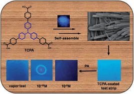
Detection of explosives is of utmost importance due to the threat to human security as a result of illegal transport and terrorist activities. Picric acid (PA) is widely used in the industrial and military fields, and is inadvertently able to contaminate the environment and groundwater, posing a threat to human health. Achieving the detection of explosives at the parts per billion (ppb) level using chemosensors is a great challenge. Herein, we demonstrate that triphenylamine based fluorescent chemosensors tris(4′-carbethoxybiphenyl) amine (TCEPA) and tris(4′-carboxybiphenyl) amine (TCPA) exhibit superior detection capability for PA in solution state at the ppb level (40 ppb and 5 ppb respectively) and efficient quenching behaviours in the vapor phase at room temperature. In addition, fluorescent test strips have been prepared by dip-coating Thin Layer Chromatography (TCL) for trace detection of PA at the attogram level.
Fluorescence signaling is one of the first choices among various methods used for detection of PA because of its high sensitivity, quick response and easy sample preparation. In the past few years, molecules based fluorescent sensors and modulated chemosensors specifically for PA have been reported. Yu and coworkers reported an optical fiber sensor for PA, designed on the basis of fluorescence energy transfer.10 S. Rajagopal group synthesized a variety of binuclear and tetranuclear complexes containing Re(I) and unearthed the first AIEE in metal-containing supramolecules for PA detection.11,12 Recently, M. Kumar and coworkers reported an (N,N-dimethylamino)cinnamaldehyde-based Hg2+ ensemble that behaves as a chemosensor for PA detection in organic media.13,14 Wang et al. have developed a colorimetric and fluorescent N-acylhydrazone based receptor for specific recognition of PA.15–17 P. S. Mukherjee group reported a variety of different types of π-electron-rich fluorophores functionalized with trimethylsilylethynyl and 3D supramolecular assemblies of metal–organic frameworks were designed for ppb level detection of PA.18,19 In addition to these, Tang, Fang and co-workers have explored fluorescent molecules in their aggregated and self-assembled states, respectively for the detection of PA among other analogues.20,21 Although there are numerous reports on fluorogenic chemosensors for PA, most detection systems are either less selective or exhibit low-to-moderate Stern–Volmer constants. As reported that due to the low vapor pressure of PA in comparison to other nitroderivatives, the vapor phase detection of PA still remains a great challenge. More than that the ppb level detection in organic solvents is also a crucial but very difficult task.
Triphenylamine (TPA) is the molecule of our favorite choice to develop chemosensors for PA detection. TPA derivatives, because of their rotatable C–C single bond and nonplanar electron-rich conjugated structure have attracted long-term interest for the formation of supramolecular self-assembled different aggregation states. Especially, the aggregation-induced emission enhancement (AIEE) offers TPA materials more diffusion channels for the exciton to migrate. Therefore, we design and synthesize two AIEE active carbonyl-based TPA derivatives tris(4′-carbethoxybiphenyl)amine (TCEPA) and tris(4′-carboxybiphenyl)amine (TCPA),22,23 selective and sensitive detection of PA.
![]() | ||
Scheme 1 Synthesis of TCEPA and TCPA, and the fluorescent image of TCEPA (a) TCPA (b) in the powder under 365 nm UV illumination. |
For vapor phase detection of PA by fluorescent paper strips, we kept the TCL strip over the mouth of the vial containing crystals of PA for 5 min. The area exposed to the vapors of PA exhibits quenching in fluorescence visible to naked eye.
Caution! The nitroaromatic compounds used in this study, specially picric acid, are very powerful explosives. They must be handled with care and also in very small quantities.
![]() | ||
Fig. 1 Normalized (a and b) absorption and (c and d) emission spectra of (a and c) TCEPA and (b and d) TCPA in varying concentrations and solvents. λex = 368 nm. |
The charge transfer (CT) state is generally sensitive to the exoteric microenvironment, so the fluorescent emission of TCEPA and TCPA may be tuned by the polarity of the solvent. From Fig. 1c and d, it is clearly demonstrate that the emission bands become broad and red-shifted significantly with increasing polarity of the solvent.25–28 For example, the emission maximum of TCPA exhibits a remarkable red-shift of 50 nm from THF (450 nm) to DMSO (500 nm). Such distinct red-shift and broadening of the emission in polar solvents indicate an ICT character for the excited state.29,30 In THF, TCEPA and TCPA exhibit strong blue emission at 455 and 450 nm with Stoke's shift of 85 and 80 nm respectively, which would de suitable for practical applications. The concentration-dependent emissive property demonstrates that the fluorescence quantum yields of TCEPA and TCPA are 6% and 18%.31 These notable differences in emission efficiency may arise from variable degrees of chromophore aggregation states.
Further, the concentration-dependent emission study of TCEPA and TCPA in THF displays a nonlinear increase in the emission intensity with an increase in the concentration, which supports the theory that emission enhancement is due to the aggregation mechanism instead of normal concentration dependent emission.32 We believe that rigidification of the molecule motion and restriction of the rotational relaxation of the excited state via nonradiative modes will occur when increasing of TCEPA and TCPA concentration, thus resulting in AIEE (Fig. 2).33–36
![]() | ||
Fig. 2 Concentration-dependant fluorescence spectra TCEPA (a) and TCPA (b) inset: non linear change in emission intensity with concentration of TCEPA and TCPA. |
In the fluorescence studies, the fluorescence titration for sensor TCEPA and TCPA with PA reveals that fluorescence emission intensity rapidly died down upon addition of increasing amounts of PA solution (Fig. 3a and b). Interestingly, the quenching efficiencies of TCEPA and TCPA toward PA are found to be 82% and 95% respectively. The appearance of intense yellow color upon addition of colorless THF solution of sensor TCEPA and TCPA to a pale yellow solution of PA in THF. This indicates the possibility of TCPA&PA complex formation.37 A similar observation could be also noted in the progress of the formation of TCEPA&PA (Fig. S1, ESI†).
The formation of aggregates of TCEPA and TCPA is supported by scanning electron microscopy (SEM) images in THF, which show the presence of flaky feathers (Fig. 4a) and divergent tubular morphology (Fig. 4c). However, in the presence of PA, the morphologies are changed into loose feathers (Fig. 4b) and short rods (Fig. 4d). These results demonstrate the interactions of TCEPA and TCPA with PA possibly through multiple hydrogen bonding led the break down of TCEPA and TCPA assembly morphology.
![]() | ||
Fig. 4 Scanning electron microscopy (SEM) images of aggregates of compounds TCEPA (a) and TCPA (c) in THF; SEM images of [TCEPA&PA] (b) and [TCPA&PA] (d). |
Fig. 5 shows the interaction of TCPA with PA investigated by 1H NMR spectroscopic titrations carried out in DMSO-d6. In the 1H NMR titration experiments, with the molar ratio of self-assembled TCEPA/TCPA and PA from 1:
1 to 1
:
35, the NMR spectra exhibit fast exchange between the PA and aggregated species. Upon increasing the molar ratio, significant downfield shifts are observed for the peaks corresponding to the phenolic hydroxy proton since the electron densities of the protons involved in H-bonds are decreased,38–40 at the same time signal of protons are extensive broadening. That is, hydrogen bonding is a crucial driving force for the fluorescence quenching process. Therefore, hydrogen-bond-mediated complex formation could be unambiguously demonstrated in DMSO-d6 (1H NMR titration of TCEPA and PA as shown in Fig. S2, ESI†).
The UV-vis absorption studies of TCEPA and TCPA with PA show the appearance of new band at 425 nm with formation of level-off tail in the visible region which further indicates interaction between TCEPA/TCPA and PA, thus, suggesting charge-transfer occurs between them in Fig. 6.41,42
To determine the detection limit, fluorescence titration of TCEPA and TCPA in THF mixture with PA is carried out by adding aliquots of PA solution and the fluorescence intensity as a function of PA added is then plotted. From Fig. 7a and b the concentration at which there was a sharp change in the fluorescence intensity multiplied with the concentration of TCEPA and TCPA gave the detection limit.43 Equation used for calculating detection limit: DL = CL × CT, (CL = concentrations of TCEPA and TCPA; CT = concentrations of Titrant at which change observed). Thus, the aggregates of TCEPA and TCPA possess detection limits of 40 ppb and 5 ppb respectively, as fluorescent sensors for PA.
![]() | ||
Fig. 7 (a and b) detection limit of PA about TCEPA and TCPA at 455 nm, (c and d) Stern–Volmer plots for sensors TCEPA and TCPA using PA as quencher at lower concentration. |
The quenching results can be quantitatively treated with the Stern–Volmer equation, I0/I = 1 + Ksv [PA],44 where I0 and I stand for the fluorescence intensity of TCEPA and TCPA aggregates in the absence and presence of PA, respectively, [PA] is concentrations of PA and Ksv is the Stern–Volmer constant. The decrease in the fluorescence intensity could be an electron transfer process on the basis of thermodynamic considerations as static and dynamic quenching. The Stern–Volmer plots showed two distinct regions: linear variation at a lower concentration of PA is mainly due to static quenching, whereas a steep curve at higher concentration of PA is presumably due to dynamic quenching.
It can be seen that the Stern–Volmer plot of the change in the intensity of aggregates of TCEPA and TCPA with respect to the PA concentration is linear at a lower concentration of PA (Fig. 7c and d) and gives a quenching constant45 (KSV) of 2.98 × 10⁻⁶ M−1 and 14.3 × 10⁻⁶ M−1, suggesting the quenching of the fluorescence of aggregates of the TCEPA and TCPA ensemble upon the addition of PA is ascribed to the static quenching through a one-to-one nonfluorescent ground state complex, which after excitation returns to the ground state without emission of light.46 Overall, the quenching constant and detection limit of the TCEPA and TCPA toward PA are noticeably higher than those reported in previous reports (as shown in Table S1, ESI†).
The selectivity of the sensors is investigated by using several aromatic/nonaromatic compounds as analytes upon addition analytes (100 equiv.) in THF into 2 mL of 1 × 10⁻⁵ M solution of sensor TCEPA (Fig. S3, ESI†) and TCPA (Fig. 8) in THF. Interestingly, the quenching efficiency of TCPA toward 35 equiv. of PA is found to be 95% indicating their effectiveness to detect PA over other aromatic compounds.
It is known that explosive particles can contaminate the human body, clothing, and other materials in the surroundings during preparation and packaging of explosive devices or during an explosion.47,48 In such cases, contact mode and vapor modes are necessary to check for residual contamination. For this purpose, test strips are prepared by transferring THF solution of TCEPA and TCPA onto Thin Layer Chromatography (TLC) by dip-coating method followed by drying under vacuum. The test strips are dipped into THF solutions of PA, and the fluorescence quenching is observed (Fig. 9b). In addition, a human thumb is rubbed with PA (caution: no direct contact, use a glove!), and then all visible PA particles are brushed off, followed by pressing the thumb against a test strip. The fingerprint of the thumb could be seen as quenched luminescence when illuminated with UV light (Fig. 9c). In another experiment, the contact-mode response to PA by the TCL strips is tested by placing PA crystals over a test strip for 5 min, resulting in black spots upon illumination with a UV lamp (Fig. 9d and e). To signify the vapor mode test, we perform a TCL test in vapor mode by placing fluorescent TCL strips over the top of the glass vial containing solid PA for 10 s at room temperature. The circular area that is exposed to the PA vapors is quenched (Fig. 9f). These images illustrate the utility of the TCEPA-coated test strips for the on-site instant visualization of trace residues of PA present on a specimen (Fig. S4, ESI†).
For detection of PA vapors, we expose the solution of aggregates of TCPA in THF to vapor of PA by inserting the vial containing solution into a sealed vial containing solid PA at room temperature. Nearly, 15% and 24% quenching efficiency for TCEPA (Fig. S5, ESI†) and TCPA (Fig. 10) solution in THF is observed within 10 s of exposure, which goes up to 39% and 42% quenching of emission intensity after 1 min at room temperature, indicating a fast response time. These results reveal that TCEPA and TCPA are responsive to PA in solution and vapor phase. Thus, TCEPA and TCPA behave as better PA sensors than many previous sensors reported in terms of selectivity, sensitivity and ease of vapor phase detection (Table S1, ESI†).
![]() | ||
Fig. 10 Time-dependent fluorescence quenching of TCPA upon PA vapor exposure. Inset shows the plot of change in emission intensity of TCPA on exposure to PA with time. |
We also check the effect of various concentrations of PA solution on the fluorescent TCL strip of TCPA (Fig. 11) and TCEPA (Fig. S6, ESI†) by applying small spots of different concentrations of PA (10 μL) on test strips. The visual fluorescence response of PA at different concentrations by contact mode detection on TCL test strips of TCPA is shown in Fig. 11. Dark spots of different strengths can be observed, which show the regulation of the quenching behavior of PA (Fig. 11(II)–(VII)), which is also practically applicable by varying the concentration of PA even up to 100 femtomolar, i.e., 230 × 10−18 gram (230 attogram) (Fig. 11VII). However, no visible change is observed by applying blank solvent (THF) over the fluorescent paper strips (Fig. 11I).
The emission spectra of quenched area on the fluorescent test strips of TCPA (Fig. 12) and TCEPA (Fig. S7, ESI†) were recorded using front face technique after applying the spots of PA (10 μL) of varying concentration. The emission spectra of test strips of TCEPA and TCPA exhibit 18 and 33% quenching respectively, with 10−13 M solution of PA. However, no effect of blank solvent is observed on the emission intensity of fluorescent test strips of TCPA and TCEPA. These results show the practical applicability of TCEPA and TCPA toward instant and economical detection of traces of PA up to attogram level by naked eye.
Footnote |
† Electronic supplementary information (ESI) available. See DOI: 10.1039/c4ra05843g |
This journal is © The Royal Society of Chemistry 2014 |